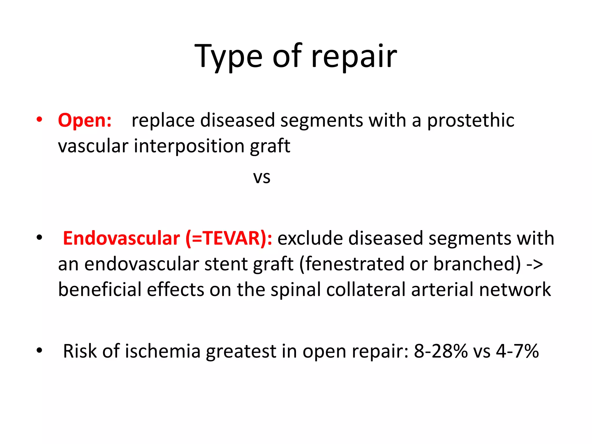
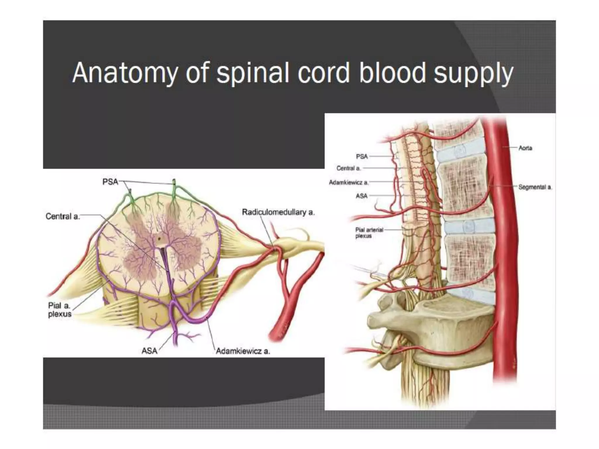
Spinal cord protection in
aortic surgeries
E. Thanigai arasu
PG in DM-Cardiac anaesthesia
Introduction
• The most feared complication following aortic surgeries –
persistent neurological deficit following Spinal cord
ischemia(SCI).
• Most common in TAAA (Mid-thoracic)
• Not common in AAA surgeries.(<1%)
Scott DJ, Denton MJ. Spinal cord protection in aortic endovascular surgery. Br J Anaesth 2016;117:26-31.
Type of repair
• Open: replace diseased segments with a prostethic
vascular interposition graft
vs
• Endovascular (=TEVAR): exclude diseased segments with
an endovascular stent graft (fenestrated or branched) ->
beneficial effects on the spinal collateral arterial network
• Risk of ischemia greatest in open repair: 8-28% vs 4-7%
Patho-physiology
• The temporary interruption of distal aortic perfusion,
• Sacrifice of segmental arteries during repair – central
events in the pathogenesis.
• Anatomy of spinal cord
• Auto regulation
• Spinal blood flow after aortic cross clamping
Anatomy
 Watershed region- Thoraco lumbar segment.
 Blood supply derived from large radicular arteries called
ARM (A of Adamkiewicz)
 Origin
T9-T12 – in 75%
T8-L3 – in 15%
L1-L2 – in 10% of patients.
• The anterior spinal artery is only one component of an
extensive paraspinous and intraspinal collateral vascular
network.
• the collateral system involves - extensive axial arterial
network in the spinal canal, the paravertebral tissues,
and the paraspinous muscles,
in which vessels anastomose with one another
and with the nutrient arteries of the spinal cord.
• The segmental arteries -3 major vessel groups.
• 1st group- intra-thecal vessels: ASA and a longitudinal
chain of epidural arcades between the spinal cord and
the vertebral bodies,
• 2nd - group of interconnecting vessels - outside the spinal
canal along the dorsal processes of the vertebral bodies,
• 3rd is a massive collection of interconnecting vessels
supplying the paraspinous muscles, including the
iliopsoas anteriorly and the erector spinae posteriorly.
Etz CD, Kari FA, Mueller CS, Silovitz D, Brenner R, Lin HM, et al. The Collateral Network Concept: A Reassessment of the
Anatomy of Spinal Cord perfusion. J Thorac Cardiovasc Surg. 2011;141:1020-28.
Auto regulation
• Neurolgical deficit
• classified as immediate or delayed.
• Immediate-direct result of hypo-perfusion and secondary
hypoxic damage.
• Delayed complications can develop between 1 & 21 days
following surgery.
• Results from reperfusion hyperemia and free radical
generation – edema of the cord –regional hypoperfusion
Wan IYP, Angelini GD, Bryan AJ, Ryder I, Underwood MJ. prevention of spinal cord ischemia during descending thoracic and
thoracoabdominal surgery. Eur J Cardio-thorac Surg 2001;19:203-13.
Svensson attributed the major cause of spinal injury is
1. The duration & degree of ischemia
2. Failure to re-establish blood flow
3. Bio-chemically mediated reperfusion injury.
svensson LG. New and future approaches for spinal cord protection, Semin Thorac Cardiovasc Surg. 1997;9:206-21.
Prevention of spinal cord injury
1) Minimize the spinal cord ischemia
2) Increase the tolerance to ischemia
3) Augmentation of perfusion
4) Early detection of ischemia
• Studies demonstrated that the duration aortic cross
clamping is a major determinant.
• < 15min -0% to 25-100% if exceeds 60min
• So different surgical approaches have been developed to
shorten the time.
i. Crawford aortic inlay technique
ii. Single clamp repair technique
iii. Sequential aortic clamping technique
iv. Staged repair( dynamic collateral vascular network)
Minimize the spinal cord ischemia
Crawford ES, Palemane AE, Saleh SA, Roebm JOE. Aortic aneursym: current status of surgical treatment.
Surg Clin North Am 1979;59:597-636
Cooley DA. Single-clamp repair of aneursyms of the descending thoracic aorta. Semin Thorac Cardiovasc Surg
1988;10:87-90.
• TEVAR- disruption of inter-costal arteries minimised –
lower risk of SCI
STS guidelines for DTA –
 >5.5cm - with significant co-morbid (class IIa LOE B)
 without co-morbid- class IIb LOE C
 <5.5cm – against TEVAR ( class III LOE C)
In setting of TAAA
• In patients with severe co-morbid class
• aortic arch with distal extensions IIB LOE C
• Reasonable surgical risk - aortic arch involvement
with distal extensions (class III LOE A)
• High quality RCT s comparing Open vs Endovascular AAA
repair demonstrated that significant peri-operative
survival benefit due to EVAR
• with no significant difference in mid-long term mortality.
• ultimately the decision for the individual pts depends on
multiple factors such as
 aortic anatomy,(Hostile proximal neck etc..)
 urgency,
 pt preference,
 surgical expertise.
Reduction of severity of ischemia
Distal aortic perfusion
• 5-6mm in Diameter
• Polyurethane PV coated with heparin bonded non-
thrombogenic material
• At pressure gradient 60mmHg- 50% of CO
• Small diameter- Resistance significantly increases
• Adv- simple, low cost, partial anticoagulation
• Disadvantage- vessel injury, dislodgement, bleeding,
athero-embolism.
Gott VF, Whiffen JD, Dutton RC.Heparin bonding on colooidal graphite surfaces. Science
1963;142:1287.
• LAFA bypass – better control of both proximal & distal
perfusion.
• Roller or centrifugal pump.
• With the use of the pump distal aortic perfusion 60-
70mmHg -71.4% CO.
• Advantage – control of aortic pressure, temp, distal
aortic perfusion, selective antegrade perfusion.
• Disadvantage – expense, complexity, anti-coagulation.
Schepens MAAM, Vermuelen FE, Morshusis WJ, Dossehe KM. Impact of left heart bypass on the results of
thoroacoabdominal aortic aneursym repair. Ann Thorac Surg 1999;67:1931034.
Increase the tolerance
• Hypothermia – one of the most effective methods – to
protect the neural tissue-ischemia
• By O2 demand, BMR, inhibit the release of neuro
transmitter (Glutamate), cell membrane stabilization.
• Mild hypothermia can confer a marked protective effect
on the spinal cord
• Intraop hypothermia can prevent delayed ischemic SCI.
• Moderate hypothermia(32-34*) during TAAA repair –
class IIa LOE B
Nakashima K, Todd MM, Warner DS. The relationship between cerebtal metabolic rate and ischemjuc depolarization. a
comparison of the effects of hypothermia, pentobarbital and isoflurane. Anaesthesilogy 1995;82:1199-1208.
• CPB with DHCA (12-14*) – when TAAA involves distal
aortic arch – precludes cross clamp site.
• Methyl-prednisolone & thiopental can be given during
cooling.
• TEE monitoring needed to look for AR.
• Acceptable peri-operative outcomes for major
reconstructions of TAA
• Disadvantage –limited safe period, risk of stroke,
bleeding, post-op enchepalopathy.
• Kouchoukos et al reported 8% 30 day mortality with
neurological deficit of 2.8%.
• However the use of CPB necessitates full heparinisation –
CSF drainage and intrathecal manoeuvers hazardous due
to bleeding.
Kouchoukos NT, Rokkas CK. Hypothermic Cardiopulmonary bypass for spinal cord protection: rationale and clinical
results. Ann Thorac Surg 1999;67:1940-42.
• Regional hypothermia – the principal component is-
Epidural catheter placed @ T10-T12 level – infusion of ice
NS 4°C
• To achieve spinal cord temp of 26-28°C.
• Second catheter into the subarachnoid space @ L3-L4
space to measure the CSF temp and allow drainage.
• With the above technique 100 pts with type I-III TAA
operated with the complication rate of 3.5%.
Cambria R, Davison J. Regional hypothermia for prevention of spinal cord ischemic complications after
thoracoabdominal aortic surgery. Experirnce with epidural cooling. Semin Thorac Cardiovasc Surg 1998;10:61-65.
• Riluzole, Memantine.
• Ca channel antagonist
• SOD-PEG
Augmentation of perfusion
• The optimization of SCPP for spinal cord protection –
class IIa LOE B.
• Done by either arterial pressure augmentation or CSF
drainage. (SCPP = MAP-CSF pr).
• SCPP should maintain 70-80mmHg -that is MAP around
80-90mmHg.
• Because SCI often involves thoraco-lumbar cord it often
accompanied by significant sympathectomy –spinal
vasodilatatory shock
• Early intervention to treat hypotension-with
vasopressors.
• Therapeutic hypertension after TAAA must be weighted
against the risk of arterial bleeding.
• Controversies exist regarding re implantation of
segmental arteries
• In view of increase cross clamp time.
• However a large no of intercostal & lumbar arteries
oversewn – risk is high.
• Re attachment of segmental arteries especially T6-L2 -
important even if the cross clamp is prolonged.
• Svensson et al, studied 99 pts & found if oversewn – 37%,
if re attched -6%.
• However no RCTS showed significant reduction.
Svensson LG, Hess KR, Coselli JS, Influence of segmental arteries, extent, and atriofemoral bypass on postoperative
paralegia after thoracoabdominal aortic perations. J Vasc Surg 1994;20:255-62.
CSF draiange
• Class I recommendation LOE B.
• Silicon elastomer ventriculostomy catheter via 14G tuohy
needle L3-L4 space. Depth 8-10cm.
• Or Epidural catheter.
• There is no clear advantage to one over other with
silastic catheter soft, larger bore, but more prone for
kinking breaking.
• Either choice should involve cath with multiple distal
orifice –mimize risk of obstruction.
McCullough JL. Hollier LH, Nugent M. Paraplegia after thoracic aortic oclusion: influence of cerebrospinal fluid drainage. J Vasc
Surg 1988;7:153-60
• The open end of catheter is attached to sterile reservoir.
• CSF is drained when the pressure exceeds
> 10mmHg @ the rate of 8-10ml/hr.
• It should be inserted before or at the time of surgery and
continued upto 24hrs after surgery.
• Subsequently capped and left in place for next 24 hrs.
After normal neurological examinations and adequate
coagulation catheter can be removed
Early detection of SCI
• Neurophysiologic monitoring of spinal cord – for the
early diagnosis of SCI - class IIb LOE B.
• This management strategy may prevent immediate onset
paraplegia.
• The primary injury during surgery & aortic cross clamp is
hypoxic damage-neurons.
• The conduction of nerve impulses is – highly sensitive.
• Stimulating electrodes-skin adjacent to peripheral nerves
in arms or legs.
• Record the evoked potentials lumbar/brachial plexus,
spinal cord, brainstem, thalamus, cortex.
• The amplitude & latency of this is affected by disruption
of blood supply.
• This concept has led to the development of SSEP.
• SSEP amplitude 40% after cross clamp with
hypotension for 3-4 min.
• If this allowed to persist – flat line response –absence of
conduction.
• Practically – posterior tibial nerve- scalp.
• Before cross clamp serves as baseline
• Tracings are recorded at every 2 min intervals.
• The potentials are amplified by 10,000 times & each
trace is avg of 200 consecutive stimulus
• A 10% increase in the signal latency usually precedes a
decrease in amplitude & its ˜ decrease in the perfusion
pressure.
• The duration of ischemia required to shift the latency
10% from its baseline is defined as L10time.
• Indicating the need to re-establish perfusion in 4-6min.
• Type 1 response- ↓amplitude & ↑latency 3-5min after
cross clamp-failure to provide adequate perfusion
pressure of up to 60mmHg distal to the clamp
• Type II response – SSEP is maintained through out the
period-adequate PP –critical segments are not located.
• Type III response – sudden loss of sensory conduction –
critical arteries are located- this is in indication for re-
implantation.
• Type IV response – gradual fadeout of normal tracings in
30-50 min – marginal distal perfusion even in the
presence of bypass with acceptable perfusion pressure –
profound vasodilatation, extensive
aneurysmal disease, failure of retrograde perfusion.
• Clinical studies have shown that SSEP is sensitive & offered an
improvement in surgical strategy.
• But only records the activity of posterior & lateral columns.
• Fails to represent the function of anterior spinal cord which is
supplied by single ASA.
• The anterior CST is the critical area which is affected by
ischemic insult.
• The specificity of SSEP is low with False positive rate of 67%
Guerit JM, Verheist R, Rubay J, Dion R. Multilelevel somatosensory evoked potential for spinal cord monitoring in
descending thoracic and thoracoabdominal aortic surgery. Eur J Cardio-thorac Surg 1996;10:93-103.
• In view of the above disadvantages the use of MEP has
been proposed.
• MEPs are elicited either trans-cranially or by cord
directly.
• Motor responses can be recorded at the level of
cord(spinal MEP), the nerve (neurogenic MEP), or the
muscle (myogenic MEP)
• Experimental studies suggest myogenic MEP may be
sensitive in predicting paraplegia.
• However pre-op anaesthetic planning is necessary as
most volatile anaesthetics will depress myogenic MEP.
• NMBD can affect the amplitude.
• The other shortcoming is axonal conduction is resistant
to ischemia and the dis-appearance of MEP is slow after
blood flow interrupted.
• Paraplegia caused by SCI significantly dampens lower
extremity evoked potentials as compared with the upper
extremity
De Hann P, Kalkmann CJ, de Mol BA. Efficacy of transcranial motor evoked myogenic potentials to detect spinal cord ischemia during
operations for thoracoabdominal aneurysms. J Thorac Cardiovasc Surg 1997;113:87-100.
• Intraoperative comparison of upper and lower extremity
evoked potentials distinguishes SCI from the generalized
effects of anesthetics, hypothermia, and/or electrical
interference .
• A recent study compared both MEPs and SSEPs for spinal
cord monitoring TAAA repairs. Both monitoring
modalities - nearly 90% correlation for spinal cord
infarction as well as a 98% negative predictive value for
immediate-onset paraplegia.
• Furthermore, reversible changes in MEPs and SSEPs had
no correlation with permanent paraplegia.
Keyhani K, Miller CC, Estrera AL, et al. Analysis of motor and somatosensory evoked potentials during thoracic and
thoracoabdominal aortic aneurysm repair. J Vasc Surg 2009;49:36-41.
• In summary, despite the theoretic advantages of MEPs
for monitoring the at-risk anterior spinal columns,
In practice, data suggest that SSEPs alone
suffice for clinical purposes, and that MEPs did not add
significantly to clinical management.
• Serial post-op neurologic examination should be started
early.
• Diagnosis should trigger a protocol for spinal cord rescue.
Field of research
• Polarographic techniques- spinal cord oxygenation.
• Oxygen content monitoring
which are sensitive to detect cord ischemia- still
limited to the field of research
Identification of critical segmental
arteries
• Because of anatomic diversity of spinal cord supply its
better to identify critical segmental arteries by preop
angiography.
• The major problem of this method is direct injection toxic
contrast via the ARM which may itself paraplegia.
Use smaller volume
less toxic agents.
• Recently MRA has been proposed as an alternative
non invasive method.
Fereshatian A, Kadir S, Kaufman SL. Digital subtraction spinal cord angiography in patients undergoing thoracic aneurysm
surgery. Cardiovasc Intervent Radiol 1989;12:7-9.
• Hydrogen Mapping method.-
A technique which allows intra-op identification
of segmental arteries based on hydrogen is dissolved in
solution with the production weak current when in
contact with platinum
• H2 –radicular artery supplying spinal cord carried to ASA.
– will pass through the membrane & wall of the artery.
This is then detected by platinum electrode placed
alongside spinal cord.
Svensson LG,Patel V, Coselli JS, Crawford ES. Preliminary report of localization of spinal cord blood supply by hydrogen
during aortic operations. Ann Thorac Surg 1990;49:528-36.
Spinal cord protection in aortic surgeries

Spinal cord protection in aortic surgeries

  • 1.
    Spinal cord protectionin aortic surgeries E. Thanigai arasu PG in DM-Cardiac anaesthesia
  • 2.
    Introduction • The mostfeared complication following aortic surgeries – persistent neurological deficit following Spinal cord ischemia(SCI). • Most common in TAAA (Mid-thoracic) • Not common in AAA surgeries.(<1%)
  • 3.
    Scott DJ, DentonMJ. Spinal cord protection in aortic endovascular surgery. Br J Anaesth 2016;117:26-31.
  • 5.
    Type of repair •Open: replace diseased segments with a prostethic vascular interposition graft vs • Endovascular (=TEVAR): exclude diseased segments with an endovascular stent graft (fenestrated or branched) -> beneficial effects on the spinal collateral arterial network • Risk of ischemia greatest in open repair: 8-28% vs 4-7%
  • 6.
    Patho-physiology • The temporaryinterruption of distal aortic perfusion, • Sacrifice of segmental arteries during repair – central events in the pathogenesis. • Anatomy of spinal cord • Auto regulation • Spinal blood flow after aortic cross clamping
  • 7.
  • 9.
     Watershed region-Thoraco lumbar segment.  Blood supply derived from large radicular arteries called ARM (A of Adamkiewicz)  Origin T9-T12 – in 75% T8-L3 – in 15% L1-L2 – in 10% of patients.
  • 11.
    • The anteriorspinal artery is only one component of an extensive paraspinous and intraspinal collateral vascular network. • the collateral system involves - extensive axial arterial network in the spinal canal, the paravertebral tissues, and the paraspinous muscles, in which vessels anastomose with one another and with the nutrient arteries of the spinal cord.
  • 12.
    • The segmentalarteries -3 major vessel groups. • 1st group- intra-thecal vessels: ASA and a longitudinal chain of epidural arcades between the spinal cord and the vertebral bodies, • 2nd - group of interconnecting vessels - outside the spinal canal along the dorsal processes of the vertebral bodies, • 3rd is a massive collection of interconnecting vessels supplying the paraspinous muscles, including the iliopsoas anteriorly and the erector spinae posteriorly. Etz CD, Kari FA, Mueller CS, Silovitz D, Brenner R, Lin HM, et al. The Collateral Network Concept: A Reassessment of the Anatomy of Spinal Cord perfusion. J Thorac Cardiovasc Surg. 2011;141:1020-28.
  • 13.
  • 15.
    • Neurolgical deficit •classified as immediate or delayed. • Immediate-direct result of hypo-perfusion and secondary hypoxic damage. • Delayed complications can develop between 1 & 21 days following surgery. • Results from reperfusion hyperemia and free radical generation – edema of the cord –regional hypoperfusion Wan IYP, Angelini GD, Bryan AJ, Ryder I, Underwood MJ. prevention of spinal cord ischemia during descending thoracic and thoracoabdominal surgery. Eur J Cardio-thorac Surg 2001;19:203-13.
  • 16.
    Svensson attributed themajor cause of spinal injury is 1. The duration & degree of ischemia 2. Failure to re-establish blood flow 3. Bio-chemically mediated reperfusion injury. svensson LG. New and future approaches for spinal cord protection, Semin Thorac Cardiovasc Surg. 1997;9:206-21.
  • 17.
    Prevention of spinalcord injury 1) Minimize the spinal cord ischemia 2) Increase the tolerance to ischemia 3) Augmentation of perfusion 4) Early detection of ischemia
  • 18.
    • Studies demonstratedthat the duration aortic cross clamping is a major determinant. • < 15min -0% to 25-100% if exceeds 60min • So different surgical approaches have been developed to shorten the time. i. Crawford aortic inlay technique ii. Single clamp repair technique iii. Sequential aortic clamping technique iv. Staged repair( dynamic collateral vascular network) Minimize the spinal cord ischemia Crawford ES, Palemane AE, Saleh SA, Roebm JOE. Aortic aneursym: current status of surgical treatment. Surg Clin North Am 1979;59:597-636 Cooley DA. Single-clamp repair of aneursyms of the descending thoracic aorta. Semin Thorac Cardiovasc Surg 1988;10:87-90.
  • 19.
    • TEVAR- disruptionof inter-costal arteries minimised – lower risk of SCI STS guidelines for DTA –  >5.5cm - with significant co-morbid (class IIa LOE B)  without co-morbid- class IIb LOE C  <5.5cm – against TEVAR ( class III LOE C) In setting of TAAA • In patients with severe co-morbid class • aortic arch with distal extensions IIB LOE C • Reasonable surgical risk - aortic arch involvement with distal extensions (class III LOE A)
  • 20.
    • High qualityRCT s comparing Open vs Endovascular AAA repair demonstrated that significant peri-operative survival benefit due to EVAR • with no significant difference in mid-long term mortality. • ultimately the decision for the individual pts depends on multiple factors such as  aortic anatomy,(Hostile proximal neck etc..)  urgency,  pt preference,  surgical expertise.
  • 21.
    Reduction of severityof ischemia Distal aortic perfusion
  • 22.
    • 5-6mm inDiameter • Polyurethane PV coated with heparin bonded non- thrombogenic material • At pressure gradient 60mmHg- 50% of CO • Small diameter- Resistance significantly increases • Adv- simple, low cost, partial anticoagulation • Disadvantage- vessel injury, dislodgement, bleeding, athero-embolism. Gott VF, Whiffen JD, Dutton RC.Heparin bonding on colooidal graphite surfaces. Science 1963;142:1287.
  • 24.
    • LAFA bypass– better control of both proximal & distal perfusion. • Roller or centrifugal pump. • With the use of the pump distal aortic perfusion 60- 70mmHg -71.4% CO. • Advantage – control of aortic pressure, temp, distal aortic perfusion, selective antegrade perfusion. • Disadvantage – expense, complexity, anti-coagulation. Schepens MAAM, Vermuelen FE, Morshusis WJ, Dossehe KM. Impact of left heart bypass on the results of thoroacoabdominal aortic aneursym repair. Ann Thorac Surg 1999;67:1931034.
  • 25.
    Increase the tolerance •Hypothermia – one of the most effective methods – to protect the neural tissue-ischemia • By O2 demand, BMR, inhibit the release of neuro transmitter (Glutamate), cell membrane stabilization. • Mild hypothermia can confer a marked protective effect on the spinal cord • Intraop hypothermia can prevent delayed ischemic SCI. • Moderate hypothermia(32-34*) during TAAA repair – class IIa LOE B Nakashima K, Todd MM, Warner DS. The relationship between cerebtal metabolic rate and ischemjuc depolarization. a comparison of the effects of hypothermia, pentobarbital and isoflurane. Anaesthesilogy 1995;82:1199-1208.
  • 26.
    • CPB withDHCA (12-14*) – when TAAA involves distal aortic arch – precludes cross clamp site. • Methyl-prednisolone & thiopental can be given during cooling. • TEE monitoring needed to look for AR. • Acceptable peri-operative outcomes for major reconstructions of TAA
  • 27.
    • Disadvantage –limitedsafe period, risk of stroke, bleeding, post-op enchepalopathy. • Kouchoukos et al reported 8% 30 day mortality with neurological deficit of 2.8%. • However the use of CPB necessitates full heparinisation – CSF drainage and intrathecal manoeuvers hazardous due to bleeding. Kouchoukos NT, Rokkas CK. Hypothermic Cardiopulmonary bypass for spinal cord protection: rationale and clinical results. Ann Thorac Surg 1999;67:1940-42.
  • 28.
    • Regional hypothermia– the principal component is- Epidural catheter placed @ T10-T12 level – infusion of ice NS 4°C • To achieve spinal cord temp of 26-28°C. • Second catheter into the subarachnoid space @ L3-L4 space to measure the CSF temp and allow drainage. • With the above technique 100 pts with type I-III TAA operated with the complication rate of 3.5%. Cambria R, Davison J. Regional hypothermia for prevention of spinal cord ischemic complications after thoracoabdominal aortic surgery. Experirnce with epidural cooling. Semin Thorac Cardiovasc Surg 1998;10:61-65.
  • 30.
    • Riluzole, Memantine. •Ca channel antagonist • SOD-PEG
  • 31.
    Augmentation of perfusion •The optimization of SCPP for spinal cord protection – class IIa LOE B. • Done by either arterial pressure augmentation or CSF drainage. (SCPP = MAP-CSF pr). • SCPP should maintain 70-80mmHg -that is MAP around 80-90mmHg. • Because SCI often involves thoraco-lumbar cord it often accompanied by significant sympathectomy –spinal vasodilatatory shock
  • 32.
    • Early interventionto treat hypotension-with vasopressors. • Therapeutic hypertension after TAAA must be weighted against the risk of arterial bleeding. • Controversies exist regarding re implantation of segmental arteries • In view of increase cross clamp time.
  • 33.
    • However alarge no of intercostal & lumbar arteries oversewn – risk is high. • Re attachment of segmental arteries especially T6-L2 - important even if the cross clamp is prolonged. • Svensson et al, studied 99 pts & found if oversewn – 37%, if re attched -6%. • However no RCTS showed significant reduction. Svensson LG, Hess KR, Coselli JS, Influence of segmental arteries, extent, and atriofemoral bypass on postoperative paralegia after thoracoabdominal aortic perations. J Vasc Surg 1994;20:255-62.
  • 34.
    CSF draiange • ClassI recommendation LOE B. • Silicon elastomer ventriculostomy catheter via 14G tuohy needle L3-L4 space. Depth 8-10cm. • Or Epidural catheter. • There is no clear advantage to one over other with silastic catheter soft, larger bore, but more prone for kinking breaking. • Either choice should involve cath with multiple distal orifice –mimize risk of obstruction. McCullough JL. Hollier LH, Nugent M. Paraplegia after thoracic aortic oclusion: influence of cerebrospinal fluid drainage. J Vasc Surg 1988;7:153-60
  • 35.
    • The openend of catheter is attached to sterile reservoir. • CSF is drained when the pressure exceeds > 10mmHg @ the rate of 8-10ml/hr. • It should be inserted before or at the time of surgery and continued upto 24hrs after surgery. • Subsequently capped and left in place for next 24 hrs. After normal neurological examinations and adequate coagulation catheter can be removed
  • 36.
    Early detection ofSCI • Neurophysiologic monitoring of spinal cord – for the early diagnosis of SCI - class IIb LOE B. • This management strategy may prevent immediate onset paraplegia. • The primary injury during surgery & aortic cross clamp is hypoxic damage-neurons. • The conduction of nerve impulses is – highly sensitive.
  • 37.
    • Stimulating electrodes-skinadjacent to peripheral nerves in arms or legs. • Record the evoked potentials lumbar/brachial plexus, spinal cord, brainstem, thalamus, cortex. • The amplitude & latency of this is affected by disruption of blood supply. • This concept has led to the development of SSEP. • SSEP amplitude 40% after cross clamp with hypotension for 3-4 min.
  • 38.
    • If thisallowed to persist – flat line response –absence of conduction. • Practically – posterior tibial nerve- scalp. • Before cross clamp serves as baseline • Tracings are recorded at every 2 min intervals. • The potentials are amplified by 10,000 times & each trace is avg of 200 consecutive stimulus
  • 39.
    • A 10%increase in the signal latency usually precedes a decrease in amplitude & its ˜ decrease in the perfusion pressure. • The duration of ischemia required to shift the latency 10% from its baseline is defined as L10time. • Indicating the need to re-establish perfusion in 4-6min. • Type 1 response- ↓amplitude & ↑latency 3-5min after cross clamp-failure to provide adequate perfusion pressure of up to 60mmHg distal to the clamp
  • 40.
    • Type IIresponse – SSEP is maintained through out the period-adequate PP –critical segments are not located. • Type III response – sudden loss of sensory conduction – critical arteries are located- this is in indication for re- implantation. • Type IV response – gradual fadeout of normal tracings in 30-50 min – marginal distal perfusion even in the presence of bypass with acceptable perfusion pressure – profound vasodilatation, extensive aneurysmal disease, failure of retrograde perfusion.
  • 41.
    • Clinical studieshave shown that SSEP is sensitive & offered an improvement in surgical strategy. • But only records the activity of posterior & lateral columns. • Fails to represent the function of anterior spinal cord which is supplied by single ASA. • The anterior CST is the critical area which is affected by ischemic insult. • The specificity of SSEP is low with False positive rate of 67% Guerit JM, Verheist R, Rubay J, Dion R. Multilelevel somatosensory evoked potential for spinal cord monitoring in descending thoracic and thoracoabdominal aortic surgery. Eur J Cardio-thorac Surg 1996;10:93-103.
  • 42.
    • In viewof the above disadvantages the use of MEP has been proposed. • MEPs are elicited either trans-cranially or by cord directly. • Motor responses can be recorded at the level of cord(spinal MEP), the nerve (neurogenic MEP), or the muscle (myogenic MEP) • Experimental studies suggest myogenic MEP may be sensitive in predicting paraplegia.
  • 43.
    • However pre-opanaesthetic planning is necessary as most volatile anaesthetics will depress myogenic MEP. • NMBD can affect the amplitude. • The other shortcoming is axonal conduction is resistant to ischemia and the dis-appearance of MEP is slow after blood flow interrupted. • Paraplegia caused by SCI significantly dampens lower extremity evoked potentials as compared with the upper extremity De Hann P, Kalkmann CJ, de Mol BA. Efficacy of transcranial motor evoked myogenic potentials to detect spinal cord ischemia during operations for thoracoabdominal aneurysms. J Thorac Cardiovasc Surg 1997;113:87-100.
  • 44.
    • Intraoperative comparisonof upper and lower extremity evoked potentials distinguishes SCI from the generalized effects of anesthetics, hypothermia, and/or electrical interference . • A recent study compared both MEPs and SSEPs for spinal cord monitoring TAAA repairs. Both monitoring modalities - nearly 90% correlation for spinal cord infarction as well as a 98% negative predictive value for immediate-onset paraplegia. • Furthermore, reversible changes in MEPs and SSEPs had no correlation with permanent paraplegia. Keyhani K, Miller CC, Estrera AL, et al. Analysis of motor and somatosensory evoked potentials during thoracic and thoracoabdominal aortic aneurysm repair. J Vasc Surg 2009;49:36-41.
  • 45.
    • In summary,despite the theoretic advantages of MEPs for monitoring the at-risk anterior spinal columns, In practice, data suggest that SSEPs alone suffice for clinical purposes, and that MEPs did not add significantly to clinical management. • Serial post-op neurologic examination should be started early. • Diagnosis should trigger a protocol for spinal cord rescue.
  • 47.
    Field of research •Polarographic techniques- spinal cord oxygenation. • Oxygen content monitoring which are sensitive to detect cord ischemia- still limited to the field of research
  • 48.
    Identification of criticalsegmental arteries • Because of anatomic diversity of spinal cord supply its better to identify critical segmental arteries by preop angiography. • The major problem of this method is direct injection toxic contrast via the ARM which may itself paraplegia. Use smaller volume less toxic agents. • Recently MRA has been proposed as an alternative non invasive method. Fereshatian A, Kadir S, Kaufman SL. Digital subtraction spinal cord angiography in patients undergoing thoracic aneurysm surgery. Cardiovasc Intervent Radiol 1989;12:7-9.
  • 49.
    • Hydrogen Mappingmethod.- A technique which allows intra-op identification of segmental arteries based on hydrogen is dissolved in solution with the production weak current when in contact with platinum • H2 –radicular artery supplying spinal cord carried to ASA. – will pass through the membrane & wall of the artery. This is then detected by platinum electrode placed alongside spinal cord. Svensson LG,Patel V, Coselli JS, Crawford ES. Preliminary report of localization of spinal cord blood supply by hydrogen during aortic operations. Ann Thorac Surg 1990;49:528-36.