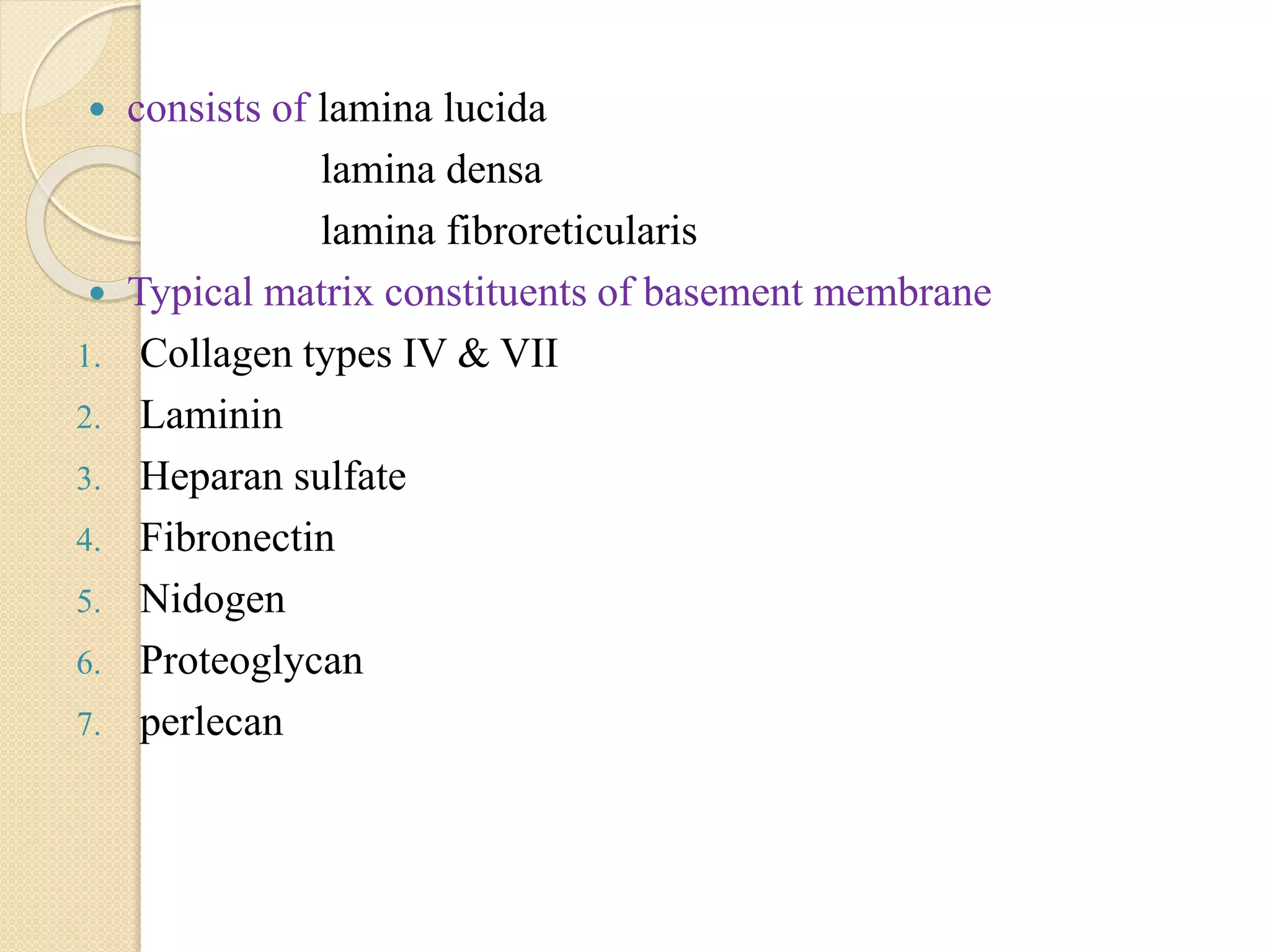
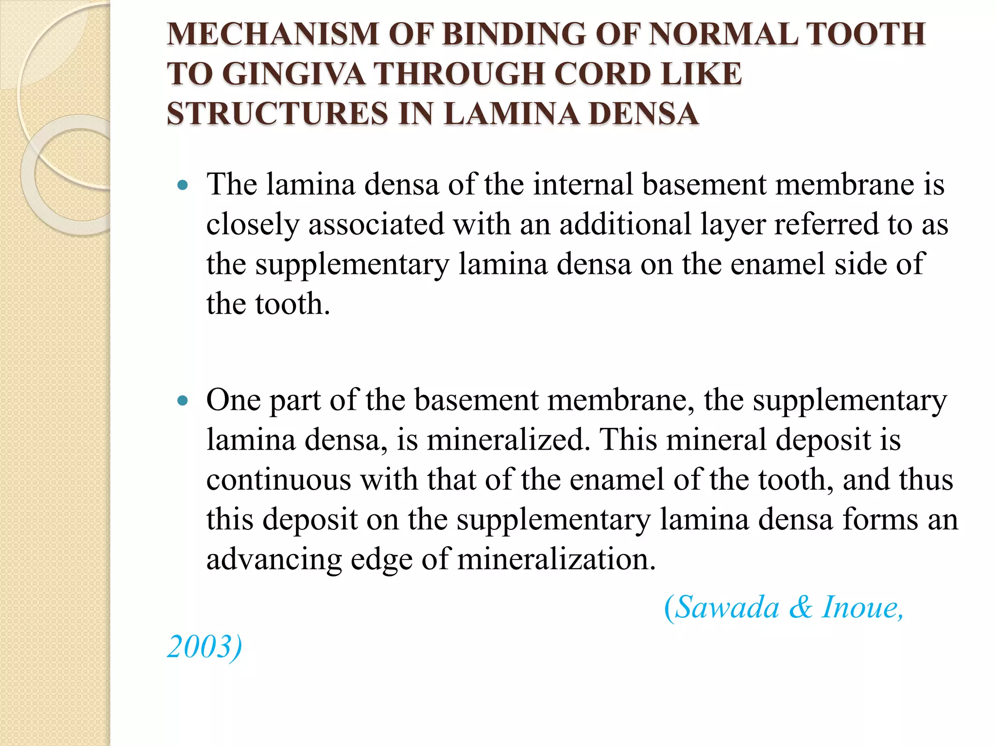
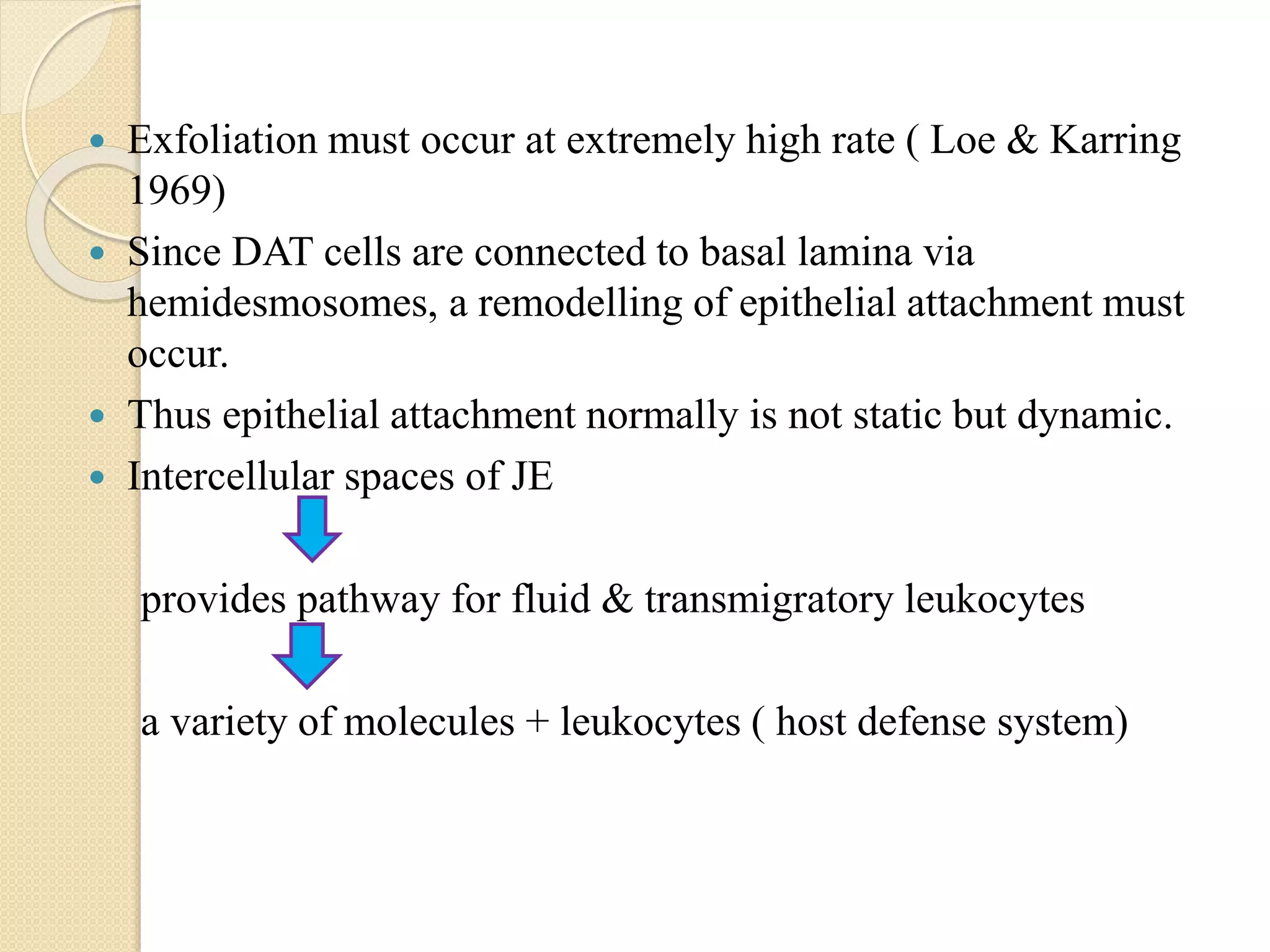
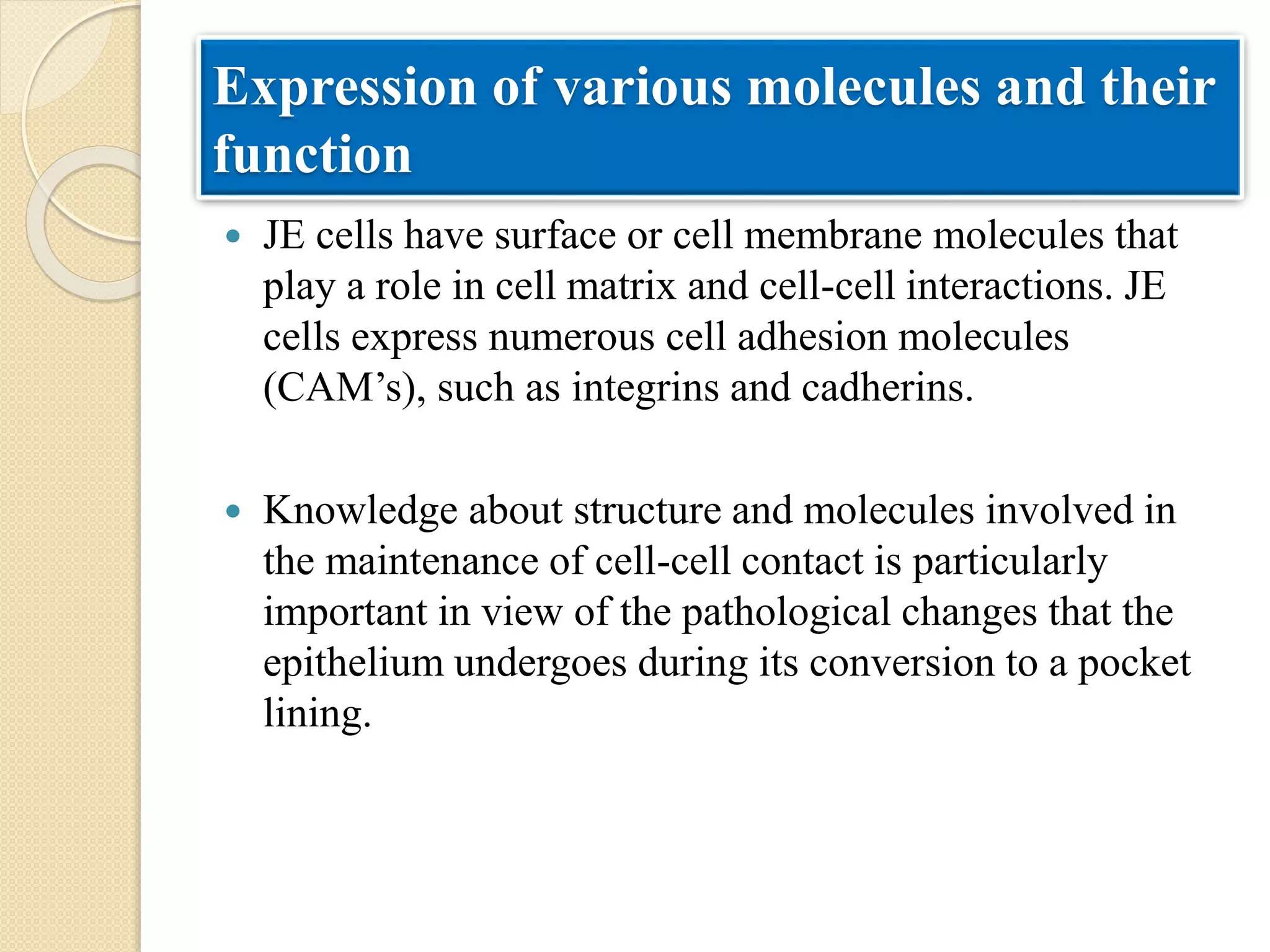
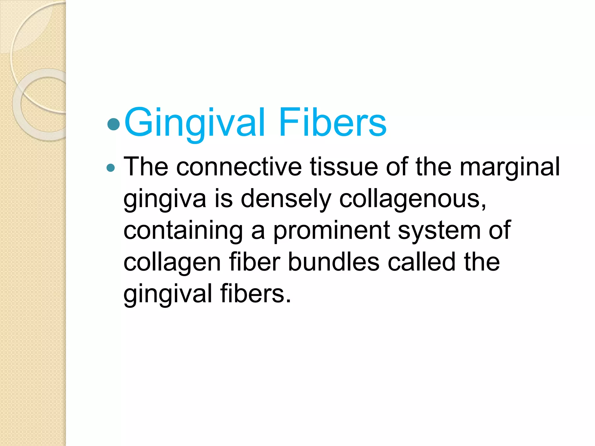
DENTO-GINGIVAL UNIT
Sangeeta Roy
Pg 1st year
 Introduction
 Definition
 Junctional epithelium
◦ Definition
◦ Development of junctional epithelium
◦ Structure
◦ Epithelial attachment
◦ Dynamic aspects of junctional epithelium
◦ Expression of various molecules and their functions
◦ Permeability
◦ Functions
◦ Regeneration
◦ role of JE in initiation of pocket formation
◦ Junctional epithelium around implants
CONTENTS
 Gingival fiber
◦ Classification
◦ Features
◦ Functions
 Biologic width
 Conclusion
INTRODUCTION
 Teeth are trans-mucosal organs.
 This is a unique association in the human
body where a hard tissue emerges through
the soft tissue.
 The tooth-epithelial interface is a specialized
structural and functional adaptation
 Sicher H, 1959:The dentogingival unit (DGU) has
been described as a functional unit composed of the
epithelial attachment and connective tissue
attachment of the gingiva – both of which afford
biological protection .
 Listgarten MA, 1970:Dento-gingival unit refers to
the functional unit comprising of junctional
epithelium and the gingival fibers.
DEFINITION
 “Physiological DGU” is the anatomical complex
formed by the gingival margin, sulcus,
junctional epithelium and connective tissue
attachment.
 The epithelial attachment measures 0.97 mm
 The connective tissue attachment 1.07 mm
Gargiulo et al. 1961
Gingival fibers
Junctional epithelium
Dento
gingiva
l unit
 The attachment of junctional epithelium to tooth is reinforced
with gingival fibers, which brace the gingiva against tooth
surface.
gingival fibers & junctional epithelium
functional unit
Dentogingival unit
 The oral epithelium around a tooth is divided into
three functional compartments– gingival, sulcular,
and junctional epithelium
 The gingival epithelium extends from the
mucogingival junction to the gingival margin
where crevicular/sulcular epithelium lines the
sulcus
 At the base of the sulcus connection between
gingiva and tooth is mediated with
JUNCTIONAL EPITHELIUM
Three zones of the gingival epithelium
Crevicular (sulcular)
epithelium -1. 8mm
Oral epithelium -
Junctional
epithelium
1.35mm
JUNCTIONAL EPITHELIUM
 The dentogingival junction is the
interface between the mineralized
tissue of a tooth and the gingiva. In
this region, an unusual stratified
integument called the junctional
epithelium makes a tight seal against
the mineralized tooth surface via a
structural complex termed the
epithelial attachment (Schroeder,
1986;Rateitschak et al., 1989).
DEFINITION
HISTORY
• Gottlieb (1921) was the first to describe the junctional
epithelium
• Schroeder and Listgarten (1977) clarified the anatomy
and histology of the dentogingival junction
 Formed from oral epithelium and reduced enamel
epithelium.
 Shortly before 1st contact a slow cell transformation
 Beginning orally and ending at CEJ,
1 to 2 yrs ( Schroeder & Listgarten 1977)
3 to 4 yrs (Tencate 1998)
 REE gradually multilayer non keratinizing
converts to squamous epithelium
DEVELOPMENT OF JUNCTIONAL
EPITHELIUM
JUNCTIONAL EPITHELIUM ON AN
ERUPTING TOOTH.
The junctional epithelium (JE) is formed by the
joining of the oral epithelium(OE) and the
reduced enamel epithelium (REE).
Afibrillar cementum, sometimes formed on
enamel after degeneration of the REE
The arrows indicate the coronal movement of the
regenerating epithelial cells, which multiply
more rapidly in the JE than in the OE.
E, Enamel; C, root cementum.
A similar cell turnover pattern exists in the fully
erupted tooth.
 During transformation process,
Reduced ameloblasts undergo structural change
short columnar flattened cells that
orient parallel to
enamel surface
Structure of junctional epithelium
 Anatomical aspects
 Junctional epithelium and interstitial cells
 Epithelial attachment
Anatomical aspects
 part of marginal free gingiva
 Forms a collar like band
 Interproximal area fuse to form
epithelial lining of interdental col
 The coronal termination of the
junctional epithelium corresponds
usually to the bottom of the
gingival sulcus.
JE and Interstitial cell
 JE is a collar-like band of nonkeratinised stratified
squamous epithelium extending from cemento-enamel
junction to bottom of gingival sulcus
 Coronally it is 3-4 cell layers thick , increase upto 10-20
layers with age and apically narrows to 1-2cells
 Its length varies from 0.25 – 1.35mm
stratum basale ( towards CT)
 Made up of 2 strata
stratum suprabasale
(facing tooth surface)
 Organelles- lysosomal bodies, free ribosome, prominent
membrane bound structure, i.e. golgi complex,
cytoplasmic vacuoles, polyribosomes, cisternae of RER
are abundant.
 Cells are connected by Desmosomes.
 Fluid filled intercellular spaces may vary in width.
 All JE cells express unique set of cytokeratins
K5, 14, 19 & occasionally K8, 18.
 Lacks K6,16
 JE is only stratified non keratinized tissue that
does not synthesize K4, K13. Morgan et al.
 Lysosomelike bodies are present, but the
absence of keratinosomes(Odland bodies)
and histochemically demonstrable acid
phosphatase, correlated with the low degree
of differentiation, cause low defense power
against microbial plaque accumulation in the
gingival sulcus.
EPITHELIAL ATTACHMENT APPARATUS
 The term epithelial attachment refers to the attachment
apparatus, i.e. internal basal lamina
& hemidesmosomes
that connects the junctional epithelium to the tooth
surface.
 Connected with gingival connective tissue by external
basal lamina.
 Hemidesmosomes at the plasma membrane of the DAT
(Directly attached cells) directly attached to the tooth
and internal basal lamina( lamina lucida), is present on
the tooth surface
N-nucleus of a DAT cell,
IF-cytoplasmic keratin filaments
The hemidesmosomes at the plasma
membrane are associated with the α6 ,β4
integrin that communicates with laminin 5
located mainly in the internal basal lamina, the
extracellular domain for B180 (collagenous
protein type VIII), that has not yet been
definitely characterized.
LL = lamina lucida,
LD = lamina densa,
SLL = sub lamina lucida,
IBL = internal basal lamina.
STRUCTURAL AND MOLECULAR COMPOSITION OF
THE EPITHELIALATTACHMENT APPARATUS AT DAT
CELL
 Hemidesmosome act as specific site of
signal transduction thus participate in
regulation of gene expression, cell
proliferation, cell differentiation.
 Organic strands from the enamel appear
to extend into the lamina densa. Stern
IB,1967
 The junctional epithelium attaches to
afibrillar cementum present on the crown
(usually restricted to an area within 1 mm
of the cementoenamel
junction,Schroeder HE, Listgarten
MA,2000) and root cementum in a
similar manner.
 Histochemical evidence for the presence
of neutral polysaccharides in the zone of
the epithelial attachment.
 The basal lamina of the junctional
epithelium resembles that of endothelial
and epithelial cells in its laminin content
but differs in its internal basal lamina,
which has type IV collagen .
Bowers,1963
 These findings indicate that the cells of
the junctional epithelium are involved in
the production of laminin and play a key
role in the adhesion mechanism.
HISTORICAL CONCEPTS OF
ATTACHMENT
 Gottlieb (1921)
 Waerhaug (1952)
 Orban (1953)
 Waerhaug (1960)
 Listgarten (1966)
 Schroeder and Listgarten (1971)
Gottlieb’s concept (1921)
 Soft tissue of gingiva is organically united to enamel
surface.
 He termed the epithelium contacting the tooth “epithelial
attachment”.
Orban’s concept (1953)
 He stated that the separation of the epithelial attachment
cells from the tooth surface involved preparatory
degenerative changes in the epithelium.
Waerhaug’s concept (1960)
 He presented the concept of epithelial cuff. This concept
was based on insertion of thin blades between the
surface of tooth and the gingiva
 Blades could be easily passed apically to the connective
tissue attachment at CEJ without resistance.
 It was concluded that gingival tissue and tooth are
closely adapted but not organically united.
Schroeder and Listgarten concept
(1971)
 The previous controversy was resolved after evolution
of transmission electron microscopy.
 Primary epithelial attachment refers to the epithelial
attachment lamina released by the REE. It lies in direct
contact with enamel and epithelial cells attached to it by
hemi-desmosomes.
 When REE cells transform into JE cells the primary
epithelial attachment becomes secondary epithelial
attachment . It is made of epithelial attachment between
basal lamina and hemi-desmosomes.
 The junctional epithelium faces both the gingival
connective tissue (i.e., the lamina propria of the gingiva)
and the tooth surface
JE
EPITHELIAL ATTACHMENT AT
MOLECULAR LEVEL
 Basement membrane – specialized extracellular matrix
 Functions-
a. Compatmentalization
b. Filtration(selective permeability barrier function)- allow access
of gingival fluid, inflammatory cells , component of
immunologic host defence to gingival margin,
c. Cell polarization, migration.
d. Cell adhesions (production of laminin)
e. Cell differentiation(rapid turn over – tissue repair, host parasite
eqquilibrium
f. Prevent pathogenic bacteria from colonizing subgingivally
g. Barrier agains plaque bacteria
h. Endolytic capacity like macrophages, neutrophils.
 consists of lamina lucida
lamina densa
lamina fibroreticularis
 Typical matrix constituents of basement membrane
1. Collagen types IV & VII
2. Laminin
3. Heparan sulfate
4. Fibronectin
5. Nidogen
6. Proteoglycan
7. perlecan
SCHEMATIC DEPICTION OF THE DETAIL
OF THE INTERNAL BASAL LAMINA
It consists of two layers: the
lamina lucida and lamina
densa.
Hemidesmosomes (HD)
originate from the lamina
lucida, and tonofilaments
splay out from each
hemidesmosome.
 The internal basement membrane is 80-120nm wide
homogeneous layer facing enamel, with an intervening
laminated or non-laminated layer of cuticles (Listgarten,
1966) or afibrillar cementum (Kobayashi et al., 1976).
 There are numerous fine strands crossing the lamina
densa of the internal basement membrane at the
hemidesmosomes. These strands may have been the
anchoring filaments of hemidesmosomes (Eady, 1994;
Garrod, 1993).
 In the cytoplasm of the cells of the junctional epithelium,
the tonofibrils are associated with hemidesmosomes.
 The internal basement membrane of the dentogingival
border is uniquely specialized for mechanical strength,
sealing off the periodontal tissues from the oral
environment (Sawada & Inoue, 1996).
 This is a band of multilayered thick basement membranes
 The finer level structure of the internal basement
membrane is, the “cord” network. The basic texture of the
lamina densa is made up of a 3-dimensional network
formed by anastomosing, irregular, thread-like structures
referred to as “cords” (Inoue, 1994; Sawada & Inoue,
2001).

INTERNAL BASEMENT MEMBRANE
Internal basement membrane is
composed of broad band like
lamina densa
Internal basement membrane is
composed of multi-layers
of lamina densa
 Lamina densa is composed of fine
network of irregular anastomosing
cords
MECHANISM OF BINDING OF NORMAL TOOTH
TO GINGIVA THROUGH CORD LIKE
STRUCTURES IN LAMINA DENSA
 The lamina densa of the internal basement membrane is
closely associated with an additional layer referred to as
the supplementary lamina densa on the enamel side of
the tooth.
 One part of the basement membrane, the supplementary
lamina densa, is mineralized. This mineral deposit is
continuous with that of the enamel of the tooth, and thus
this deposit on the supplementary lamina densa forms an
advancing edge of mineralization.
(Sawada & Inoue,
2003)
 In the mineralized portion of the lamina densa, mineral
crystals were arranged in a network pattern which was
comparable to the pattern of the cord network.
 This may facilitate more powerful gripping, and
demonstrates the elaborate mechanism by which firm
binding of the mineral and organic phases is achieved.
DENTO-GINGIVAL BORDER OF
TOOTH FROM DEMINERALIZED AND
NON-DEMINERALIZED SAMPLES
DEMINERALIZED SECTION
SHOWING THE EMPTY SPACE
OF SUBLAMINA DENSA
MINERALISED SECTION SHOWING
MINERALISED SUBLAMINA DENSA
LUCIDA CONTINUOUS WITH
ENAMEL
 Cells and extracellular dynamics of JE
– essential for its protective &
regenerative function.
 Exfoliation must occur at extremely high rate ( Loe & Karring
1969)
 Since DAT cells are connected to basal lamina via
hemidesmosomes, a remodelling of epithelial attachment must
occur.
 Thus epithelial attachment normally is not static but dynamic.
 Intercellular spaces of JE
provides pathway for fluid & transmigratory leukocytes
a variety of molecules + leukocytes ( host defense system)
Expression of various molecules and their
function
 JE cells have surface or cell membrane molecules that
play a role in cell matrix and cell-cell interactions. JE
cells express numerous cell adhesion molecules
(CAM’s), such as integrins and cadherins.
 Knowledge about structure and molecules involved in
the maintenance of cell-cell contact is particularly
important in view of the pathological changes that the
epithelium undergoes during its conversion to a pocket
lining.
 Integrins – are cell surface receptors that mediate interactions
between cell and extracellular matrix, and also contribute to
cell to cell adhesion.
 The cadherins are responsible for tight contacts between cells.
 E-cadherin, an epithelium specific cell adhesion molecule,
plays a crucial role in maintaining the structural integrity.
 Intercellular adhesion molecule-1(ICAM-1 or CD-54) and
lymphocytic function antigen- 3(LFA-3) are additional cell
adhesion molecules.
 Cells in contact with the internal basal lamina express the
integrins.
 (CEACAM1)—a transmembrane cell-adhesion molecule that
is expressed on leukocytes, epithelia, and blood vessel
endothelia .
 high expression of interleukin-8 (IL-8), a chemotactic
cytokine, is seen in the coronal-most cells of the junctional
epithelium
 interleukin-1α (IL-1α),
 interleukin-1β (IL-1β),
 tumor necrosis factor-α (TNF-α)—are strongly expressed in
the coronal half of the junctional epithelium
 N-acetyllactosamine—the type 2 chain H precursor of the
blood group A-specific carbohydrate, which is usually
associated with the lowest level of cell differentiation.
 Antimicrobial molecules--- α and β defensins
cathelicidin family
calprotectin
DYNAMICS (TURNOVER RATE) OF JE
 The turnover rate of JE cells is exceptionally rapid. In
non-human primates it is about 5 days (twice that of oral
epithelium).
 The DAT cells express a high density of transferrin
receptors supporting the idea of active metabolism and
high turnover.
 DAT cells have an important role in tissue dynamics and
reparative capacity of the JE.
 The existence of a dividing population of DAT cells in a
suprabasal location in several layers from connective
tissue is a unique feature of JE.
Mechanism of JE cells turnover
(1)The daughter cells are produced by
dividing DAT cells and replace
degenerating cells on the tooth surface.
(2) The daughter cells enter the exfoliation
pathway and gradually migrate coronally
between the basal cells and the DAT cells
to eventually break off into the sulcus, or
(3)Epithelial cells move/migrate in the
coronal direction along the tooth surface
and are replaced by basal cells migrating
round the apical termination of the
junctional epithelium.
E
PERMEABILITY OF JUNCTIONAL
EPITHELIUM
• The bi-directional arrows indicate that
the junctional epithelium is the most
permeable portion of the gingival
epithelia.
• Because of its permeability to
bacterial products and other assorted
antigens, the connective tissue
adjacent to the junctional epithelium
tends to become infiltrated with
chronic inflammatory cells, primarily
lymphocytes and plasma cells.
FUNCTIONS OF JUNCTIONAL
EPITHELIUM
 Has attachment role and protective role.
 Permeability allows GCF and defence cells to pass
across to protect underlying tissues from disease
processes (periodontal disease).
 GCF contains gamma globulins and poly-
morphonuclear leukocytes (PMNs) giving it
immunological /phagocytic properties to combat disease
processes.
 Such molecules pass readily across JE to underlying
tissues.
 JE may contain neutrophils & other inflammatory cells
indicating disease & state of health of periodontium.
 Cell polarization, migration.
 Cell adhesions (production of laminin)
 Cell differentiation –Rapid turn over
 Helps maintaing integrity of tooth/periodontium
structure
 The junctional epithelium plays a crucial role since it
essentially seals off periodontal tissues from the oral
environment.
 Its integrity is thus essential for maintaining a healthy
periodontium.
JE in antimicrobial defense
(1) JE cells exfoliate because of rapid cell
division
(2) Funnelling of junctional epithelial cells
towards the sulcus hinder bacterial
colonization.
(3) Active antimicrobial substances are
produced in junctional epithelial cells.
(4) Epithelial cells activated by microbial
substances secrete chemokines,
(5) Chemokines attract and activate
professional defense cells, PMN.
 ROLE OF JE IN POCKET
FORMATION
Role of JE in the initiation of pocket formation
 Conversion of the JE to pocket epithelium is regarded as
a hallmark in the development of periodontitis.
 Schroeder – 1996 pointed to a biologically relevant and
clinically important question that still awaits resolution:
‘what happens to the JE under conditions of sub-
gingival microbial attack i.e. in context with pocket
formation and deepening?’
 Schluger et al 1977: Pocket formation is attributed to a loss
of cellular continuity in the coronal most portion of the JE
 Thus the initiation of pocket formation may be attributed to
the detachment of the DAT cells from the tooth surface or to
the development of intraepithelial split.
 Schroeder and Listgarten 1977: An increased number of
mononuclear leukocytes (T and B cells, macrophages)
together with PMNs are considered as factors contributing to
the disintegration of the JE.
 Takata and Donath (1988) observed degenerative changes in
the second or third layer of the DAT cells in the coronal most
portion of the JE cells facing the biofilm.
The degeneration and
detachment of DAT cells
exposes tooth surface and
creates a sub-gingival niche
suitable for the colonization
of anaerobic gram-negative
bacteria and apical growth of
dental plaque.
 Hintermann et al 2002: Gingipains degrade the epithelial
cell-cell junctional complexes and cells exposed to
proteinases derived from P.gingivalis showed reduced
adhesion to extracellular matrix.
 Destruction of cell-cell and cell to ECM attachment
degrade the structural and functional integrity of the JE.
 REGENERATION OF JE
• Injury to JE may occur due to intentional or
accidental trauma.
• Accidental trauma can occur during probing,
flossing or tooth margin preparations for
restorations.
• Intentional trauma occurs during periodontal
surgeries where the JE is completely lost.
 Many studies have been done to investigate the
renewal of JE.
 These include - renewal of JE on tooth and
implant surface after mechanical detachment by
probing.
 Mechanical trauma during flossing and on
regeneration of JE after gingivectomy procedure
which completely removes JE.
 Taylor and Campbell 1972: A new and complete
attachment indistinguishable from that in control was
established 5 days after complete separation of the JE
from the tooth surface.
 Frank et al 1972: A study demonstrated that newly
differentiated attachment apparatus with normal
hemidesmosomal attachment is possible following
surgery. This new attachment apparatus was seen on
cementum as well as dentin.
 Listgarten 1972: Hemidesmosomes appeared to form prior to
the basal lamina. The basal lamina is initially formed in close
proximity to the hemidesmosomes at both the tooth and
connective tissue interface.
 At 4 to 7 weeks, the basal lamina appeared complete.
 Studies have shown that regeneration of JE after procedure
usually occurs within 20 days.
 JE AROUND IMPLANTS
 The junctional epithelium around implants always
originates from epithelial cells of the oral mucosa, as
opposed to the junctional epithelium around teeth which
originates from the reduced enamel epithelium.
 Despite different origins of the 2 epithelia, a functional
adaptation occurs when oral epithelia form an epithelial
attachment around implants.
NATURAL TOOTH
 Epithelium tapers
towards the depth
 Large number of
cell organelles
 Fibers are
arranged
perpendicular
IMPLANT
• Epithelium is
thicker
•Few organelles
•Fibers are arranged
parallely
•Numerous kerato-
hyalin granules
NATURAL TOOTH IMPLANT
GINGIVAL FIBERS
Gingival Fibers
 The connective tissue of the marginal
gingiva is densely collagenous,
containing a prominent system of
collagen fiber bundles called the
gingival fibers.
THE GINGIVAL FIBER APPARATUS
 The gingival lamina propria consists mainly of a
dense network of collagen fiber bundles that
account for about 55.43% of the connective
tissue volume. This network is called the
supragingival fiber apparatus.
 On the basis of their preferential orientation,
architectural arrangement and sites of insertion,
these bundles have been classified.
 These fiber bundles are densely populated by fibroblasts
and consist mainly of collagen Type I and III.
 Collagen Type I represents mainly dense fibers; Type III is
related to loose connective tissue, subepithelially and
around blood vessels.
 Mast cells are also regular residents, whereas
lymphocytes, monocytes and macrophages vary in
number with the need for and degree of protective
activity.
CLASSIFICATION OF GINGIVAL FIBER GROUPS
based on their orientation , sites of insertion, the
structures that they connect gingival fibers are
classified as-
GINGIVAL FIBERS
PRIMARY FIBERS SECONDARY FIBERS
PRIMARY FIBERS:
• Alveologingival fibers
• Circular fibers.
• Dentogingival fibers
 Alveologingival fibers—extend from the periosteum of
the alveolar crest into the gingival connective tissue.
These fiber bundles attach the gingiva to the bone. (The
periosteum is a dense membrane composed of fibrous
connective tissue that closely wraps around the outer
surface of the alveolar bone.)
 Circular fibers—encircle the tooth in a ring-like
manner coronal to the alveolar crest and are not
attached to the cementum of the tooth. These fiber
bundles connect adjacent teeth to one another.
 Dentogingival fibers— are embedded in the cementum
near the CEJ and fan out into the gingival connective
tissue. These fibers attach the gingiva to teeth.
SECONDARY FIBERS
 Periostogingival fibers.
 Intergingival fibers
 Intercircular fibers
 Interpapillary fibers
 Transseptal Fibers-
1. Transgingival fibers
2. Semicircular fibers
- Page et al(1972)
Transseptal fibers
 Located interproximally,
 The transseptal fibers form horizontal
bundles that extend between the
cementum of approximating teeth into
which they are embedded.
 They lie in the area between the
epithelium at the base of the gingival
sulcus and the crest of the interdental
bone
 Page et al1972 described
 (1) a group of semicircular fibers, which is
attached at the proximal surface of a tooth,
immediately below the cementoenamel junction,
go around the facial or lingual marginal gingiva of
the tooth, and attach on the other proximal surface
of the same tooth; and
 (2) a group of transgingival fibers, that attach in
the proximal surface of one tooth, traverse the
interdental space diagonally, go around the facial
or lingual surface of the adjacent tooth, again
traverse diagonally the interdental space, and
attach in the proximal surface of the next tooth.
Functions of the Gingival Fiber Bundles
1. Brace the marginal gingiva firmly against the tooth
and reinforce the attachment of the JE to the tooth.
2. Provide the rigidity necessary to withstand the
forces of mastication without being deflected away
from the tooth surface
3. Unite the free marginal gingiva with the cementum
of the root and adjacent attaced gingiva.
4. Connect adjacent teeth to one another to control
tooth positioning within the dental arch.
THE SUPRAGINGIVAL FIBER APPARATUS
 The formation and insertion of transseptal and
dentogingival fibers is not specific to certain
anatomic surfaces, but is functionally rather than
anatomically determined.
 The supragingival fiber apparatus not only attaches
the gingiva to teeth and bone but also provides a
dense framework that accounts for the rigidity and
biomechanical resistance of the gingiva.
 The fiber apparatus also controls the positioning of
teeth within the dental arch and also protects the
very sophisticated cellular defenses located at the
dentogingival interface.
SUPRAGINGIVAL FIBERS AROUND
IMPLANTS
BIOLOGICAL WIDTH
 BIOLOGIC WIDTH is defined as the dimension of the soft
tissue which is attached to the portion of the tooth coronal to
the crest of the alveolar bone
 JUNCTIONAL EPITHELIUM CONNECTIVE TISSUE
BIOLOGIC WIDTH
 It is important from the restorative point of view because its
violation leads to complications like gingival enlargement
alveolar bone loss and improper fit of the restoration.
 The average combined histological width of connective tissue
attachment and junctional epithelium was 2.04mm, which is
referred to as the BIOLOGIC WIDTH.
Average human biologic width: Connective tissue attachment
1 mm in height; epithelial attachment 1 mm in height;
sulcus depth of approximately 1 mm. The combined connective
tissue attachment and epithelial attachment or biologic width
equals 2 mm.
 Gargiulo et al (1961) in their study described the
dimensions and relations of dentogingival junction in
humans. The average histological width of connective
tissue attachment was 1.07mm. The mean average length
of epithelial attachment was 0.97mm with the range of
0.71mm-1.35mm.
 They reported biologic width as 2mm but found as
narrow as 0.75mm to even 4.3mm in some patient.
 Vacek et al, 1994 found average biologic width as 2mm
CONCLUSION
 DENTOGINGIVAL UNIT is important because of its
anatomical location.
 It is the site of host-bacterial interaction in initiation of
periodontal disease.
 There is a constant presence of bacteria and their
products in the gingival sulcus which makes this an
important structural component of periodontal defense
mechanism.
 The conversion of the junctional epithelium to pocket
epithelium is regarded as hallmark in the development of
periodontitis.
THANK YOU

Dento gingival unit

  • 1.
  • 2.
     Introduction  Definition Junctional epithelium ◦ Definition ◦ Development of junctional epithelium ◦ Structure ◦ Epithelial attachment ◦ Dynamic aspects of junctional epithelium ◦ Expression of various molecules and their functions ◦ Permeability ◦ Functions ◦ Regeneration ◦ role of JE in initiation of pocket formation ◦ Junctional epithelium around implants CONTENTS
  • 3.
     Gingival fiber ◦Classification ◦ Features ◦ Functions  Biologic width  Conclusion
  • 4.
    INTRODUCTION  Teeth aretrans-mucosal organs.  This is a unique association in the human body where a hard tissue emerges through the soft tissue.  The tooth-epithelial interface is a specialized structural and functional adaptation
  • 5.
     Sicher H,1959:The dentogingival unit (DGU) has been described as a functional unit composed of the epithelial attachment and connective tissue attachment of the gingiva – both of which afford biological protection .  Listgarten MA, 1970:Dento-gingival unit refers to the functional unit comprising of junctional epithelium and the gingival fibers. DEFINITION
  • 6.
     “Physiological DGU”is the anatomical complex formed by the gingival margin, sulcus, junctional epithelium and connective tissue attachment.  The epithelial attachment measures 0.97 mm  The connective tissue attachment 1.07 mm Gargiulo et al. 1961 Gingival fibers Junctional epithelium Dento gingiva l unit
  • 7.
     The attachmentof junctional epithelium to tooth is reinforced with gingival fibers, which brace the gingiva against tooth surface. gingival fibers & junctional epithelium functional unit Dentogingival unit
  • 8.
     The oralepithelium around a tooth is divided into three functional compartments– gingival, sulcular, and junctional epithelium  The gingival epithelium extends from the mucogingival junction to the gingival margin where crevicular/sulcular epithelium lines the sulcus  At the base of the sulcus connection between gingiva and tooth is mediated with JUNCTIONAL EPITHELIUM
  • 9.
    Three zones ofthe gingival epithelium Crevicular (sulcular) epithelium -1. 8mm Oral epithelium - Junctional epithelium 1.35mm
  • 10.
  • 11.
     The dentogingivaljunction is the interface between the mineralized tissue of a tooth and the gingiva. In this region, an unusual stratified integument called the junctional epithelium makes a tight seal against the mineralized tooth surface via a structural complex termed the epithelial attachment (Schroeder, 1986;Rateitschak et al., 1989). DEFINITION
  • 12.
    HISTORY • Gottlieb (1921)was the first to describe the junctional epithelium • Schroeder and Listgarten (1977) clarified the anatomy and histology of the dentogingival junction
  • 13.
     Formed fromoral epithelium and reduced enamel epithelium.  Shortly before 1st contact a slow cell transformation  Beginning orally and ending at CEJ, 1 to 2 yrs ( Schroeder & Listgarten 1977) 3 to 4 yrs (Tencate 1998)  REE gradually multilayer non keratinizing converts to squamous epithelium DEVELOPMENT OF JUNCTIONAL EPITHELIUM
  • 14.
    JUNCTIONAL EPITHELIUM ONAN ERUPTING TOOTH. The junctional epithelium (JE) is formed by the joining of the oral epithelium(OE) and the reduced enamel epithelium (REE). Afibrillar cementum, sometimes formed on enamel after degeneration of the REE The arrows indicate the coronal movement of the regenerating epithelial cells, which multiply more rapidly in the JE than in the OE. E, Enamel; C, root cementum. A similar cell turnover pattern exists in the fully erupted tooth.
  • 15.
     During transformationprocess, Reduced ameloblasts undergo structural change short columnar flattened cells that orient parallel to enamel surface
  • 16.
    Structure of junctionalepithelium  Anatomical aspects  Junctional epithelium and interstitial cells  Epithelial attachment
  • 17.
    Anatomical aspects  partof marginal free gingiva  Forms a collar like band  Interproximal area fuse to form epithelial lining of interdental col  The coronal termination of the junctional epithelium corresponds usually to the bottom of the gingival sulcus.
  • 18.
    JE and Interstitialcell  JE is a collar-like band of nonkeratinised stratified squamous epithelium extending from cemento-enamel junction to bottom of gingival sulcus  Coronally it is 3-4 cell layers thick , increase upto 10-20 layers with age and apically narrows to 1-2cells  Its length varies from 0.25 – 1.35mm
  • 19.
    stratum basale (towards CT)  Made up of 2 strata stratum suprabasale (facing tooth surface)  Organelles- lysosomal bodies, free ribosome, prominent membrane bound structure, i.e. golgi complex, cytoplasmic vacuoles, polyribosomes, cisternae of RER are abundant.  Cells are connected by Desmosomes.  Fluid filled intercellular spaces may vary in width.
  • 20.
     All JEcells express unique set of cytokeratins K5, 14, 19 & occasionally K8, 18.  Lacks K6,16  JE is only stratified non keratinized tissue that does not synthesize K4, K13. Morgan et al.  Lysosomelike bodies are present, but the absence of keratinosomes(Odland bodies) and histochemically demonstrable acid phosphatase, correlated with the low degree of differentiation, cause low defense power against microbial plaque accumulation in the gingival sulcus.
  • 21.
    EPITHELIAL ATTACHMENT APPARATUS The term epithelial attachment refers to the attachment apparatus, i.e. internal basal lamina & hemidesmosomes that connects the junctional epithelium to the tooth surface.  Connected with gingival connective tissue by external basal lamina.  Hemidesmosomes at the plasma membrane of the DAT (Directly attached cells) directly attached to the tooth and internal basal lamina( lamina lucida), is present on the tooth surface
  • 22.
    N-nucleus of aDAT cell, IF-cytoplasmic keratin filaments The hemidesmosomes at the plasma membrane are associated with the α6 ,β4 integrin that communicates with laminin 5 located mainly in the internal basal lamina, the extracellular domain for B180 (collagenous protein type VIII), that has not yet been definitely characterized. LL = lamina lucida, LD = lamina densa, SLL = sub lamina lucida, IBL = internal basal lamina. STRUCTURAL AND MOLECULAR COMPOSITION OF THE EPITHELIALATTACHMENT APPARATUS AT DAT CELL
  • 23.
     Hemidesmosome actas specific site of signal transduction thus participate in regulation of gene expression, cell proliferation, cell differentiation.  Organic strands from the enamel appear to extend into the lamina densa. Stern IB,1967  The junctional epithelium attaches to afibrillar cementum present on the crown (usually restricted to an area within 1 mm of the cementoenamel junction,Schroeder HE, Listgarten MA,2000) and root cementum in a similar manner.
  • 24.
     Histochemical evidencefor the presence of neutral polysaccharides in the zone of the epithelial attachment.  The basal lamina of the junctional epithelium resembles that of endothelial and epithelial cells in its laminin content but differs in its internal basal lamina, which has type IV collagen . Bowers,1963  These findings indicate that the cells of the junctional epithelium are involved in the production of laminin and play a key role in the adhesion mechanism.
  • 25.
    HISTORICAL CONCEPTS OF ATTACHMENT Gottlieb (1921)  Waerhaug (1952)  Orban (1953)  Waerhaug (1960)  Listgarten (1966)  Schroeder and Listgarten (1971)
  • 26.
    Gottlieb’s concept (1921) Soft tissue of gingiva is organically united to enamel surface.  He termed the epithelium contacting the tooth “epithelial attachment”.
  • 27.
    Orban’s concept (1953) He stated that the separation of the epithelial attachment cells from the tooth surface involved preparatory degenerative changes in the epithelium.
  • 28.
    Waerhaug’s concept (1960) He presented the concept of epithelial cuff. This concept was based on insertion of thin blades between the surface of tooth and the gingiva  Blades could be easily passed apically to the connective tissue attachment at CEJ without resistance.  It was concluded that gingival tissue and tooth are closely adapted but not organically united.
  • 29.
    Schroeder and Listgartenconcept (1971)  The previous controversy was resolved after evolution of transmission electron microscopy.  Primary epithelial attachment refers to the epithelial attachment lamina released by the REE. It lies in direct contact with enamel and epithelial cells attached to it by hemi-desmosomes.  When REE cells transform into JE cells the primary epithelial attachment becomes secondary epithelial attachment . It is made of epithelial attachment between basal lamina and hemi-desmosomes.
  • 30.
     The junctionalepithelium faces both the gingival connective tissue (i.e., the lamina propria of the gingiva) and the tooth surface JE EPITHELIAL ATTACHMENT AT MOLECULAR LEVEL
  • 31.
     Basement membrane– specialized extracellular matrix  Functions- a. Compatmentalization b. Filtration(selective permeability barrier function)- allow access of gingival fluid, inflammatory cells , component of immunologic host defence to gingival margin, c. Cell polarization, migration. d. Cell adhesions (production of laminin) e. Cell differentiation(rapid turn over – tissue repair, host parasite eqquilibrium f. Prevent pathogenic bacteria from colonizing subgingivally g. Barrier agains plaque bacteria h. Endolytic capacity like macrophages, neutrophils.
  • 32.
     consists oflamina lucida lamina densa lamina fibroreticularis  Typical matrix constituents of basement membrane 1. Collagen types IV & VII 2. Laminin 3. Heparan sulfate 4. Fibronectin 5. Nidogen 6. Proteoglycan 7. perlecan
  • 33.
    SCHEMATIC DEPICTION OFTHE DETAIL OF THE INTERNAL BASAL LAMINA It consists of two layers: the lamina lucida and lamina densa. Hemidesmosomes (HD) originate from the lamina lucida, and tonofilaments splay out from each hemidesmosome.
  • 34.
     The internalbasement membrane is 80-120nm wide homogeneous layer facing enamel, with an intervening laminated or non-laminated layer of cuticles (Listgarten, 1966) or afibrillar cementum (Kobayashi et al., 1976).  There are numerous fine strands crossing the lamina densa of the internal basement membrane at the hemidesmosomes. These strands may have been the anchoring filaments of hemidesmosomes (Eady, 1994; Garrod, 1993).  In the cytoplasm of the cells of the junctional epithelium, the tonofibrils are associated with hemidesmosomes.
  • 35.
     The internalbasement membrane of the dentogingival border is uniquely specialized for mechanical strength, sealing off the periodontal tissues from the oral environment (Sawada & Inoue, 1996).  This is a band of multilayered thick basement membranes  The finer level structure of the internal basement membrane is, the “cord” network. The basic texture of the lamina densa is made up of a 3-dimensional network formed by anastomosing, irregular, thread-like structures referred to as “cords” (Inoue, 1994; Sawada & Inoue, 2001). 
  • 36.
    INTERNAL BASEMENT MEMBRANE Internalbasement membrane is composed of broad band like lamina densa Internal basement membrane is composed of multi-layers of lamina densa
  • 37.
     Lamina densais composed of fine network of irregular anastomosing cords
  • 38.
    MECHANISM OF BINDINGOF NORMAL TOOTH TO GINGIVA THROUGH CORD LIKE STRUCTURES IN LAMINA DENSA  The lamina densa of the internal basement membrane is closely associated with an additional layer referred to as the supplementary lamina densa on the enamel side of the tooth.  One part of the basement membrane, the supplementary lamina densa, is mineralized. This mineral deposit is continuous with that of the enamel of the tooth, and thus this deposit on the supplementary lamina densa forms an advancing edge of mineralization. (Sawada & Inoue, 2003)
  • 39.
     In themineralized portion of the lamina densa, mineral crystals were arranged in a network pattern which was comparable to the pattern of the cord network.  This may facilitate more powerful gripping, and demonstrates the elaborate mechanism by which firm binding of the mineral and organic phases is achieved.
  • 40.
    DENTO-GINGIVAL BORDER OF TOOTHFROM DEMINERALIZED AND NON-DEMINERALIZED SAMPLES DEMINERALIZED SECTION SHOWING THE EMPTY SPACE OF SUBLAMINA DENSA MINERALISED SECTION SHOWING MINERALISED SUBLAMINA DENSA LUCIDA CONTINUOUS WITH ENAMEL
  • 41.
     Cells andextracellular dynamics of JE – essential for its protective & regenerative function.
  • 42.
     Exfoliation mustoccur at extremely high rate ( Loe & Karring 1969)  Since DAT cells are connected to basal lamina via hemidesmosomes, a remodelling of epithelial attachment must occur.  Thus epithelial attachment normally is not static but dynamic.  Intercellular spaces of JE provides pathway for fluid & transmigratory leukocytes a variety of molecules + leukocytes ( host defense system)
  • 43.
    Expression of variousmolecules and their function  JE cells have surface or cell membrane molecules that play a role in cell matrix and cell-cell interactions. JE cells express numerous cell adhesion molecules (CAM’s), such as integrins and cadherins.  Knowledge about structure and molecules involved in the maintenance of cell-cell contact is particularly important in view of the pathological changes that the epithelium undergoes during its conversion to a pocket lining.
  • 44.
     Integrins –are cell surface receptors that mediate interactions between cell and extracellular matrix, and also contribute to cell to cell adhesion.  The cadherins are responsible for tight contacts between cells.  E-cadherin, an epithelium specific cell adhesion molecule, plays a crucial role in maintaining the structural integrity.  Intercellular adhesion molecule-1(ICAM-1 or CD-54) and lymphocytic function antigen- 3(LFA-3) are additional cell adhesion molecules.  Cells in contact with the internal basal lamina express the integrins.
  • 45.
     (CEACAM1)—a transmembranecell-adhesion molecule that is expressed on leukocytes, epithelia, and blood vessel endothelia .  high expression of interleukin-8 (IL-8), a chemotactic cytokine, is seen in the coronal-most cells of the junctional epithelium  interleukin-1α (IL-1α),  interleukin-1β (IL-1β),  tumor necrosis factor-α (TNF-α)—are strongly expressed in the coronal half of the junctional epithelium  N-acetyllactosamine—the type 2 chain H precursor of the blood group A-specific carbohydrate, which is usually associated with the lowest level of cell differentiation.
  • 46.
     Antimicrobial molecules---α and β defensins cathelicidin family calprotectin
  • 47.
    DYNAMICS (TURNOVER RATE)OF JE  The turnover rate of JE cells is exceptionally rapid. In non-human primates it is about 5 days (twice that of oral epithelium).  The DAT cells express a high density of transferrin receptors supporting the idea of active metabolism and high turnover.  DAT cells have an important role in tissue dynamics and reparative capacity of the JE.  The existence of a dividing population of DAT cells in a suprabasal location in several layers from connective tissue is a unique feature of JE.
  • 48.
    Mechanism of JEcells turnover (1)The daughter cells are produced by dividing DAT cells and replace degenerating cells on the tooth surface. (2) The daughter cells enter the exfoliation pathway and gradually migrate coronally between the basal cells and the DAT cells to eventually break off into the sulcus, or (3)Epithelial cells move/migrate in the coronal direction along the tooth surface and are replaced by basal cells migrating round the apical termination of the junctional epithelium. E
  • 49.
    PERMEABILITY OF JUNCTIONAL EPITHELIUM •The bi-directional arrows indicate that the junctional epithelium is the most permeable portion of the gingival epithelia. • Because of its permeability to bacterial products and other assorted antigens, the connective tissue adjacent to the junctional epithelium tends to become infiltrated with chronic inflammatory cells, primarily lymphocytes and plasma cells.
  • 50.
    FUNCTIONS OF JUNCTIONAL EPITHELIUM Has attachment role and protective role.  Permeability allows GCF and defence cells to pass across to protect underlying tissues from disease processes (periodontal disease).  GCF contains gamma globulins and poly- morphonuclear leukocytes (PMNs) giving it immunological /phagocytic properties to combat disease processes.  Such molecules pass readily across JE to underlying tissues.
  • 51.
     JE maycontain neutrophils & other inflammatory cells indicating disease & state of health of periodontium.  Cell polarization, migration.  Cell adhesions (production of laminin)  Cell differentiation –Rapid turn over
  • 52.
     Helps maintaingintegrity of tooth/periodontium structure  The junctional epithelium plays a crucial role since it essentially seals off periodontal tissues from the oral environment.  Its integrity is thus essential for maintaining a healthy periodontium.
  • 53.
    JE in antimicrobialdefense (1) JE cells exfoliate because of rapid cell division (2) Funnelling of junctional epithelial cells towards the sulcus hinder bacterial colonization. (3) Active antimicrobial substances are produced in junctional epithelial cells. (4) Epithelial cells activated by microbial substances secrete chemokines, (5) Chemokines attract and activate professional defense cells, PMN.
  • 54.
     ROLE OFJE IN POCKET FORMATION
  • 55.
    Role of JEin the initiation of pocket formation  Conversion of the JE to pocket epithelium is regarded as a hallmark in the development of periodontitis.  Schroeder – 1996 pointed to a biologically relevant and clinically important question that still awaits resolution: ‘what happens to the JE under conditions of sub- gingival microbial attack i.e. in context with pocket formation and deepening?’
  • 56.
     Schluger etal 1977: Pocket formation is attributed to a loss of cellular continuity in the coronal most portion of the JE  Thus the initiation of pocket formation may be attributed to the detachment of the DAT cells from the tooth surface or to the development of intraepithelial split.  Schroeder and Listgarten 1977: An increased number of mononuclear leukocytes (T and B cells, macrophages) together with PMNs are considered as factors contributing to the disintegration of the JE.  Takata and Donath (1988) observed degenerative changes in the second or third layer of the DAT cells in the coronal most portion of the JE cells facing the biofilm.
  • 57.
    The degeneration and detachmentof DAT cells exposes tooth surface and creates a sub-gingival niche suitable for the colonization of anaerobic gram-negative bacteria and apical growth of dental plaque.
  • 58.
     Hintermann etal 2002: Gingipains degrade the epithelial cell-cell junctional complexes and cells exposed to proteinases derived from P.gingivalis showed reduced adhesion to extracellular matrix.  Destruction of cell-cell and cell to ECM attachment degrade the structural and functional integrity of the JE.
  • 59.
     REGENERATION OFJE • Injury to JE may occur due to intentional or accidental trauma. • Accidental trauma can occur during probing, flossing or tooth margin preparations for restorations. • Intentional trauma occurs during periodontal surgeries where the JE is completely lost.
  • 60.
     Many studieshave been done to investigate the renewal of JE.  These include - renewal of JE on tooth and implant surface after mechanical detachment by probing.  Mechanical trauma during flossing and on regeneration of JE after gingivectomy procedure which completely removes JE.
  • 61.
     Taylor andCampbell 1972: A new and complete attachment indistinguishable from that in control was established 5 days after complete separation of the JE from the tooth surface.  Frank et al 1972: A study demonstrated that newly differentiated attachment apparatus with normal hemidesmosomal attachment is possible following surgery. This new attachment apparatus was seen on cementum as well as dentin.
  • 62.
     Listgarten 1972:Hemidesmosomes appeared to form prior to the basal lamina. The basal lamina is initially formed in close proximity to the hemidesmosomes at both the tooth and connective tissue interface.  At 4 to 7 weeks, the basal lamina appeared complete.  Studies have shown that regeneration of JE after procedure usually occurs within 20 days.
  • 63.
     JE AROUNDIMPLANTS
  • 64.
     The junctionalepithelium around implants always originates from epithelial cells of the oral mucosa, as opposed to the junctional epithelium around teeth which originates from the reduced enamel epithelium.  Despite different origins of the 2 epithelia, a functional adaptation occurs when oral epithelia form an epithelial attachment around implants.
  • 65.
    NATURAL TOOTH  Epitheliumtapers towards the depth  Large number of cell organelles  Fibers are arranged perpendicular IMPLANT • Epithelium is thicker •Few organelles •Fibers are arranged parallely •Numerous kerato- hyalin granules
  • 66.
  • 67.
  • 68.
    Gingival Fibers  Theconnective tissue of the marginal gingiva is densely collagenous, containing a prominent system of collagen fiber bundles called the gingival fibers.
  • 69.
    THE GINGIVAL FIBERAPPARATUS  The gingival lamina propria consists mainly of a dense network of collagen fiber bundles that account for about 55.43% of the connective tissue volume. This network is called the supragingival fiber apparatus.  On the basis of their preferential orientation, architectural arrangement and sites of insertion, these bundles have been classified.
  • 70.
     These fiberbundles are densely populated by fibroblasts and consist mainly of collagen Type I and III.  Collagen Type I represents mainly dense fibers; Type III is related to loose connective tissue, subepithelially and around blood vessels.  Mast cells are also regular residents, whereas lymphocytes, monocytes and macrophages vary in number with the need for and degree of protective activity.
  • 72.
    CLASSIFICATION OF GINGIVALFIBER GROUPS based on their orientation , sites of insertion, the structures that they connect gingival fibers are classified as- GINGIVAL FIBERS PRIMARY FIBERS SECONDARY FIBERS
  • 74.
    PRIMARY FIBERS: • Alveologingivalfibers • Circular fibers. • Dentogingival fibers
  • 75.
     Alveologingival fibers—extendfrom the periosteum of the alveolar crest into the gingival connective tissue. These fiber bundles attach the gingiva to the bone. (The periosteum is a dense membrane composed of fibrous connective tissue that closely wraps around the outer surface of the alveolar bone.)  Circular fibers—encircle the tooth in a ring-like manner coronal to the alveolar crest and are not attached to the cementum of the tooth. These fiber bundles connect adjacent teeth to one another.  Dentogingival fibers— are embedded in the cementum near the CEJ and fan out into the gingival connective tissue. These fibers attach the gingiva to teeth.
  • 76.
    SECONDARY FIBERS  Periostogingivalfibers.  Intergingival fibers  Intercircular fibers  Interpapillary fibers  Transseptal Fibers- 1. Transgingival fibers 2. Semicircular fibers - Page et al(1972)
  • 78.
    Transseptal fibers  Locatedinterproximally,  The transseptal fibers form horizontal bundles that extend between the cementum of approximating teeth into which they are embedded.  They lie in the area between the epithelium at the base of the gingival sulcus and the crest of the interdental bone
  • 79.
     Page etal1972 described  (1) a group of semicircular fibers, which is attached at the proximal surface of a tooth, immediately below the cementoenamel junction, go around the facial or lingual marginal gingiva of the tooth, and attach on the other proximal surface of the same tooth; and  (2) a group of transgingival fibers, that attach in the proximal surface of one tooth, traverse the interdental space diagonally, go around the facial or lingual surface of the adjacent tooth, again traverse diagonally the interdental space, and attach in the proximal surface of the next tooth.
  • 80.
    Functions of theGingival Fiber Bundles 1. Brace the marginal gingiva firmly against the tooth and reinforce the attachment of the JE to the tooth. 2. Provide the rigidity necessary to withstand the forces of mastication without being deflected away from the tooth surface 3. Unite the free marginal gingiva with the cementum of the root and adjacent attaced gingiva. 4. Connect adjacent teeth to one another to control tooth positioning within the dental arch.
  • 81.
    THE SUPRAGINGIVAL FIBERAPPARATUS  The formation and insertion of transseptal and dentogingival fibers is not specific to certain anatomic surfaces, but is functionally rather than anatomically determined.  The supragingival fiber apparatus not only attaches the gingiva to teeth and bone but also provides a dense framework that accounts for the rigidity and biomechanical resistance of the gingiva.  The fiber apparatus also controls the positioning of teeth within the dental arch and also protects the very sophisticated cellular defenses located at the dentogingival interface.
  • 82.
  • 83.
  • 84.
     BIOLOGIC WIDTHis defined as the dimension of the soft tissue which is attached to the portion of the tooth coronal to the crest of the alveolar bone  JUNCTIONAL EPITHELIUM CONNECTIVE TISSUE BIOLOGIC WIDTH  It is important from the restorative point of view because its violation leads to complications like gingival enlargement alveolar bone loss and improper fit of the restoration.  The average combined histological width of connective tissue attachment and junctional epithelium was 2.04mm, which is referred to as the BIOLOGIC WIDTH.
  • 85.
    Average human biologicwidth: Connective tissue attachment 1 mm in height; epithelial attachment 1 mm in height; sulcus depth of approximately 1 mm. The combined connective tissue attachment and epithelial attachment or biologic width equals 2 mm.
  • 86.
     Gargiulo etal (1961) in their study described the dimensions and relations of dentogingival junction in humans. The average histological width of connective tissue attachment was 1.07mm. The mean average length of epithelial attachment was 0.97mm with the range of 0.71mm-1.35mm.  They reported biologic width as 2mm but found as narrow as 0.75mm to even 4.3mm in some patient.  Vacek et al, 1994 found average biologic width as 2mm
  • 87.
    CONCLUSION  DENTOGINGIVAL UNITis important because of its anatomical location.  It is the site of host-bacterial interaction in initiation of periodontal disease.  There is a constant presence of bacteria and their products in the gingival sulcus which makes this an important structural component of periodontal defense mechanism.  The conversion of the junctional epithelium to pocket epithelium is regarded as hallmark in the development of periodontitis.
  • 88.

Editor's Notes

  • #20 K6,16 ,1,2,10,12 ortho, 5,14 stratification specific; 19 parakeratinized ;4,13 nonkeratinized Keratinosome-ODLAND BODIES are absent
  • #59 Gingipains- trypsin like cysteine proteinases produced by porphyromonas gingivalis