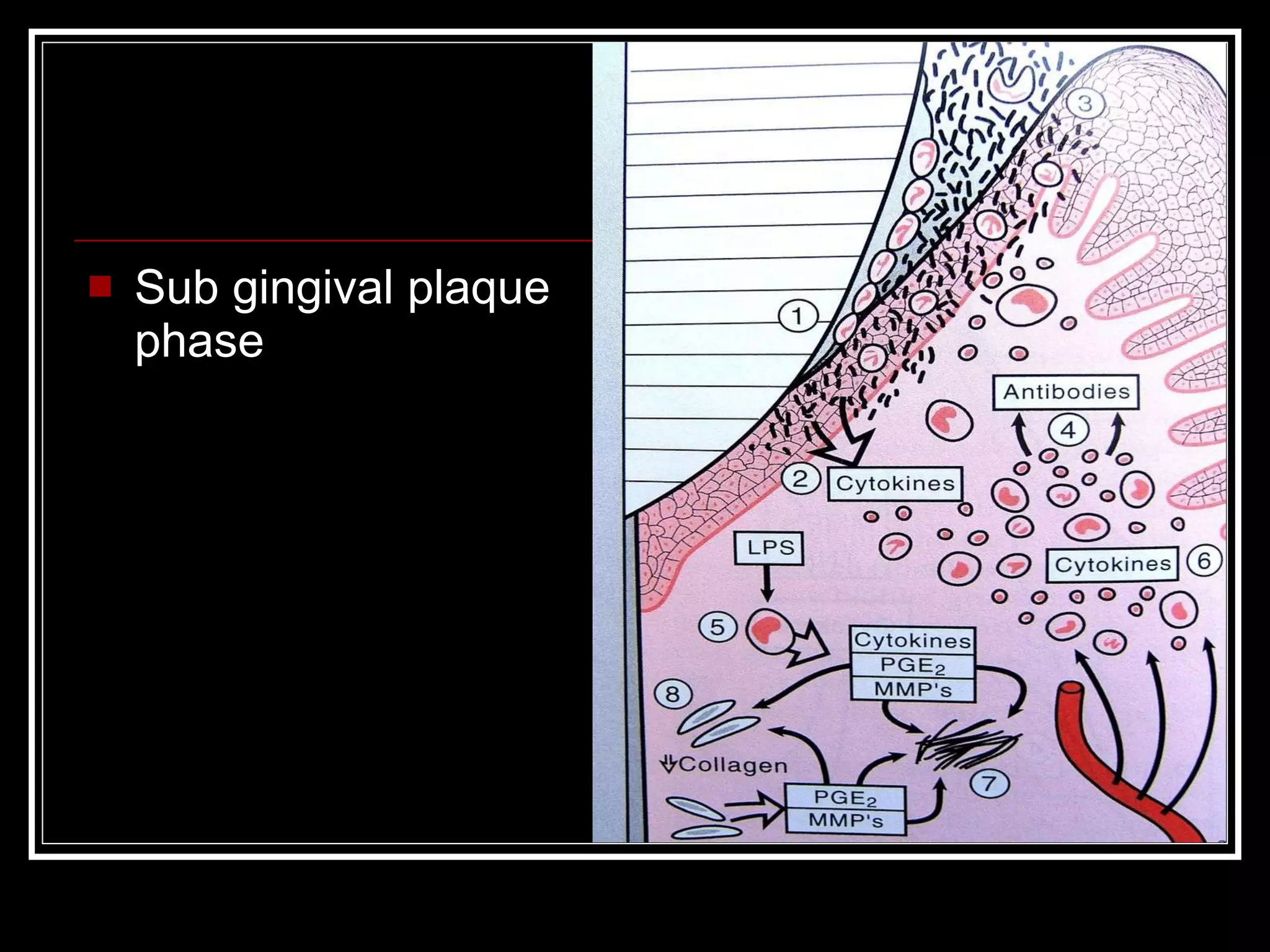
Dental plaque
INTRODUCTION 1 mm 3  of dental plaque, weighing about 1 mg contains more than 200 million bacteria
Index  Definition Classification Composition Clinical feature Formation Clinical significance Diagnosis & treatment Conclusion References
Definition IT  IS  A  MICROBIAL  ECOSYSTEM OR BIOFILM  COMPOSED  OF  DENSELY  PACKED MICROBIAL  STRUCTURE  INSOLUBLE SALIVARYGLYCOPROTEIN , MICROBIAL INTRACELLULAR  PRODUCT & TO  SOME EXTENT  EPITHELIAL CELLS  &  DEBRIES ARRANGED  IN  AN  ORGANISED  COMPLEX INTER  CELLULAR  MATRIX. - BY WHO
by location on tooth supra gingival  plaque  sub gingival plaque  coronal  marginal  attached  unattached  tooth asso.  tissue asso. CLASSIFICATION
 
By pathogenic effects Cariogenic  periodontal ds’  calculogenic plaque  producing plaque  plaque
PRIMARILY OF MICRO-ORGANISMS Gram +ve  -  s.mutans s.Sanguis s.Milleri s.Salivarius Gram –ve  - a.Viscosus a.Naslundi a.Israelli Gram –ve anaerobic cocci – Vellionellae v.parvula COMPOSITION
 
Organic : -  Polysaccharide Protein Glycoprotein Lipid Albumin Inorganic : - Phosphorous Sodium Pottasium Fluoride calculus
PLAQUE MICRO-ORGANISMS DAY :- 1-2 Early plaque is consisted of streptococci which dominates bacterial population include,  s. mutans, s.sanguis Day :- 2-4 cocci r still dominate & increase in no of filamentous may be seen. gradually filamentous form grows into cocci layer & replace them.
Day : - 6-10 filamentous increase in no.along with rods, spirilia, & fusobacteria. Plaque near the gingival margin is thicker & develops more mature flora earlier with spirochete & vibrios As plaque matures- more gram –ve & anaerobic organism appear. During the period when this is happening, signs of inflmmation begin to observe in the gingiva. Older plaque :- spirochete & vibrios r prevalent along with cocci & filamentous m.o arranged themselves perpendicular to the tooth surface in a palisade
DENTAL PLAQUE IS A MICROBIAL BIOFILM. Biofilms – “ defined as matrix enclosed bacterial populations adherent to each other and/or to surfaces or interfaces.” ( costerton,1994 )  FORMATION
Biofilm can be formed by a single bacterial species or multiple bacterial species as well as other organisms & debris. It can form on any surfaces that is wet. It can exist on any solid surfaces that is exposed to bacteria-containing fluid.
Biofilm structure The bacteria in a biofilm r not distributed evenly, they cluster to gether to form sessile mushroom shaped microcolonies. Each microcolony is an independent community with its own customised living environment.
A protective extra cellular slime layer surrounds the microcolonies.  A series of fluid channels penetrate the slime layer & facilitate the movement of nutrients & bacterial products throughout the biofilm A primitive communication system of chemical signals allows communication bt. the bacterial microcolonies.
Bacteria in the center of a microcolony may live in a strict anaerobic environment, while other bacteria at the edges of the fluid channel may live in an aerobic environment.
Fluid channels provide nutrients & oxygen for the bacterial microcolonies, waste products & enzymes within the biofilm structure
The bacterial microcolonies use “chemical signals” to communicate with each other.
Bacterial microcolonies r protected by one another or by extracellular slime layer & r usually resistant to antibiotics & antimicrobials, & the body’s defense system. Can be destroyed by simply wipping off them.
FORMATION OF DENTAL PLAQUE BOIFILMS THE PATTERN OF PLAQUE BIOFILM CAN BE DIVIDED INTO 3 PHASES : Attchment of bacteria to the solid surface Formation of microcolonies on the surface Formation of the mature subgingival plaque biofilms
1.Initial attachment of bacteria Pellicle formation – defined as “an acellular layer of salivary proteins & other macromolecules, aproximately 2 to 10 micrometer thick, adsorbed on to the enamel surface.”  It has important role in protecting the enamel from abrasion and attrition but it also serves as diffusion barrier.
A thin, bacteria free layer forms within minutes on a cleaned tooth surface.
The purpose is to protect the enamel from acidic activity. Acts like a double sided adhesive tape with their amino- terminal segments to the tooth surface,leaving their carboxy-terminal regions directed towards the  oral cavity,where they may interact with oral micro-organisms
Initial colonization of the tooth surface :  within few hours bacteria connect to the pellicle & each other with hundreds of hair like structures called fimbriae. Stimulate other free floating bacteria to join the community
4 hrs. after cleaning, there r 103 to 104 bacteria / 1 mm 2  of tooth surface,predominently streptococci & actinomycetes. Within a day a no. of bacteria increases, due to growth of streptococci The initial bacteria r called “pioneer bacteria” It is interection bt. Pr. Adhesion on the surface of colonizing bacteria & carbo. Receptor on the salivary components adsorbed to the tooth surface.
If within 2 days no further cleaning is undertaken, the tooth surface is colonized predominantly by gram +ve facultative cocci, which r primarily streptococci. Extra cellular slime layer formation : The act of attaching to the solid surface stimulate the bacteria to excrete a slimy glue like substance that helps to anchor them to the surface & provides protection for attached bacteria
2.Microcolony formation Bacterial growth :- primarily through cell division of the adherent bacteria(rather than attachment of new bacteria) Next proliferating bacteria begin to grow away from the tooth. Plaque doubling time are rapid in early development & slow in more mature biofilms. Bacterial blooms -  r periods when specific species or grp. Of species grow at rapidly accelerated rate
Coaggregation into mushroom shaped microcolonies It is ability of new bacterial colonizer to adheres to previously attached cells  Forms sessile, mushroom shaped microcolonies that r attached to tooth surface at a narrow base & gives “corn cob” appearance or “taste tube brushes.”
Central rod shaped bacterium becomes surrounded by many round cocci  “ bristles” of taste tube brush formation r composed of gram –ve rods.
3. Formation of mature subgingival plaque biofilm
Sub gingival plaque biofilm
Relationship of plaque biofilm to alveolar bone The distance bt. Apical edge of calculus & crest of alveolar bone in human pd pocket is constant, having mean length of 1.97 mm.  (indicates that bacteria r capable of producing bone resorbing activity only in alv. Bone no further than 2.7mm away from biofilm.
Microbial interactions
Although bacteria initiate the periodontal disease process, it is the body’s own immune response that is actually responsible for the breakdown of periodontal tissues.  Immune system complex in periodontitis Phagocytes  ( the army ) Lymphocytes ( the air force ) The complement system ( the navy )
Neutrophil
Macrophage
Lymphocyte
Complementary system 3 major function Recruitment of phagocytic cells Facilitation of phagocytosis by opsonization Direct killing of bacteria by puncturing the bacterial cell membrane
Phagocytosis
Sequence of events in host response
Early bacterial accumulation phase
Plaque over growth phase : acute inflammation
Sub gingival plaque phase
Tissue destruction phase
Tissue destruction in periodontitis
Direct vision : - Thin plaque – may be translucent & therefore not visible Stained plaque – may be acquired e.g tobacco stained Thick plaque – tooth may appear dull & durty DETECTION OF PLAQUE
USE OF EXPLORER : - Tactile Examination – when calcification has started it appears slightly rough, otherwise it may feel slippery due to coating of soft , slimy plaque Removal Of Plaque – when no plaque is visible , an explorer can be passed over the tooth surface & when plaque is present it will adhere to explorer tip.this technique is used when evaluating plaque index. Use of disclosing solutions -
SUPRA GINGIVAL PLAQUE Occlusal surface leads to caries Gingivitis SUB GINGIVAL PLAQUE Periodontitis Other periodontal disease CLINICAL SIGNIFICANCE
Treatment  Several investigations r being carried out to develop plaque inhibiting agents which could be applied as mouth rinse, tooth paste,& chewing gums. They interfere in plaque formation by Prevention of plaque formation or alteration of plaque microflora by anti microbial substance Prevention or absorption of organic matrix of the teeth By means of enzymes capable of decomposing the inter microbial substance
Surface treatment Chemical plaque inhibition is very little successful Antibiotics Other antimicrobial substance like chemical disinfectant Chlorhexidine longer time use is not preferable because it causes discoloration of mucus membrane
CONCLUSION PLAQUE + TIME = GINGIVITIS GINGIVITIS +TIME = PERIODONTITIS PERIODONTITIS + TIME = TOOTH LOSS
REFERENCES FOUNDATION OF PERIODONTICS - By donald E. willmann DIAGNOSIS & RISK PREDICTION OF PERIODONTAL DISEASE - per axelson,dds,odont dr CLINICAL PERIODONTOLOGY  - caranza ( 9 th edition)

Dental plaque

  • 1.
  • 2.
    INTRODUCTION 1 mm3 of dental plaque, weighing about 1 mg contains more than 200 million bacteria
  • 3.
    Index DefinitionClassification Composition Clinical feature Formation Clinical significance Diagnosis & treatment Conclusion References
  • 4.
    Definition IT IS A MICROBIAL ECOSYSTEM OR BIOFILM COMPOSED OF DENSELY PACKED MICROBIAL STRUCTURE INSOLUBLE SALIVARYGLYCOPROTEIN , MICROBIAL INTRACELLULAR PRODUCT & TO SOME EXTENT EPITHELIAL CELLS & DEBRIES ARRANGED IN AN ORGANISED COMPLEX INTER CELLULAR MATRIX. - BY WHO
  • 5.
    by location ontooth supra gingival plaque sub gingival plaque coronal marginal attached unattached tooth asso. tissue asso. CLASSIFICATION
  • 6.
  • 7.
    By pathogenic effectsCariogenic periodontal ds’ calculogenic plaque producing plaque plaque
  • 8.
    PRIMARILY OF MICRO-ORGANISMSGram +ve - s.mutans s.Sanguis s.Milleri s.Salivarius Gram –ve - a.Viscosus a.Naslundi a.Israelli Gram –ve anaerobic cocci – Vellionellae v.parvula COMPOSITION
  • 9.
  • 10.
    Organic : - Polysaccharide Protein Glycoprotein Lipid Albumin Inorganic : - Phosphorous Sodium Pottasium Fluoride calculus
  • 11.
    PLAQUE MICRO-ORGANISMS DAY:- 1-2 Early plaque is consisted of streptococci which dominates bacterial population include, s. mutans, s.sanguis Day :- 2-4 cocci r still dominate & increase in no of filamentous may be seen. gradually filamentous form grows into cocci layer & replace them.
  • 12.
    Day : -6-10 filamentous increase in no.along with rods, spirilia, & fusobacteria. Plaque near the gingival margin is thicker & develops more mature flora earlier with spirochete & vibrios As plaque matures- more gram –ve & anaerobic organism appear. During the period when this is happening, signs of inflmmation begin to observe in the gingiva. Older plaque :- spirochete & vibrios r prevalent along with cocci & filamentous m.o arranged themselves perpendicular to the tooth surface in a palisade
  • 13.
    DENTAL PLAQUE ISA MICROBIAL BIOFILM. Biofilms – “ defined as matrix enclosed bacterial populations adherent to each other and/or to surfaces or interfaces.” ( costerton,1994 ) FORMATION
  • 14.
    Biofilm can beformed by a single bacterial species or multiple bacterial species as well as other organisms & debris. It can form on any surfaces that is wet. It can exist on any solid surfaces that is exposed to bacteria-containing fluid.
  • 15.
    Biofilm structure Thebacteria in a biofilm r not distributed evenly, they cluster to gether to form sessile mushroom shaped microcolonies. Each microcolony is an independent community with its own customised living environment.
  • 16.
    A protective extracellular slime layer surrounds the microcolonies. A series of fluid channels penetrate the slime layer & facilitate the movement of nutrients & bacterial products throughout the biofilm A primitive communication system of chemical signals allows communication bt. the bacterial microcolonies.
  • 17.
    Bacteria in thecenter of a microcolony may live in a strict anaerobic environment, while other bacteria at the edges of the fluid channel may live in an aerobic environment.
  • 18.
    Fluid channels providenutrients & oxygen for the bacterial microcolonies, waste products & enzymes within the biofilm structure
  • 19.
    The bacterial microcoloniesuse “chemical signals” to communicate with each other.
  • 20.
    Bacterial microcolonies rprotected by one another or by extracellular slime layer & r usually resistant to antibiotics & antimicrobials, & the body’s defense system. Can be destroyed by simply wipping off them.
  • 21.
    FORMATION OF DENTALPLAQUE BOIFILMS THE PATTERN OF PLAQUE BIOFILM CAN BE DIVIDED INTO 3 PHASES : Attchment of bacteria to the solid surface Formation of microcolonies on the surface Formation of the mature subgingival plaque biofilms
  • 22.
    1.Initial attachment ofbacteria Pellicle formation – defined as “an acellular layer of salivary proteins & other macromolecules, aproximately 2 to 10 micrometer thick, adsorbed on to the enamel surface.” It has important role in protecting the enamel from abrasion and attrition but it also serves as diffusion barrier.
  • 23.
    A thin, bacteriafree layer forms within minutes on a cleaned tooth surface.
  • 24.
    The purpose isto protect the enamel from acidic activity. Acts like a double sided adhesive tape with their amino- terminal segments to the tooth surface,leaving their carboxy-terminal regions directed towards the oral cavity,where they may interact with oral micro-organisms
  • 25.
    Initial colonization ofthe tooth surface : within few hours bacteria connect to the pellicle & each other with hundreds of hair like structures called fimbriae. Stimulate other free floating bacteria to join the community
  • 26.
    4 hrs. aftercleaning, there r 103 to 104 bacteria / 1 mm 2 of tooth surface,predominently streptococci & actinomycetes. Within a day a no. of bacteria increases, due to growth of streptococci The initial bacteria r called “pioneer bacteria” It is interection bt. Pr. Adhesion on the surface of colonizing bacteria & carbo. Receptor on the salivary components adsorbed to the tooth surface.
  • 27.
    If within 2days no further cleaning is undertaken, the tooth surface is colonized predominantly by gram +ve facultative cocci, which r primarily streptococci. Extra cellular slime layer formation : The act of attaching to the solid surface stimulate the bacteria to excrete a slimy glue like substance that helps to anchor them to the surface & provides protection for attached bacteria
  • 28.
    2.Microcolony formation Bacterialgrowth :- primarily through cell division of the adherent bacteria(rather than attachment of new bacteria) Next proliferating bacteria begin to grow away from the tooth. Plaque doubling time are rapid in early development & slow in more mature biofilms. Bacterial blooms - r periods when specific species or grp. Of species grow at rapidly accelerated rate
  • 29.
    Coaggregation into mushroomshaped microcolonies It is ability of new bacterial colonizer to adheres to previously attached cells Forms sessile, mushroom shaped microcolonies that r attached to tooth surface at a narrow base & gives “corn cob” appearance or “taste tube brushes.”
  • 30.
    Central rod shapedbacterium becomes surrounded by many round cocci “ bristles” of taste tube brush formation r composed of gram –ve rods.
  • 31.
    3. Formation ofmature subgingival plaque biofilm
  • 32.
  • 33.
    Relationship of plaquebiofilm to alveolar bone The distance bt. Apical edge of calculus & crest of alveolar bone in human pd pocket is constant, having mean length of 1.97 mm. (indicates that bacteria r capable of producing bone resorbing activity only in alv. Bone no further than 2.7mm away from biofilm.
  • 34.
  • 35.
    Although bacteria initiatethe periodontal disease process, it is the body’s own immune response that is actually responsible for the breakdown of periodontal tissues. Immune system complex in periodontitis Phagocytes ( the army ) Lymphocytes ( the air force ) The complement system ( the navy )
  • 36.
  • 37.
  • 38.
  • 39.
    Complementary system 3major function Recruitment of phagocytic cells Facilitation of phagocytosis by opsonization Direct killing of bacteria by puncturing the bacterial cell membrane
  • 40.
  • 41.
    Sequence of eventsin host response
  • 42.
  • 43.
    Plaque over growthphase : acute inflammation
  • 44.
  • 45.
  • 46.
    Tissue destruction inperiodontitis
  • 47.
    Direct vision :- Thin plaque – may be translucent & therefore not visible Stained plaque – may be acquired e.g tobacco stained Thick plaque – tooth may appear dull & durty DETECTION OF PLAQUE
  • 48.
    USE OF EXPLORER: - Tactile Examination – when calcification has started it appears slightly rough, otherwise it may feel slippery due to coating of soft , slimy plaque Removal Of Plaque – when no plaque is visible , an explorer can be passed over the tooth surface & when plaque is present it will adhere to explorer tip.this technique is used when evaluating plaque index. Use of disclosing solutions -
  • 49.
    SUPRA GINGIVAL PLAQUEOcclusal surface leads to caries Gingivitis SUB GINGIVAL PLAQUE Periodontitis Other periodontal disease CLINICAL SIGNIFICANCE
  • 50.
    Treatment Severalinvestigations r being carried out to develop plaque inhibiting agents which could be applied as mouth rinse, tooth paste,& chewing gums. They interfere in plaque formation by Prevention of plaque formation or alteration of plaque microflora by anti microbial substance Prevention or absorption of organic matrix of the teeth By means of enzymes capable of decomposing the inter microbial substance
  • 51.
    Surface treatment Chemicalplaque inhibition is very little successful Antibiotics Other antimicrobial substance like chemical disinfectant Chlorhexidine longer time use is not preferable because it causes discoloration of mucus membrane
  • 52.
    CONCLUSION PLAQUE +TIME = GINGIVITIS GINGIVITIS +TIME = PERIODONTITIS PERIODONTITIS + TIME = TOOTH LOSS
  • 53.
    REFERENCES FOUNDATION OFPERIODONTICS - By donald E. willmann DIAGNOSIS & RISK PREDICTION OF PERIODONTAL DISEASE - per axelson,dds,odont dr CLINICAL PERIODONTOLOGY - caranza ( 9 th edition)