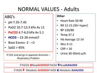
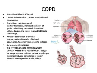
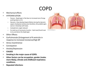
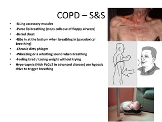
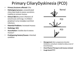
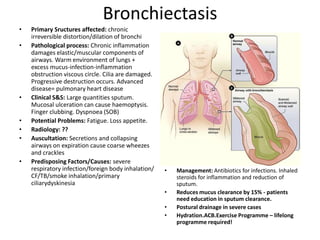
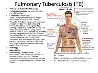
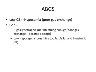
CVR Conditions
           Wiki/You tube: bronchoscopy/ABG
      demo/CPAP/BIPAP/AUSCULTATION/Incentive
    spirometry/IPPB Bird/Couch Assist/Chest Physical
Therapy/Suctioning Mobility & ICU/COPD Living/Cardiac or
                pulmonary rehabilitation
          Blackboard: Differences in Children
NORMAL VALUES - Adults
ABG’s                                          Other
                                               • Heart Rate 50-90
•    pH 7.35-7.45
                                               • RR 12-15 (30+ hyper)
•    PaO2 10.7-13.3 kPa Av 11
                                               • BP 120/80
•    PaCO2 4.7-6.0 kPa Av 5.1
                                               • Temp 37.2
•    HCO3 – 22-26 mmol/l
                                               • Hb Average 12-14
•    Base Excess -2 - +2                       • Wcc 4-11
•    Sa02 > 95%                                • CRP < 10
    If CO2 and pH go in opposite direction –   • Urine 30-50mls p/h
             Respiratory Problem

                If PaCO2 RespACIDOSISIf PaC02 RespALKALOSIS
          If HC03  Metabolic ACIDOSISIf HC03  Metabolic ALKALOSIS
COPD
•   Bronchi and Alveoli Afffected
•   Chronic inflammation - chronic bronchitis and
    emphysema
•   Bronchioles – destruction of
    elastin/decilliation/increased numbers of
    goblet cells - lining becomes irritated and
    inflamed producing excess mucus that blocks
    the airways.
•   Alveoli – destruction of alveolar
    septum, reduced transfer of O2 and
    CO2, bullae, floppy airways prone to collapse
•   Slow progressive disease
•   THE EFFECTS OF COPD MEAN THAT LESS
    OXYGEN PASSES INTO YOUR BLOOD . less gas
    exchange area and reduced surface area for gas
    exchange/reduced compliance of lungs?
    Alveolar interdependence affected too
COPD
•   Mechanical effects
•   HYPERINFLATION
     –   Passive - Diaphragm is flat (due to increased size of lungs
         pushing down on it)
     –   Dynamic- they develop hyperinflation during the dynamic
         aspect of physical activity and, therefore, the problem gets
         worse or develops with tasks.
     –   Costophrenic angle reduced
     –   So need use of accessory muscles – bent over/hunch over
         to try and dome the diaphragm
•   Other Effects:
•   CorPulmonale (Enlargement of R ventricle in
    response to increased resistance/High BP
•   Stress incontinence
•   Constipation
•   Anxiety/Depression
•   CAUSES
•   Smoking is the major cause of COPD.
•   Other factors can be occupation, gender (males
    more likely), climate and childhood respiratory
    conditions.
•   Repeated infections
COPD – S&S
•   -Using accessory muscles
•   -Purse lip breathing (stops collapse of floppy airways)
•   -Barrel chest
•   -Ribs in at the bottom when breathing in (paradoxical
    breathing)
•   -Chronic dirty phlegm
•   -Wheezing or a whistling sound when breathing
•   -Feeling tired / Losing weight without trying
•   Hypercapnia (Hich PaCo2 in advanced disease) use hypoxic
    drive to trigger breathing
COPD- Complications/Treatment
TREATMENT
• Quit smoking – advice on smoking cessation
• Adopt a healthy lifestyle to prevent infections
Medications-
• Bronchodilators (inhaler) treat SOB
• corticosteroid pills prevent and treat COPD flare ups
• antibitotics treat infections that cause flare ups
• flu and pneumonia vaccines
• supplemental oxygen
COPD- Physio
•   PHYSIOTHERAPY
•   -Pulmonary rehabilitation therapy
•   Postural/gravity assisted position
•   -Exercise is very important to help stay
    fitter for longer and improve
    breathlessness
•   -Diaphragmatic breathing
•   -Active cycle of breathing
•   -Controlled coughing to get rid of mucus
    and prevent infections
•   -Percussion techniques
•   Long term O2
•   EXACERBATION – clear secretions and
    reduce WOB. Non – invasive ventilation
•   BIPAP
•   Bear in mind what their NORMAL o2 sat is
    (some use low O2 sats to trigger breathing)
•   MRC Dysponea scale
Primary CiliaryDyskinesia (PCD)
•   Primary Sructures affected: Cilia
•   Pathological process: uncoordinated
    ciliary beat leads to excess secretions.
    Inability to remove mucous
•   Clinical S&S:Recurrent infections of
    ears/sinuses and lungs. In children-
    perpetual runny nose/glue ear/chest
    infections
•   Potential Problems: Increased mucous
•   Radiology: N/A
•   Auscultation: Crackles due to excess
    mucous?
•   Predisposing Factors/Causes: Inherited
    condition

                                               •   Management: Not a progressive disorder.
                                                   Antibiotics and physio needed to delay onset of
                                                   bronchiectasis.
                                               •   Chest Physical Therapy to aid mucous removal
                                               •   Cough Assist?????
Bronchiectasis
•   Primary Sructures affected: chronic
    irreversible distortion/dilation of bronchi
•   Pathological process: Chronic inflammation
    damages elastic/muscular components of
    airways. Warm environment of lungs +
    excess mucus-infection-inflammation
    obstruction viscous circle. Cilia are damaged.
    Progressive destruction occurs. Advanced
    disease= pulmonary heart disease
•   Clinical S&S: Large quantities sputum.
    Mucosal ulceration can cause haemoptysis.
    Finger clubbing. Dyspnoea (SOB)
•   Potential Problems: Fatigue. Loss appetite.
•   Radiology: ??
•   Auscultation: Secretions and collapsing
    airways on expiration cause coarse wheezes
    and crackles
•   Predisposing Factors/Causes: severe
    respiratory infection/foreign body inhalation/   •   Management: Antibiotics for infections. Inhaled
    CF/TB/smoke inhalation/primary                       steroids for inflammation and reduction of
    ciliarydyskinesia                                    sputum.
                                                     •   Reduces mucus clearance by 15% - patients
                                                         need education in sputum clearance.
                                                     •   Postural drainage in severe cases
                                                     •   Hydration.ACB.Exercise Programme – lifelong
                                                         programme required!
Pulmonary Tuberculosis (TB)
•   Primary Sructures affected: Lungs
•   Pathological process: bacterial infection
    from TB bacillus.
•   Clinical S&S: Fever/Night
    Sweats/Cough/Chest wall pain/Weight
    Loss/Haemoptysis and SOB, fatigue
•   Potential Problems: Tissue destruction and
    necrosis are balanced by healing and
    fibrosis.Affected tissue is replaced by
    scarring and cavities filled with cheese-like
    white necrotic material. During active
    disease, some of these cavities are joined to
    the air passages bronchi and this material
    can be coughed up. It contains living bacteria
    and can therefore pass on infection.
    Treatment with appropriate antibiotics kills
    bacteria and allows healing to take place.
    Upon cure, affected areas are eventually
    replaced by scar tissue.
•   Radiology:
•   Auscultation:
•   Predisposing Factors/Causes: 1/3 of              •   Management: Antibiotics for up to 6 months –
    population is infected with TB, which can            very resilient
    become active if the hosts defence
    mechanisms are compromised due to poor           •   Devise exercise regimes within isolations rooms
    living/drug dependency/HIV. Diabetes.Low         •   Air – filtered mask to be worn at all times!!
    Weight
Asthma
•   Asthma is a long-term condition
•   Bronchioles are affected in asthma
•   Chronic inflammatory with airway narrowing
    and mucus plugging
•   Pathological process:
•   an asthma trigger stimulates excess
    immunoglobulin E (IgE) in serum. This fixes to
    Mast cells which then react to antigens and
    release bronchorestrictor substances-
    histamine
•   the muscles around bronchioles tighten
    airways become narrower and the lining of the
    airways becomes inflamed and starts to swell.
•   air flow can further be decreased by
    inflammation or excess mucus secretion
•   PERSISTENT INFLAMMATION leads to fibrosis
    of airway walls and irreversibility
•   Common triggers include house dust
    mitesanimal fur, pollen, tobacco
    smoke, exercise, cold air and chest infections.
Asthma – S& S
•   Clinical S&S: :
•   SOB
•   Tightening of the chest
•   Wheezing which can make a whistling sound when you breathe.
•   Coughing
•   Features of acute asthma:
•   PaO2 ↓ RR >25 PaCO2 ↓ Pulse>110 BP↑
•   DIAGNOSIS
•   The spirometer takes two measurements: the volume of air you can breathe out in
    one second (called the forced expiratory volume in one second or FEV1) and the
    total amount of air you breathe out (called the forced vital capacity or FVC). This
    can be done by the GP.

•   Predisposing Factors/Causes: A family history, Prolonged exposure to tabacco
    smoke as a child, Food allergy, Bronchiolitis as a child, Prem baby, Anxiety
Asthma - Management
Management: . Asthma can be well controlled in most
  people most of the time.
• Inhalers
• Inhaling a drug is an effective way of taking an asthma
  medicine as it goes straight to the lungs, with very little
  ending up elsewhere in the body. The drug used is
  Salbutomol – widens airways
• Steroids are also used but these can have side effects in
  the long term.
• PHYSIOTHERAPY MANAGEMENT
• Breathing techniques can be used to help the patient to
  relax and feel in control - simple gentle abdominal
  breathing is used.
• Exercise can help if aerobic training is used.
Asthma
Cystic Fibrosis
Cystic Fibrosis
• Primary Structures affected:
• It is a chronic progressive obstructive disorder affecting
  the exocrine glands. It is an inherited disease. Cystic
  Fibrosis affects the internal organs, especially the lungs
  and digestive system, by clogging them with thick sticky
  mucus. This makes it hard to breathe and digest food.

• Pathological process: CF is caused by a mutation in the
  gene for required to regulate the components of
  sweat, digestive juices, and mucus.
Cystic Fibrosis- S&S
• Clinical S&S: Symptoms of CF can include a
  troublesome cough, repeated chest
  infections, prolonged diarrhoea and poor weight
  gain. These symptoms are not unique to Cystic
  Fibrosis – difficult diagnosis
• CF affects the pancreas, which makes it difficult for
  people with CF to digest food. This can cause
  malnutrition, which can lead to poor growth, physical
  weakness and delayed puberty.
Cystic Fibrosis- Physio
•   Predisposing Factors/Causes: Genetic
•   Management:
•   Chest physiotherapy is important
    because helps to prevent the
    thick, sticky lung secretions from
    blocking the air tubes. This helps to
    reduce infection and prevent lung
    damage.
•   ACTIVE CYCLE OF BREATHING
•   POSITIVE EXPIRATORY PRESSURE
•   OSCILLATING POSITIVE EXPIRATORY
    PRESSURE e.g. acapella, flutter.
•   Teach family how to do percussions
    etc
•   The patient needs to do exercise to
    keep the lungs from deteriorating
    and to improve physical strength and
    bullk.
•   Stretching exercises and being taught
    correct posture can help.
Acute Renal Failure
• Primary Sructures affected: rapid loss of kidney function
Acute Renal Failure
Acute Renal Failure
• S&S: Puffy ankles as kidneys not removing proteins so BCOP goes
  up – more fluid pushed into interstitial fluid space?? Also fluid
  retention causes oedema
• Potential Problems: metabolic acidosis ( Kidneys retaining acid),
  high potassium levels, uremia, changes in body fluid balance, and
  effects to other organ systems.
• Management: hinges on identification and treatment of the
  underlying cause. In addition to treatment of the underlying
  disorder, management of AKI routinely includes the avoidance of
  substances that are toxic to the kidneys, called nephrotoxins. These
  include NSAIDs such as ibuprofen, iodinated contrasts such as those
  used for CT scans, and others.Monitoring of renal function, by serial
  serum creatinine measurements and monitoring of urine output, is
  routinely performed. In the hospital, insertion of a urinary catheter
  helps monitor urine output and relieves possible bladder outlet
  obstruction, such as with an enlarged prostate.
Diabetes
• Primary Sructures
  affected:
• Pathological process:
• Clinical S&S
• Potential Problems
• Radiology
• Auscultation
• Predisposing
  Factors/Causes
• Management
Pulmonary Fibrosis/ idiopathic pulmonary fibrosis
Primary structures effected:               In severe cases, symptoms can be
Lung tissue                                significantly worse and may include
Pathological progress:                     congestive heart failure, respiratory
                                           failure, and strokes
is caused by repeated injury to small
    areas of lung. Results in
    inflammation of lungs causing
    scarring and stiffness.
Clinical signs and symptoms:
Thickened lung tissues cause
    decreased oxygen uptake from
    blood, brain and other organs don’t
    get enough oxygen.
SOB, clubbing of nails, shallow
    breathing . Aching muscles and
    joints, decreased exercise tolerance
Potential problems :decreased o2 to
    the brain, chronic dry cough.
Radiograpy:                                       Management:
•   10% of patients with IPF have normal      •   Allergen avoidance.
    chest x-rays. X-ray usually reveals       •   At present for IPF, there is no treatment
    shadows, mostly in the lower part of          available which has been shown to
    the lungs. In addition, lung size tends       improve the condition.
    to appear smaller than normal.            •   Counselling may be required in view of the
Auscultation: may present with                    poor prognosis.
   crackles heard late in inspiration.        •   Supportive therapy may be helpful in the
                                                  form of oxygen therapy and physiotherapy
Predisposing factors/Causes:                      if bronchiectasisis present.
No common cause known, can be a               •   Patients should be encouraged to take
                                                  regular exercise to maintain their exercise
   side effect of
                                                  tolerance.
   medication, expositor to
                                              •   Patients should be encouraged to be
   pollution, smoking, viral                      vaccinated against influenza and
   infections.                                •   Lung transplant is the only option which
                                                  improves long-term survival.3
                                              •                                    (patient.uk)
• The pulmonary fibrosis organisation suggests:

• Pulmonary rehabilitation :exercise training;
  breathing exercises and retraining;
  anxiety, stress and depression management;
  and nutritional counseling, to name a few.
Pneumonia
Primary Sructures affected:
lung tissue

Pathological process:
• infection of one or both lungs which is usually caused by
    bacteria, viruses, or fungi. contracted by breathing in small droplets that
    contain the organisms that can cause pneumonia. Normally immune
    system would suppress virus. if a person is in a weakened condition from
    another illness, a severe pneumonia can develop.
• Alveoli fill with fluid
Clinical S&S:
• symptoms of cold, high fever, shaking, discolored sputum
    production, sharp chest pain, skin color may change due to deoxygenating
Potential Problems:
• ifthe spongy tissue of the lungs that contain the air sacs is more
    involved., oxygenation can be impaired, along with stiffening of the
    lung, which results in SOB. if low immune response pneumonia may be life
    threatening.
A)Normal chest, B) pneumonia
• Radiology:
• Auscultation: finecrackles,
  sometimes pleural rub.
• Predisposing
  Factors/Causes: very young,
  very old, if immune system
  is weakened by any
  predisposing factors like
  long term illness
• Management: oral or
  intervenous fluids
• Antiviral/antibacterial drugs
• Oxygen if indicated
• In acute stage physio is limited to positioning to much Va/Q
  and CPAP if hypoxemia persists despite 40% oxygen.

• Modified postural drainage - this allows gravity to drain
  secretions from specific segments of the lungs
• IPPB Machine
• Shaking and vibes - to mobilize secretions
• Coughing and huffing exercises - to expectorate secretions
• Administer humidification - to mobilize secretions
• Breathing exercises - Localized and Diaphragmatic
• IPPB administration - to increase lung volumes
• Mobilization of the patient - done to increase air entry,
  increase chest expansion, and to loosen secretions
Pneumothorax
Primary Sructures affected: pleural cavity
    between lung and chest wall
Pathological process: may occur spontaneously
    (primary) or in people with lung conditions
    (secondary). If either pleural layer is ruptured
    gas rushes into the pleural space either from
    inside the lung or from outside. the tear
    often occurs at the site of a tiny 'bullae' on
    the edge of a lung
• Clinical S&S:
• The typical symptom is a sharp, stabbing pain
    on one side of the chest which suddenly
    develops.
• The pain is usually made worse by breathing
    in (inspiration).
• Pt may become breathless. As a rule, the
    larger the pneumothorax, the more
    breathless .
• Pt may have other symptoms if an injury or a
    lung disease is the cause. For example, cough
    or fever.
Potential Problems:
   breathlessness, collapsed lung
Radiology:
Auscultation: decreased or absent
   breath sounds
Predisposing Factors/Causes: may
   occur spontaneously in
   healthy fit people(primary) or
   in people with lung conditions
   (secondary)
Management:
• small pneumothorax is left to
   heal
• Larger pneumothorax can be
   treated with a ‘Heimlich valve’
   which allows air to escape but
   not to re-enter.
• Chest drains may be used
• Physio is based on education in chest drain management
• Positioning: lying on good side often most comfortable and
  best for VA/Q , lying of affected side my speed absorbition of
  air.
• Precautions include avoidance of positive pressure
  techniques.
Pleural Effusion
Primary Sructures affected:
Pleural space
Pathological process: excess
   fluid in pleural cavity
   caused by disturbed
   osmotic or hydrostatic
   pressure in plasma or
   changes in membrane
   permeability.
Clinical S&S: dull percussion
   note, SOB, sometimes
   chest pain
Potential Problems: those of
   underlying illness, decrease
   lung function
Radiology: ?fluide line tracking up
   the pleura laterally?
Auscultation: decreassed breath
   sounds, bronchial breathing
Predisposing Factors/Causes:
   malignancy causes 25% of
   pleural effusion other causes
   are heart, kidney, or liver
   failure, abdominal or cardiac
   surgery, pneumonia or TB
Management:
On large effusions, drainage of
   chest wall.
Smaller effusions may just left
   under observation.
• Deep breathing exercises can not expand the
  lungs under pressure of fluid.
• People with moderate unilateral effusion may
  benefit from side-lying affected side upmost
  as both ventilation and perfusion are greater
  in lower lung.
• Large effusions are likely to show improved
  PaC2 with effusion downwards to minimize
  compression of unaffected lung.
Pulmonary Embolus
Primary Sructures affected: blood vessels in the
    lung
Pathological process: blockage of an artery in the
    lungs by fat, air, a blood clot

Clinical S&S:include unexplained shortness of
     breath, problems breathing, chest
     pain, coughing, or coughing up blood. An
     arrhythmia (an irregular heartbeat) also may
     indicate PE. In some cases, the only signs and
     symptoms are related to deep vein
     thrombosis (DVT). These include swelling of
     the leg or along the vein in the leg, pain or
     tenderness in the leg,

•   Potential Problems: Severe breathlessness.
•   Feeling faint, feeling unwell, or a collapse.
    This is because a large blood clot interferes
    with the heart and blood circulation, causing
    the blood pressure to drop dramatically.
•   Rarely, in extreme cases, a massive PE can
    cause cardiac arrest, where the heart stops
    pumping due to the clot. This can result in
    death, even if resuscitation is attempted
Radiology:?
Auscultation: decrease in breath sounds
Predisposing Factors/Causes:
• most commonly deep vain thrombosis, blood clot breaks off and travels
   to the lung.
• Other causes : Fatty material from the marrow of a broken bone (if a
   large, long bone is broken - such as the femur (thigh bone).
• Foreign material from an impure injection. For example, with drug
   misuse.
• A small piece of tumour (cancer) that has broken off from a larger tumour
   in the body.
Management:
• Anticoagulant treatment.
• Oxygen given in the early stages to help with breathlessness and low
   oxygen levels.
Hough ( p. 114, 2001) ‘..any active physiotherapy is considered risky until
   anticoagulation therapy is established.’
CCF/LVF/RVF
• CCF                     • LVF               •   RVF
• Both left and right     • Usually caused •      Can be caused
  heart failure with
  congestions in            by coronary           by LVF or
  pulmonary and             heart disease –       Chronic
  systemic                  failing LV forces     pulmonary
  circulation               up pressure in        hypertension
• Pulmonary                 left atrim and        caused by
  Oedema                    pulmonary             hypoxic
  (extravascular
  water in lungs)           vasculature =         conditions
  caused by back            pulmonary             (pulmonary
  pressure from             oedema                vasculature
  failing Left side                               constrict if 02
                          • Pitting Oedema
                                                  reduced)
Smoking
• Primary Sructuresaffected:Lungs!
• Pathological process: inflammation/damage to
  cilia/surfactant/Alveoli /Smooth muscle hypertrophy
• Increase in secretions but decreased ability of
  mucocilliary escalator
• Alveolar gas exchange decreased/ V/Q Matching
• Fibrotic changes from ongoing inflammation
• Increase in IgE and IgM – markers of increased sensitivity
• Immune responses decreased – more infections
• LUNG CLOSING VOLUMES INCREASED – more airway
  collapse will result
• Narrowing/Clogging of arteries/atherosclerosis/increase
  cholesterol/stiffening arteries
Peripheral Vascular Disease
• Chain of events leading to      • Risk factors:
  amputation:                     • smoking- roughening
   – Obstruction/resistance/sta     means lipids stick more
     sis of blood – CLOT            easily
   – Pain
                                  • BP- stress on
   – Skin Breakdown
                                    vessels/perpetuates
   – Infection                      atherosclerosis
   – Amputation
                                  • Diabetes-glucose causes
                                    vessel damage/ Plasma
                                    viscosity
                                    increases/diabetic
                                    neuropathy – can’t feel -
                                    infections
Peripheral Vascular Disease
• Thrombus – occludes       • Buerger’s disease –
  vessel reducing blood       vessels become
  flow= ischaemia             inflamed/ clots
• Vessel wall                 form/lumen
  changes/blood flow          obstructed/fibrous
  stasis/changes in blood     tissue forms
  composition
Ischaemia
• Pain                 • INVESTIGATIONS
• Altered sensation      – Dopler
• Loss of peripheral     – Pulses
  pulses
• Decreased muscular
  strength
• Necrosis
• Intermittent
  Claudication
Atherosclerosis
Primary Sructures affected: medium and large
    arteries
Pathological process: become clogged up by
    fatty substances, such as cholesterol. These
    substances are called plaques or
    atheromas.
Clinical S&S
• Atherosclerosis does not usually produce
    any symptoms until your blood circulation
    becomes restricted, or blocked, leading to
    the onset of cardiovascular disease (CVD).
    The type of CVD and its associated
    symptoms will depend on where the
    blockage occurs.
Atherosclerosis
Potential Problems:
• peripheral arterial disease: where the blood supply to your legs is blocked, causing
   muscle pain (intermittent caludication?)
• coronary heart disease: where the main arteries that supply your heart (the
   coronary arteries) become clogged up with plaques
• stroke: a very serious condition where the blood supply to your brain is interrupted
• heart attack: a very serious condition where the blood supply to your heart is
   blocked
• Angina
• Aneurysm
Predisposing Factors/Causes:High fat diet/cholesterol/High Blood
   Pressure/Smoking/Diabetes/no exercise/Alcohol/family history
Management: Medication for BP/cholesterol. Lifestyle changes/Diet/Smoking
   cessation/Alcohol
Arteriosclerosis and atherosclerosis

  Arteriosclerosis and atherosclerosis are two terms that are
sometimes used interchangeably. However, they are different.

 Arteriosclerosis is a general term that describes any group of
diseases where there is a thickening, and a loss of elasticity, of
           the walls of the arteries it is degenerative

   Diabetes is a major factor leading to to it arteriosclerosis
Diaphragmatic Breathing
              This deep breathing is marked
              by expansion of the abdomen
               rather than the chest when
                 breathing. It is generally
               considered a healthier and
               fuller way to ingest oxygen
ABGS
• Low 02 - -Hypoxemia (poor gas exchange)
• Co2 –
  – High Hypercapnia (not breathing enough/poor gas
    exchange – become acidotic)
  – Low Hypocapnia (breathing too fast/a lot and blowing it
    off)

Cvr revision copy

  • 1.
    CVR Conditions Wiki/You tube: bronchoscopy/ABG demo/CPAP/BIPAP/AUSCULTATION/Incentive spirometry/IPPB Bird/Couch Assist/Chest Physical Therapy/Suctioning Mobility & ICU/COPD Living/Cardiac or pulmonary rehabilitation Blackboard: Differences in Children
  • 2.
    NORMAL VALUES -Adults ABG’s Other • Heart Rate 50-90 • pH 7.35-7.45 • RR 12-15 (30+ hyper) • PaO2 10.7-13.3 kPa Av 11 • BP 120/80 • PaCO2 4.7-6.0 kPa Av 5.1 • Temp 37.2 • HCO3 – 22-26 mmol/l • Hb Average 12-14 • Base Excess -2 - +2 • Wcc 4-11 • Sa02 > 95% • CRP < 10 If CO2 and pH go in opposite direction – • Urine 30-50mls p/h Respiratory Problem If PaCO2 RespACIDOSISIf PaC02 RespALKALOSIS If HC03  Metabolic ACIDOSISIf HC03  Metabolic ALKALOSIS
  • 3.
    COPD • Bronchi and Alveoli Afffected • Chronic inflammation - chronic bronchitis and emphysema • Bronchioles – destruction of elastin/decilliation/increased numbers of goblet cells - lining becomes irritated and inflamed producing excess mucus that blocks the airways. • Alveoli – destruction of alveolar septum, reduced transfer of O2 and CO2, bullae, floppy airways prone to collapse • Slow progressive disease • THE EFFECTS OF COPD MEAN THAT LESS OXYGEN PASSES INTO YOUR BLOOD . less gas exchange area and reduced surface area for gas exchange/reduced compliance of lungs? Alveolar interdependence affected too
  • 4.
    COPD • Mechanical effects • HYPERINFLATION – Passive - Diaphragm is flat (due to increased size of lungs pushing down on it) – Dynamic- they develop hyperinflation during the dynamic aspect of physical activity and, therefore, the problem gets worse or develops with tasks. – Costophrenic angle reduced – So need use of accessory muscles – bent over/hunch over to try and dome the diaphragm • Other Effects: • CorPulmonale (Enlargement of R ventricle in response to increased resistance/High BP • Stress incontinence • Constipation • Anxiety/Depression • CAUSES • Smoking is the major cause of COPD. • Other factors can be occupation, gender (males more likely), climate and childhood respiratory conditions. • Repeated infections
  • 5.
    COPD – S&S • -Using accessory muscles • -Purse lip breathing (stops collapse of floppy airways) • -Barrel chest • -Ribs in at the bottom when breathing in (paradoxical breathing) • -Chronic dirty phlegm • -Wheezing or a whistling sound when breathing • -Feeling tired / Losing weight without trying • Hypercapnia (Hich PaCo2 in advanced disease) use hypoxic drive to trigger breathing
  • 6.
    COPD- Complications/Treatment TREATMENT • Quitsmoking – advice on smoking cessation • Adopt a healthy lifestyle to prevent infections Medications- • Bronchodilators (inhaler) treat SOB • corticosteroid pills prevent and treat COPD flare ups • antibitotics treat infections that cause flare ups • flu and pneumonia vaccines • supplemental oxygen
  • 7.
    COPD- Physio • PHYSIOTHERAPY • -Pulmonary rehabilitation therapy • Postural/gravity assisted position • -Exercise is very important to help stay fitter for longer and improve breathlessness • -Diaphragmatic breathing • -Active cycle of breathing • -Controlled coughing to get rid of mucus and prevent infections • -Percussion techniques • Long term O2 • EXACERBATION – clear secretions and reduce WOB. Non – invasive ventilation • BIPAP • Bear in mind what their NORMAL o2 sat is (some use low O2 sats to trigger breathing) • MRC Dysponea scale
  • 8.
    Primary CiliaryDyskinesia (PCD) • Primary Sructures affected: Cilia • Pathological process: uncoordinated ciliary beat leads to excess secretions. Inability to remove mucous • Clinical S&S:Recurrent infections of ears/sinuses and lungs. In children- perpetual runny nose/glue ear/chest infections • Potential Problems: Increased mucous • Radiology: N/A • Auscultation: Crackles due to excess mucous? • Predisposing Factors/Causes: Inherited condition • Management: Not a progressive disorder. Antibiotics and physio needed to delay onset of bronchiectasis. • Chest Physical Therapy to aid mucous removal • Cough Assist?????
  • 9.
    Bronchiectasis • Primary Sructures affected: chronic irreversible distortion/dilation of bronchi • Pathological process: Chronic inflammation damages elastic/muscular components of airways. Warm environment of lungs + excess mucus-infection-inflammation obstruction viscous circle. Cilia are damaged. Progressive destruction occurs. Advanced disease= pulmonary heart disease • Clinical S&S: Large quantities sputum. Mucosal ulceration can cause haemoptysis. Finger clubbing. Dyspnoea (SOB) • Potential Problems: Fatigue. Loss appetite. • Radiology: ?? • Auscultation: Secretions and collapsing airways on expiration cause coarse wheezes and crackles • Predisposing Factors/Causes: severe respiratory infection/foreign body inhalation/ • Management: Antibiotics for infections. Inhaled CF/TB/smoke inhalation/primary steroids for inflammation and reduction of ciliarydyskinesia sputum. • Reduces mucus clearance by 15% - patients need education in sputum clearance. • Postural drainage in severe cases • Hydration.ACB.Exercise Programme – lifelong programme required!
  • 10.
    Pulmonary Tuberculosis (TB) • Primary Sructures affected: Lungs • Pathological process: bacterial infection from TB bacillus. • Clinical S&S: Fever/Night Sweats/Cough/Chest wall pain/Weight Loss/Haemoptysis and SOB, fatigue • Potential Problems: Tissue destruction and necrosis are balanced by healing and fibrosis.Affected tissue is replaced by scarring and cavities filled with cheese-like white necrotic material. During active disease, some of these cavities are joined to the air passages bronchi and this material can be coughed up. It contains living bacteria and can therefore pass on infection. Treatment with appropriate antibiotics kills bacteria and allows healing to take place. Upon cure, affected areas are eventually replaced by scar tissue. • Radiology: • Auscultation: • Predisposing Factors/Causes: 1/3 of • Management: Antibiotics for up to 6 months – population is infected with TB, which can very resilient become active if the hosts defence mechanisms are compromised due to poor • Devise exercise regimes within isolations rooms living/drug dependency/HIV. Diabetes.Low • Air – filtered mask to be worn at all times!! Weight
  • 11.
    Asthma • Asthma is a long-term condition • Bronchioles are affected in asthma • Chronic inflammatory with airway narrowing and mucus plugging • Pathological process: • an asthma trigger stimulates excess immunoglobulin E (IgE) in serum. This fixes to Mast cells which then react to antigens and release bronchorestrictor substances- histamine • the muscles around bronchioles tighten airways become narrower and the lining of the airways becomes inflamed and starts to swell. • air flow can further be decreased by inflammation or excess mucus secretion • PERSISTENT INFLAMMATION leads to fibrosis of airway walls and irreversibility • Common triggers include house dust mitesanimal fur, pollen, tobacco smoke, exercise, cold air and chest infections.
  • 12.
    Asthma – S&S • Clinical S&S: : • SOB • Tightening of the chest • Wheezing which can make a whistling sound when you breathe. • Coughing • Features of acute asthma: • PaO2 ↓ RR >25 PaCO2 ↓ Pulse>110 BP↑ • DIAGNOSIS • The spirometer takes two measurements: the volume of air you can breathe out in one second (called the forced expiratory volume in one second or FEV1) and the total amount of air you breathe out (called the forced vital capacity or FVC). This can be done by the GP. • Predisposing Factors/Causes: A family history, Prolonged exposure to tabacco smoke as a child, Food allergy, Bronchiolitis as a child, Prem baby, Anxiety
  • 13.
    Asthma - Management Management:. Asthma can be well controlled in most people most of the time. • Inhalers • Inhaling a drug is an effective way of taking an asthma medicine as it goes straight to the lungs, with very little ending up elsewhere in the body. The drug used is Salbutomol – widens airways • Steroids are also used but these can have side effects in the long term. • PHYSIOTHERAPY MANAGEMENT • Breathing techniques can be used to help the patient to relax and feel in control - simple gentle abdominal breathing is used. • Exercise can help if aerobic training is used.
  • 14.
  • 15.
  • 16.
    Cystic Fibrosis • PrimaryStructures affected: • It is a chronic progressive obstructive disorder affecting the exocrine glands. It is an inherited disease. Cystic Fibrosis affects the internal organs, especially the lungs and digestive system, by clogging them with thick sticky mucus. This makes it hard to breathe and digest food. • Pathological process: CF is caused by a mutation in the gene for required to regulate the components of sweat, digestive juices, and mucus.
  • 17.
    Cystic Fibrosis- S&S •Clinical S&S: Symptoms of CF can include a troublesome cough, repeated chest infections, prolonged diarrhoea and poor weight gain. These symptoms are not unique to Cystic Fibrosis – difficult diagnosis • CF affects the pancreas, which makes it difficult for people with CF to digest food. This can cause malnutrition, which can lead to poor growth, physical weakness and delayed puberty.
  • 18.
    Cystic Fibrosis- Physio • Predisposing Factors/Causes: Genetic • Management: • Chest physiotherapy is important because helps to prevent the thick, sticky lung secretions from blocking the air tubes. This helps to reduce infection and prevent lung damage. • ACTIVE CYCLE OF BREATHING • POSITIVE EXPIRATORY PRESSURE • OSCILLATING POSITIVE EXPIRATORY PRESSURE e.g. acapella, flutter. • Teach family how to do percussions etc • The patient needs to do exercise to keep the lungs from deteriorating and to improve physical strength and bullk. • Stretching exercises and being taught correct posture can help.
  • 19.
    Acute Renal Failure •Primary Sructures affected: rapid loss of kidney function
  • 20.
  • 21.
    Acute Renal Failure •S&S: Puffy ankles as kidneys not removing proteins so BCOP goes up – more fluid pushed into interstitial fluid space?? Also fluid retention causes oedema • Potential Problems: metabolic acidosis ( Kidneys retaining acid), high potassium levels, uremia, changes in body fluid balance, and effects to other organ systems. • Management: hinges on identification and treatment of the underlying cause. In addition to treatment of the underlying disorder, management of AKI routinely includes the avoidance of substances that are toxic to the kidneys, called nephrotoxins. These include NSAIDs such as ibuprofen, iodinated contrasts such as those used for CT scans, and others.Monitoring of renal function, by serial serum creatinine measurements and monitoring of urine output, is routinely performed. In the hospital, insertion of a urinary catheter helps monitor urine output and relieves possible bladder outlet obstruction, such as with an enlarged prostate.
  • 22.
    Diabetes • Primary Sructures affected: • Pathological process: • Clinical S&S • Potential Problems • Radiology • Auscultation • Predisposing Factors/Causes • Management
  • 23.
    Pulmonary Fibrosis/ idiopathicpulmonary fibrosis Primary structures effected: In severe cases, symptoms can be Lung tissue significantly worse and may include Pathological progress: congestive heart failure, respiratory failure, and strokes is caused by repeated injury to small areas of lung. Results in inflammation of lungs causing scarring and stiffness. Clinical signs and symptoms: Thickened lung tissues cause decreased oxygen uptake from blood, brain and other organs don’t get enough oxygen. SOB, clubbing of nails, shallow breathing . Aching muscles and joints, decreased exercise tolerance Potential problems :decreased o2 to the brain, chronic dry cough.
  • 25.
    Radiograpy: Management: • 10% of patients with IPF have normal • Allergen avoidance. chest x-rays. X-ray usually reveals • At present for IPF, there is no treatment shadows, mostly in the lower part of available which has been shown to the lungs. In addition, lung size tends improve the condition. to appear smaller than normal. • Counselling may be required in view of the Auscultation: may present with poor prognosis. crackles heard late in inspiration. • Supportive therapy may be helpful in the form of oxygen therapy and physiotherapy Predisposing factors/Causes: if bronchiectasisis present. No common cause known, can be a • Patients should be encouraged to take regular exercise to maintain their exercise side effect of tolerance. medication, expositor to • Patients should be encouraged to be pollution, smoking, viral vaccinated against influenza and infections. • Lung transplant is the only option which improves long-term survival.3 • (patient.uk)
  • 26.
    • The pulmonaryfibrosis organisation suggests: • Pulmonary rehabilitation :exercise training; breathing exercises and retraining; anxiety, stress and depression management; and nutritional counseling, to name a few.
  • 27.
    Pneumonia Primary Sructures affected: lungtissue Pathological process: • infection of one or both lungs which is usually caused by bacteria, viruses, or fungi. contracted by breathing in small droplets that contain the organisms that can cause pneumonia. Normally immune system would suppress virus. if a person is in a weakened condition from another illness, a severe pneumonia can develop. • Alveoli fill with fluid Clinical S&S: • symptoms of cold, high fever, shaking, discolored sputum production, sharp chest pain, skin color may change due to deoxygenating Potential Problems: • ifthe spongy tissue of the lungs that contain the air sacs is more involved., oxygenation can be impaired, along with stiffening of the lung, which results in SOB. if low immune response pneumonia may be life threatening.
  • 28.
    A)Normal chest, B)pneumonia • Radiology: • Auscultation: finecrackles, sometimes pleural rub. • Predisposing Factors/Causes: very young, very old, if immune system is weakened by any predisposing factors like long term illness • Management: oral or intervenous fluids
  • 29.
    • Antiviral/antibacterial drugs •Oxygen if indicated • In acute stage physio is limited to positioning to much Va/Q and CPAP if hypoxemia persists despite 40% oxygen. • Modified postural drainage - this allows gravity to drain secretions from specific segments of the lungs • IPPB Machine • Shaking and vibes - to mobilize secretions • Coughing and huffing exercises - to expectorate secretions • Administer humidification - to mobilize secretions • Breathing exercises - Localized and Diaphragmatic • IPPB administration - to increase lung volumes • Mobilization of the patient - done to increase air entry, increase chest expansion, and to loosen secretions
  • 30.
    Pneumothorax Primary Sructures affected:pleural cavity between lung and chest wall Pathological process: may occur spontaneously (primary) or in people with lung conditions (secondary). If either pleural layer is ruptured gas rushes into the pleural space either from inside the lung or from outside. the tear often occurs at the site of a tiny 'bullae' on the edge of a lung • Clinical S&S: • The typical symptom is a sharp, stabbing pain on one side of the chest which suddenly develops. • The pain is usually made worse by breathing in (inspiration). • Pt may become breathless. As a rule, the larger the pneumothorax, the more breathless . • Pt may have other symptoms if an injury or a lung disease is the cause. For example, cough or fever.
  • 31.
    Potential Problems: breathlessness, collapsed lung Radiology: Auscultation: decreased or absent breath sounds Predisposing Factors/Causes: may occur spontaneously in healthy fit people(primary) or in people with lung conditions (secondary) Management: • small pneumothorax is left to heal • Larger pneumothorax can be treated with a ‘Heimlich valve’ which allows air to escape but not to re-enter.
  • 32.
    • Chest drainsmay be used • Physio is based on education in chest drain management • Positioning: lying on good side often most comfortable and best for VA/Q , lying of affected side my speed absorbition of air. • Precautions include avoidance of positive pressure techniques.
  • 33.
    Pleural Effusion Primary Sructuresaffected: Pleural space Pathological process: excess fluid in pleural cavity caused by disturbed osmotic or hydrostatic pressure in plasma or changes in membrane permeability. Clinical S&S: dull percussion note, SOB, sometimes chest pain
  • 34.
    Potential Problems: thoseof underlying illness, decrease lung function Radiology: ?fluide line tracking up the pleura laterally? Auscultation: decreassed breath sounds, bronchial breathing Predisposing Factors/Causes: malignancy causes 25% of pleural effusion other causes are heart, kidney, or liver failure, abdominal or cardiac surgery, pneumonia or TB Management: On large effusions, drainage of chest wall. Smaller effusions may just left under observation.
  • 35.
    • Deep breathingexercises can not expand the lungs under pressure of fluid. • People with moderate unilateral effusion may benefit from side-lying affected side upmost as both ventilation and perfusion are greater in lower lung. • Large effusions are likely to show improved PaC2 with effusion downwards to minimize compression of unaffected lung.
  • 36.
    Pulmonary Embolus Primary Sructuresaffected: blood vessels in the lung Pathological process: blockage of an artery in the lungs by fat, air, a blood clot Clinical S&S:include unexplained shortness of breath, problems breathing, chest pain, coughing, or coughing up blood. An arrhythmia (an irregular heartbeat) also may indicate PE. In some cases, the only signs and symptoms are related to deep vein thrombosis (DVT). These include swelling of the leg or along the vein in the leg, pain or tenderness in the leg, • Potential Problems: Severe breathlessness. • Feeling faint, feeling unwell, or a collapse. This is because a large blood clot interferes with the heart and blood circulation, causing the blood pressure to drop dramatically. • Rarely, in extreme cases, a massive PE can cause cardiac arrest, where the heart stops pumping due to the clot. This can result in death, even if resuscitation is attempted
  • 37.
    Radiology:? Auscultation: decrease inbreath sounds Predisposing Factors/Causes: • most commonly deep vain thrombosis, blood clot breaks off and travels to the lung. • Other causes : Fatty material from the marrow of a broken bone (if a large, long bone is broken - such as the femur (thigh bone). • Foreign material from an impure injection. For example, with drug misuse. • A small piece of tumour (cancer) that has broken off from a larger tumour in the body. Management: • Anticoagulant treatment. • Oxygen given in the early stages to help with breathlessness and low oxygen levels. Hough ( p. 114, 2001) ‘..any active physiotherapy is considered risky until anticoagulation therapy is established.’
  • 38.
    CCF/LVF/RVF • CCF • LVF • RVF • Both left and right • Usually caused • Can be caused heart failure with congestions in by coronary by LVF or pulmonary and heart disease – Chronic systemic failing LV forces pulmonary circulation up pressure in hypertension • Pulmonary left atrim and caused by Oedema pulmonary hypoxic (extravascular water in lungs) vasculature = conditions caused by back pulmonary (pulmonary pressure from oedema vasculature failing Left side constrict if 02 • Pitting Oedema reduced)
  • 39.
    Smoking • Primary Sructuresaffected:Lungs! •Pathological process: inflammation/damage to cilia/surfactant/Alveoli /Smooth muscle hypertrophy • Increase in secretions but decreased ability of mucocilliary escalator • Alveolar gas exchange decreased/ V/Q Matching • Fibrotic changes from ongoing inflammation • Increase in IgE and IgM – markers of increased sensitivity • Immune responses decreased – more infections • LUNG CLOSING VOLUMES INCREASED – more airway collapse will result • Narrowing/Clogging of arteries/atherosclerosis/increase cholesterol/stiffening arteries
  • 40.
    Peripheral Vascular Disease •Chain of events leading to • Risk factors: amputation: • smoking- roughening – Obstruction/resistance/sta means lipids stick more sis of blood – CLOT easily – Pain • BP- stress on – Skin Breakdown vessels/perpetuates – Infection atherosclerosis – Amputation • Diabetes-glucose causes vessel damage/ Plasma viscosity increases/diabetic neuropathy – can’t feel - infections
  • 41.
    Peripheral Vascular Disease •Thrombus – occludes • Buerger’s disease – vessel reducing blood vessels become flow= ischaemia inflamed/ clots • Vessel wall form/lumen changes/blood flow obstructed/fibrous stasis/changes in blood tissue forms composition
  • 42.
    Ischaemia • Pain • INVESTIGATIONS • Altered sensation – Dopler • Loss of peripheral – Pulses pulses • Decreased muscular strength • Necrosis • Intermittent Claudication
  • 43.
    Atherosclerosis Primary Sructures affected:medium and large arteries Pathological process: become clogged up by fatty substances, such as cholesterol. These substances are called plaques or atheromas. Clinical S&S • Atherosclerosis does not usually produce any symptoms until your blood circulation becomes restricted, or blocked, leading to the onset of cardiovascular disease (CVD). The type of CVD and its associated symptoms will depend on where the blockage occurs.
  • 44.
    Atherosclerosis Potential Problems: • peripheralarterial disease: where the blood supply to your legs is blocked, causing muscle pain (intermittent caludication?) • coronary heart disease: where the main arteries that supply your heart (the coronary arteries) become clogged up with plaques • stroke: a very serious condition where the blood supply to your brain is interrupted • heart attack: a very serious condition where the blood supply to your heart is blocked • Angina • Aneurysm Predisposing Factors/Causes:High fat diet/cholesterol/High Blood Pressure/Smoking/Diabetes/no exercise/Alcohol/family history Management: Medication for BP/cholesterol. Lifestyle changes/Diet/Smoking cessation/Alcohol
  • 45.
    Arteriosclerosis and atherosclerosis Arteriosclerosis and atherosclerosis are two terms that are sometimes used interchangeably. However, they are different. Arteriosclerosis is a general term that describes any group of diseases where there is a thickening, and a loss of elasticity, of the walls of the arteries it is degenerative Diabetes is a major factor leading to to it arteriosclerosis
  • 46.
    Diaphragmatic Breathing This deep breathing is marked by expansion of the abdomen rather than the chest when breathing. It is generally considered a healthier and fuller way to ingest oxygen
  • 47.
    ABGS • Low 02- -Hypoxemia (poor gas exchange) • Co2 – – High Hypercapnia (not breathing enough/poor gas exchange – become acidotic) – Low Hypocapnia (breathing too fast/a lot and blowing it off)