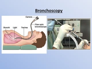
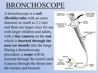
PROCEDURE ON
BRONCHOSCOPY
GUIDED BY:
MRS. MANISHA SAMUDRE
ASSISTANT PROFESSOR
HOD OF MEDICAL- SURGICAL
NURSING
WHAT IS BRONCHOSCOPY
• Bronchoscopy is a technique of visualizing the
inside of the airways for diagnostic and therapeutic
purposes.
• An instrument (bronchoscope) is inserted into the
airways, usually through the nose or mouth, or
occasionally through a tracheostomy.
• This allows the practitioner to examine the patient's
airways for abnormalities such as foreign bodies,
bleeding, tumors, or inflammation. Specimens may
be taken from inside the lungs.
BRONCHOSCOPE
A bronchoscope is a soft
(flexible) tube with an outer
diameter as small as 2.2 mm
and there are larger sizes for use
with larger children and adults,
with a tiny camera on the end
which is inserted through the
nose (or mouth) into the lungs.
During a bronchoscopy
procedure, a scope will be
inserted through the nostril until
it passes through the throat into
the trachea and bronchi.
The diagnostic procedures of bronchoalveolar
lavage, transbronchial needle aspiration
(TBNA), and transbronchial biopsy can be
performed during a bronchoscopy.
THERAPEUTIC
BRONCHOSCOPY
• Bronchoscopy can also play a therapeutic role.
For example, bronchoscope can be used in the
treatment of airway obstruction by tumors
or foreign bodies, during the removal of
secretions
TYPES OF BRONCHOSCOPY
 Flexible Bronchoscopy - Flexible
bronchoscope uses a long, thin, lighted tube
to look at your airway.
• The flexible bronchoscope is used more often
than the rigid bronchoscope because it usually
require local oral anesthesia is more
comfortable for the person, and offers a
better view of the smaller airways. It also
allows to remove small samples of tissue
(biopsy).
• The flexible tube actually contains a fiber-optic
system which attaches to a video camera and a source
of light. The light travels through the scope and lights
up the inside of the airway. The image seen at the tip
of the scope is transmitted back again through the
fiber-optic system to the video camera.
• Using Bowden cables connected to a lever at the
hand piece, the tip of the instrument can be
oriented, allowing the practitioner to navigate the
instrument into individual lobe or segment bronchi.
• Most flexible bronchoscopes also include a channel
for suctioning or instrumentation, but these are
significantly smaller than those in a rigid
bronchoscope.
FLEXIBLE
BRONCHOSCOPY
Rigid Bronchscopy - Rigid bronchoscopy is
usually done with general anesthesia and uses a
straight, hollow metal tube. It is used for:
• When there is bleeding in the airway that could
block the flexible scope's view.
• To remove large tissue samples for biopsy.
• To clear the airway of objects (such as a piece of
food) that cannot be removed using a flexible
bronchoscope.
RIGID
BROCHOSCOPY
INDICATIONS
• Lesions requiring biopsy seen on chest X-ray.
• Hemoptysis & Stridor.
• Positive sputum cytology for malignant cells
• Collection of bronchial secretions for
bacteriology, especially Tuberculosis.
• Recurrent laryngeal nerve paralysis of unknown
etiology.
• Infiltrative lung disease (to obtain a
transbronchial biopsy).
• Investigation of collapsed lobes or segments and
aspiration of mucus plugs.
PRE PROCEDURAL
• The patient is starved overnight.
• Explain the procedure to the patient and relatives
• Obtain written consent of the patient or relatives
• Remove dentures, contact lenses and other
prostheses
• Ask the patient not tosmoke for at least 24 hours.
PROCEDURE
• All patients require sedation to tolerate the
procedure (risk of cardiopulmonary arrest)
• Minor and transient cardiac dysrhythmias occur
in up to 40% of patients on passage of the
bronchoscope through the larynx. Monitoring is
required.
• Oxygen supplementation is required
• Atropine 0.6 mg IM is given 30 min before the
procedure, as an anticholinergic (muscarinic) to
decrease the respiratory secretions, and to prevent
oral secretions from obstructing the view
• Topical anesthesia (lidocaine 2% gel) is applied to
the nose, nasopharynx and pharynx, to anesthetize
the mucous membranes of the pharynx, larynx,
and trachea
• Intravenous sedation (e.g. diazepam 10 mg or
midazolam 2.5–10 mg) is given.
• You may be asked to take a deep breath &
bronchoscope is passed through the nose,
nasopharynx and pharynx under direct vision
to minimize trauma.
• Lidocaine (2 mL of 4%) is dropped through
the instrument on to the vocal cords.
• The patient is monitored during the procedure
with periodic blood pressure checks,
continuous ECG monitoring of the heart, and
pulse oximetry.
• An X-ray machine (fluoroscope) may be
placed above you to provide a picture that
helps you to see any devices, such as forceps
to collect a biopsy sample, that are being
moved into your lung.
• All segmental and subsegmental orifices
should be identified.
• After finishing the scopy A salt (saline) fluid
may be used to wash your airway, and then the
samples are collected and sent to the lab to be
studied.
AFTER CARE
• Although most patients tolerate bronchoscopy well, a
brief period of observation is required after the
procedure, So Close monitoring for 2-4 hours
• Eating and drinking is not allowed until whole anesthesia
is worn off & and gag reflex has returned.
• Advise for an post X-ray to see possible complications of
post-bronchoscopy for e.g. pneumothorax
• Asses the patient with vital signs and level of
consciousness
• Request patient strictly not to drive for at least 8 hours
after the procedure.
• Spit out your saliva until you are able to swallow without
choking.

Bronchoscopy

  • 1.
    PROCEDURE ON BRONCHOSCOPY GUIDED BY: MRS.MANISHA SAMUDRE ASSISTANT PROFESSOR HOD OF MEDICAL- SURGICAL NURSING
  • 2.
    WHAT IS BRONCHOSCOPY •Bronchoscopy is a technique of visualizing the inside of the airways for diagnostic and therapeutic purposes. • An instrument (bronchoscope) is inserted into the airways, usually through the nose or mouth, or occasionally through a tracheostomy. • This allows the practitioner to examine the patient's airways for abnormalities such as foreign bodies, bleeding, tumors, or inflammation. Specimens may be taken from inside the lungs.
  • 4.
    BRONCHOSCOPE A bronchoscope isa soft (flexible) tube with an outer diameter as small as 2.2 mm and there are larger sizes for use with larger children and adults, with a tiny camera on the end which is inserted through the nose (or mouth) into the lungs. During a bronchoscopy procedure, a scope will be inserted through the nostril until it passes through the throat into the trachea and bronchi.
  • 5.
    The diagnostic proceduresof bronchoalveolar lavage, transbronchial needle aspiration (TBNA), and transbronchial biopsy can be performed during a bronchoscopy.
  • 6.
    THERAPEUTIC BRONCHOSCOPY • Bronchoscopy canalso play a therapeutic role. For example, bronchoscope can be used in the treatment of airway obstruction by tumors or foreign bodies, during the removal of secretions
  • 7.
    TYPES OF BRONCHOSCOPY Flexible Bronchoscopy - Flexible bronchoscope uses a long, thin, lighted tube to look at your airway. • The flexible bronchoscope is used more often than the rigid bronchoscope because it usually require local oral anesthesia is more comfortable for the person, and offers a better view of the smaller airways. It also allows to remove small samples of tissue (biopsy).
  • 8.
    • The flexibletube actually contains a fiber-optic system which attaches to a video camera and a source of light. The light travels through the scope and lights up the inside of the airway. The image seen at the tip of the scope is transmitted back again through the fiber-optic system to the video camera. • Using Bowden cables connected to a lever at the hand piece, the tip of the instrument can be oriented, allowing the practitioner to navigate the instrument into individual lobe or segment bronchi. • Most flexible bronchoscopes also include a channel for suctioning or instrumentation, but these are significantly smaller than those in a rigid bronchoscope.
  • 9.
  • 10.
    Rigid Bronchscopy -Rigid bronchoscopy is usually done with general anesthesia and uses a straight, hollow metal tube. It is used for: • When there is bleeding in the airway that could block the flexible scope's view. • To remove large tissue samples for biopsy. • To clear the airway of objects (such as a piece of food) that cannot be removed using a flexible bronchoscope.
  • 11.
  • 12.
    INDICATIONS • Lesions requiringbiopsy seen on chest X-ray. • Hemoptysis & Stridor. • Positive sputum cytology for malignant cells • Collection of bronchial secretions for bacteriology, especially Tuberculosis. • Recurrent laryngeal nerve paralysis of unknown etiology. • Infiltrative lung disease (to obtain a transbronchial biopsy). • Investigation of collapsed lobes or segments and aspiration of mucus plugs.
  • 13.
    PRE PROCEDURAL • Thepatient is starved overnight. • Explain the procedure to the patient and relatives • Obtain written consent of the patient or relatives • Remove dentures, contact lenses and other prostheses • Ask the patient not tosmoke for at least 24 hours.
  • 14.
    PROCEDURE • All patientsrequire sedation to tolerate the procedure (risk of cardiopulmonary arrest) • Minor and transient cardiac dysrhythmias occur in up to 40% of patients on passage of the bronchoscope through the larynx. Monitoring is required. • Oxygen supplementation is required
  • 15.
    • Atropine 0.6mg IM is given 30 min before the procedure, as an anticholinergic (muscarinic) to decrease the respiratory secretions, and to prevent oral secretions from obstructing the view • Topical anesthesia (lidocaine 2% gel) is applied to the nose, nasopharynx and pharynx, to anesthetize the mucous membranes of the pharynx, larynx, and trachea • Intravenous sedation (e.g. diazepam 10 mg or midazolam 2.5–10 mg) is given.
  • 16.
    • You maybe asked to take a deep breath & bronchoscope is passed through the nose, nasopharynx and pharynx under direct vision to minimize trauma. • Lidocaine (2 mL of 4%) is dropped through the instrument on to the vocal cords. • The patient is monitored during the procedure with periodic blood pressure checks, continuous ECG monitoring of the heart, and pulse oximetry.
  • 17.
    • An X-raymachine (fluoroscope) may be placed above you to provide a picture that helps you to see any devices, such as forceps to collect a biopsy sample, that are being moved into your lung. • All segmental and subsegmental orifices should be identified. • After finishing the scopy A salt (saline) fluid may be used to wash your airway, and then the samples are collected and sent to the lab to be studied.
  • 18.
    AFTER CARE • Althoughmost patients tolerate bronchoscopy well, a brief period of observation is required after the procedure, So Close monitoring for 2-4 hours • Eating and drinking is not allowed until whole anesthesia is worn off & and gag reflex has returned. • Advise for an post X-ray to see possible complications of post-bronchoscopy for e.g. pneumothorax • Asses the patient with vital signs and level of consciousness • Request patient strictly not to drive for at least 8 hours after the procedure. • Spit out your saliva until you are able to swallow without choking.