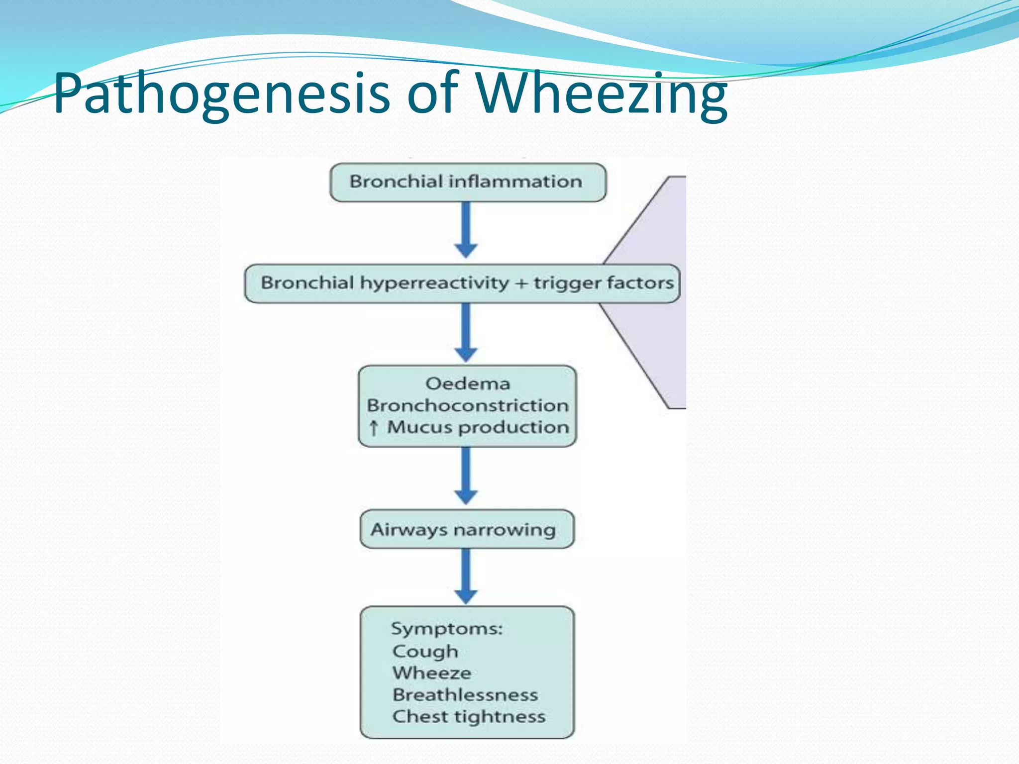
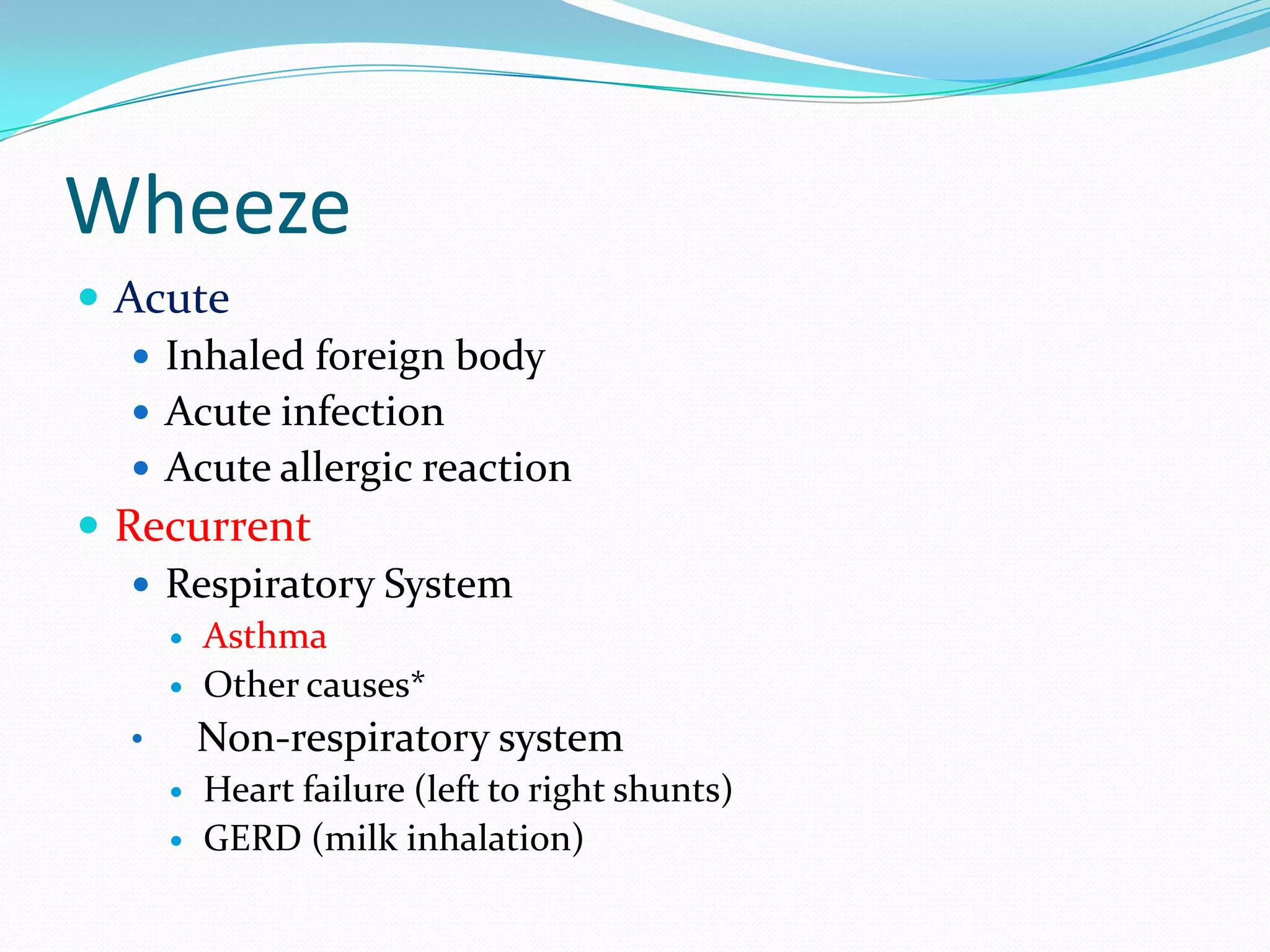
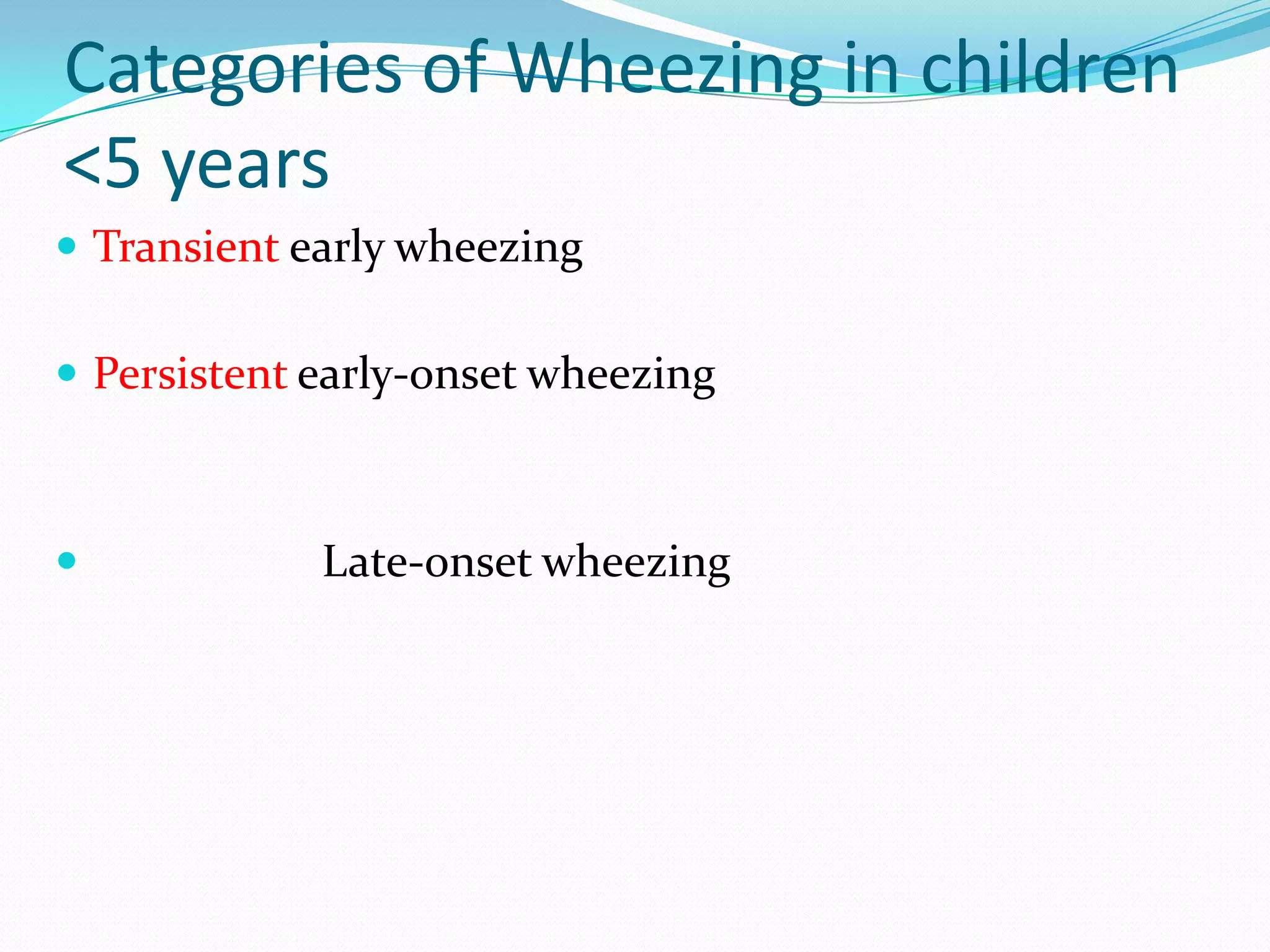
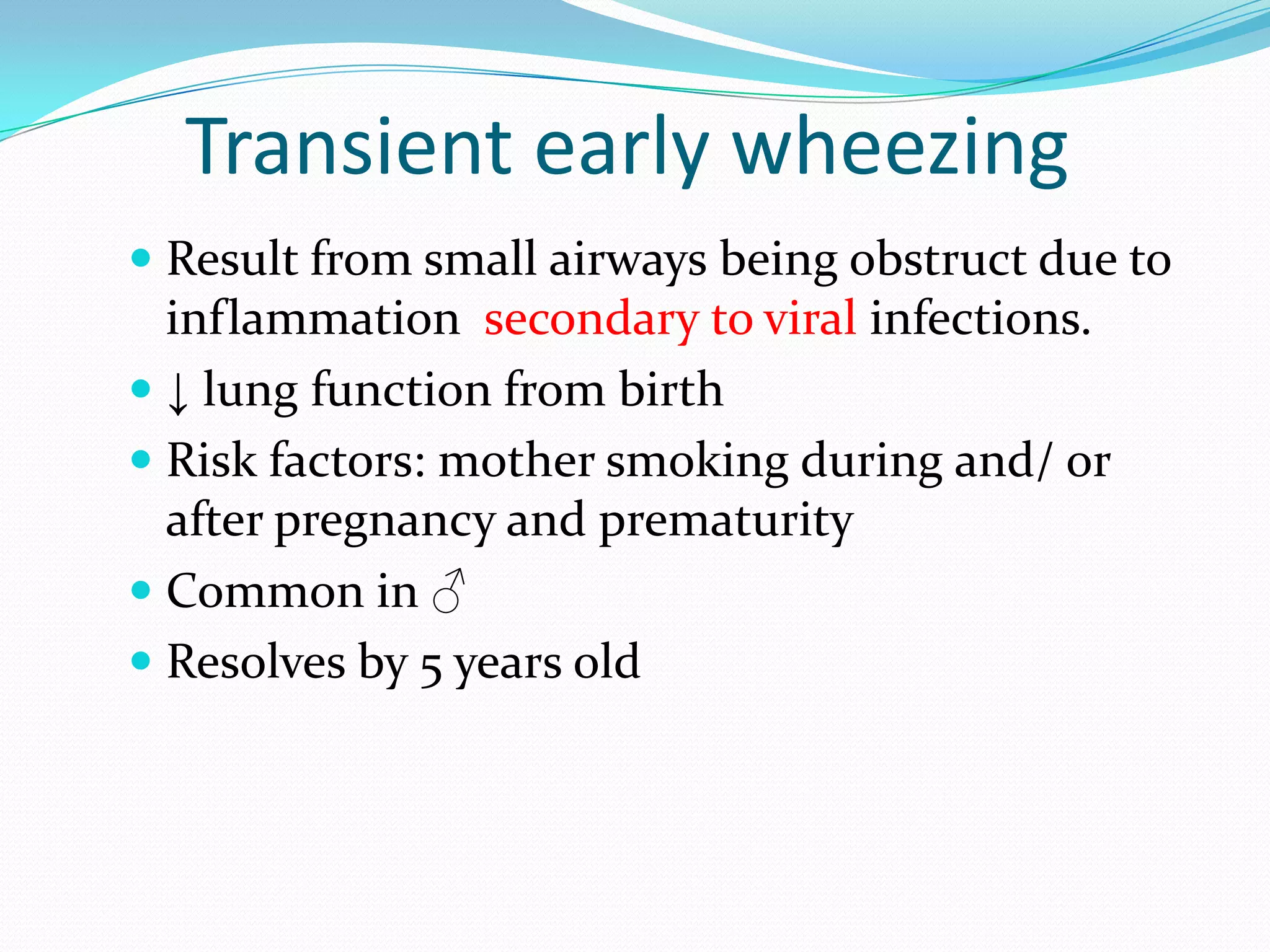
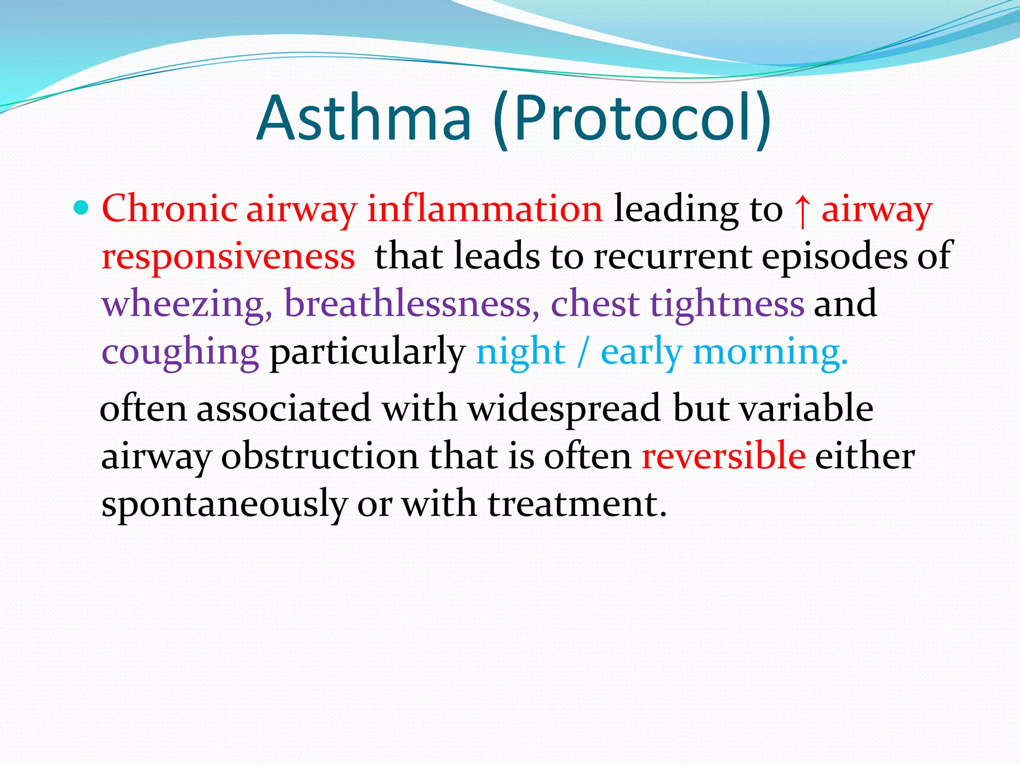
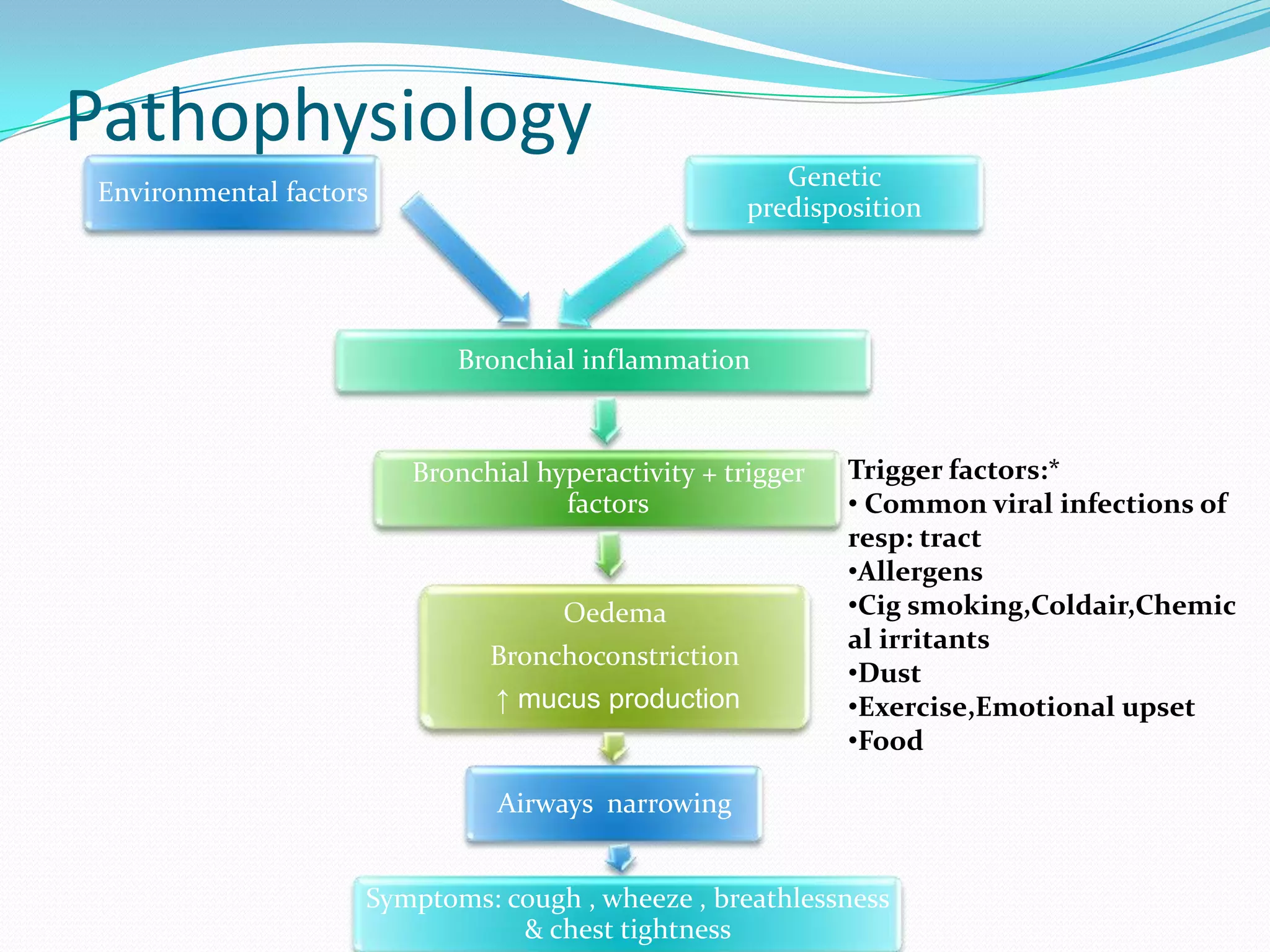
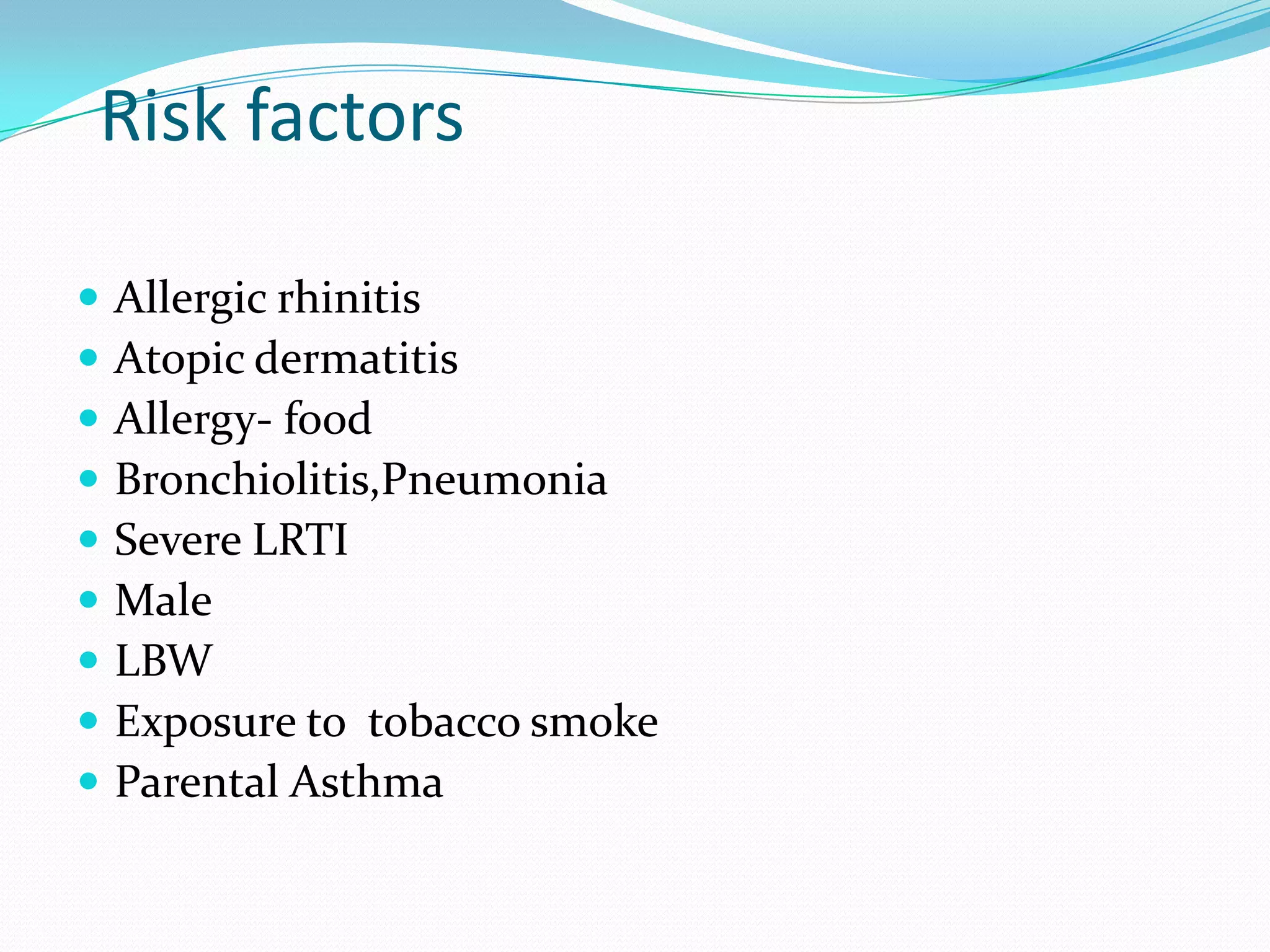
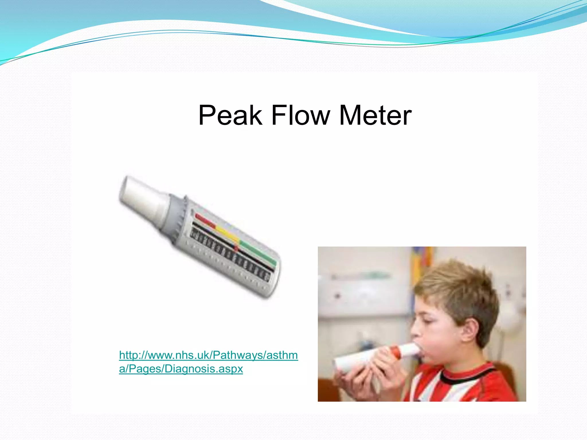
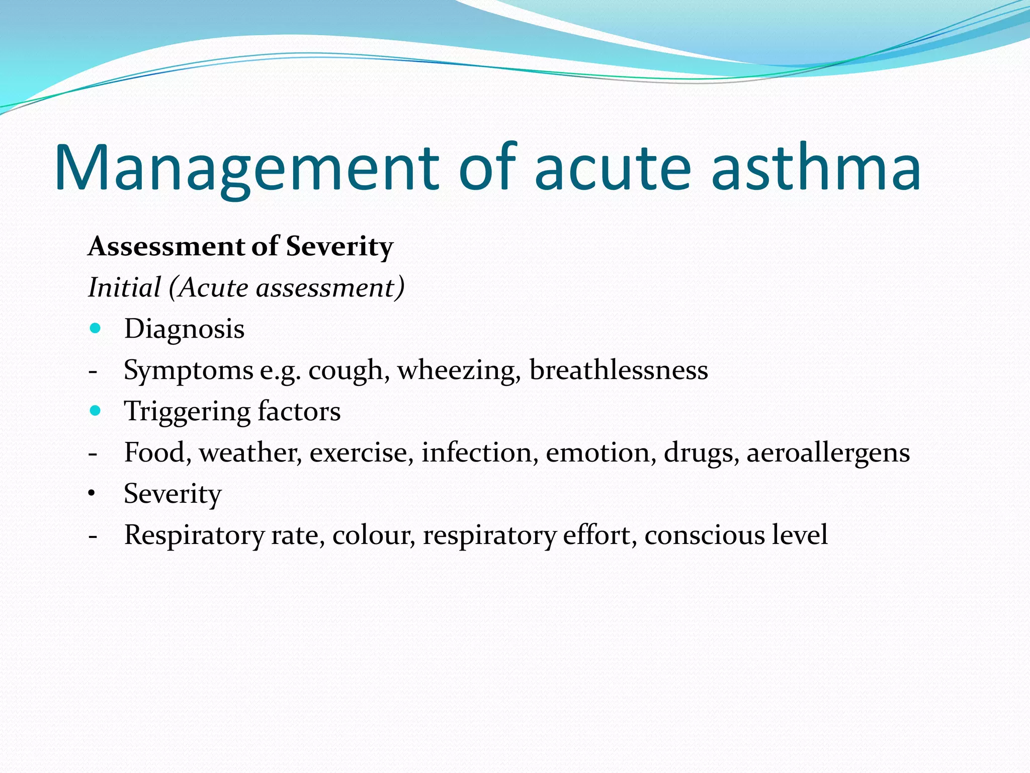
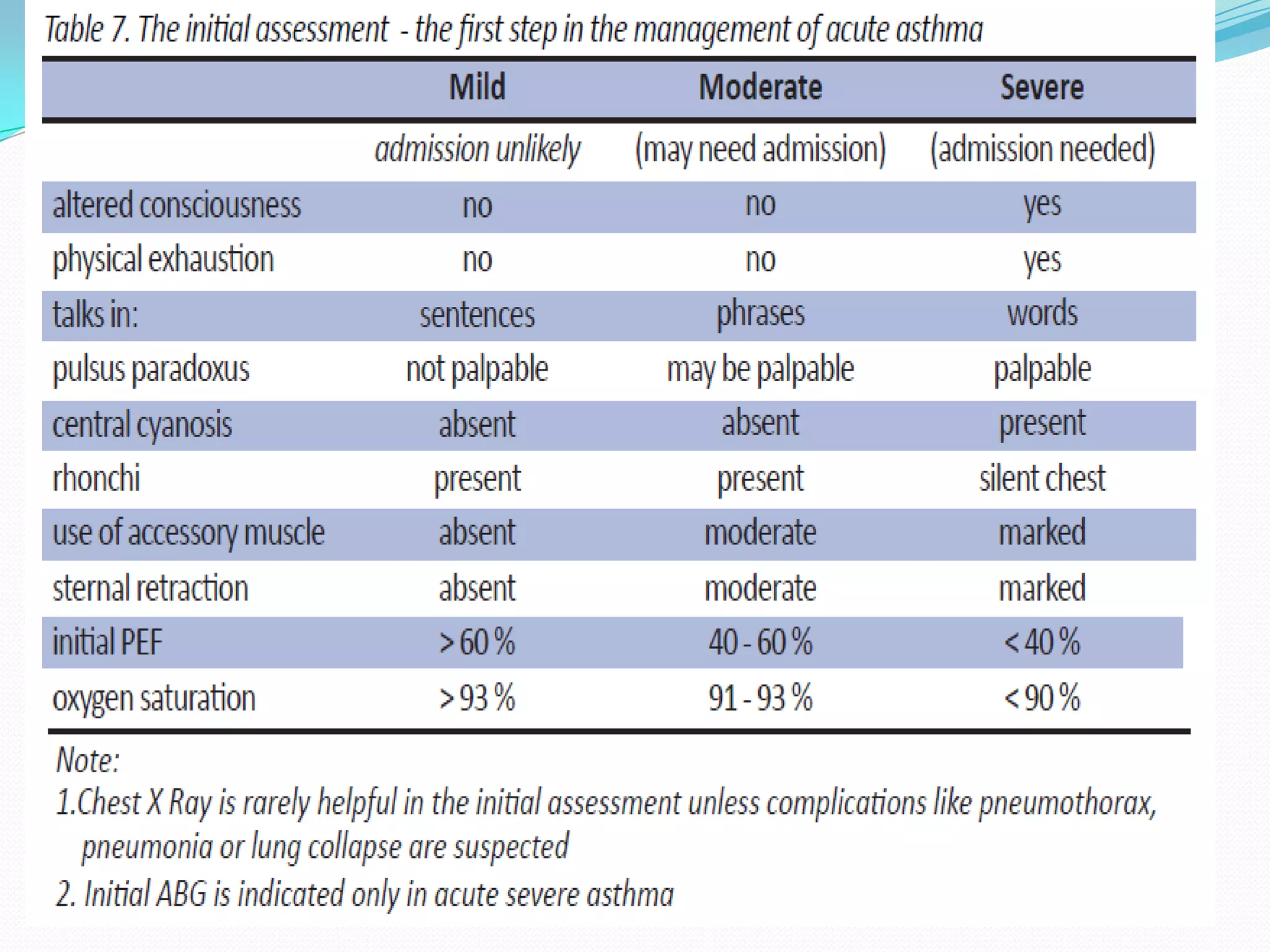
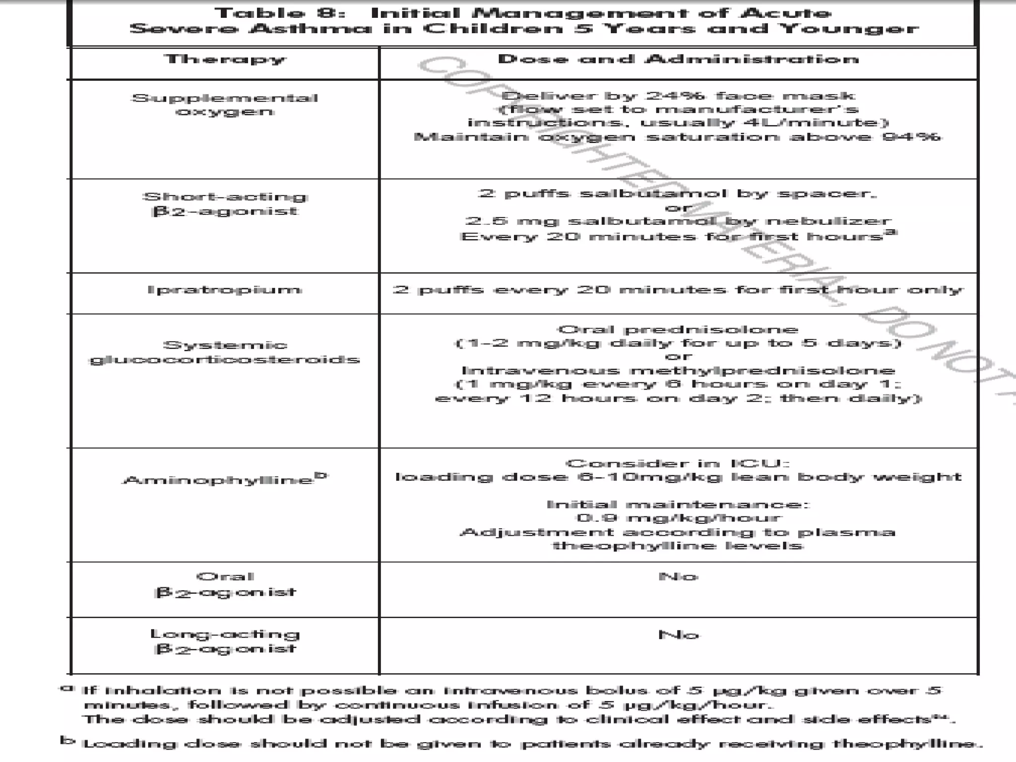
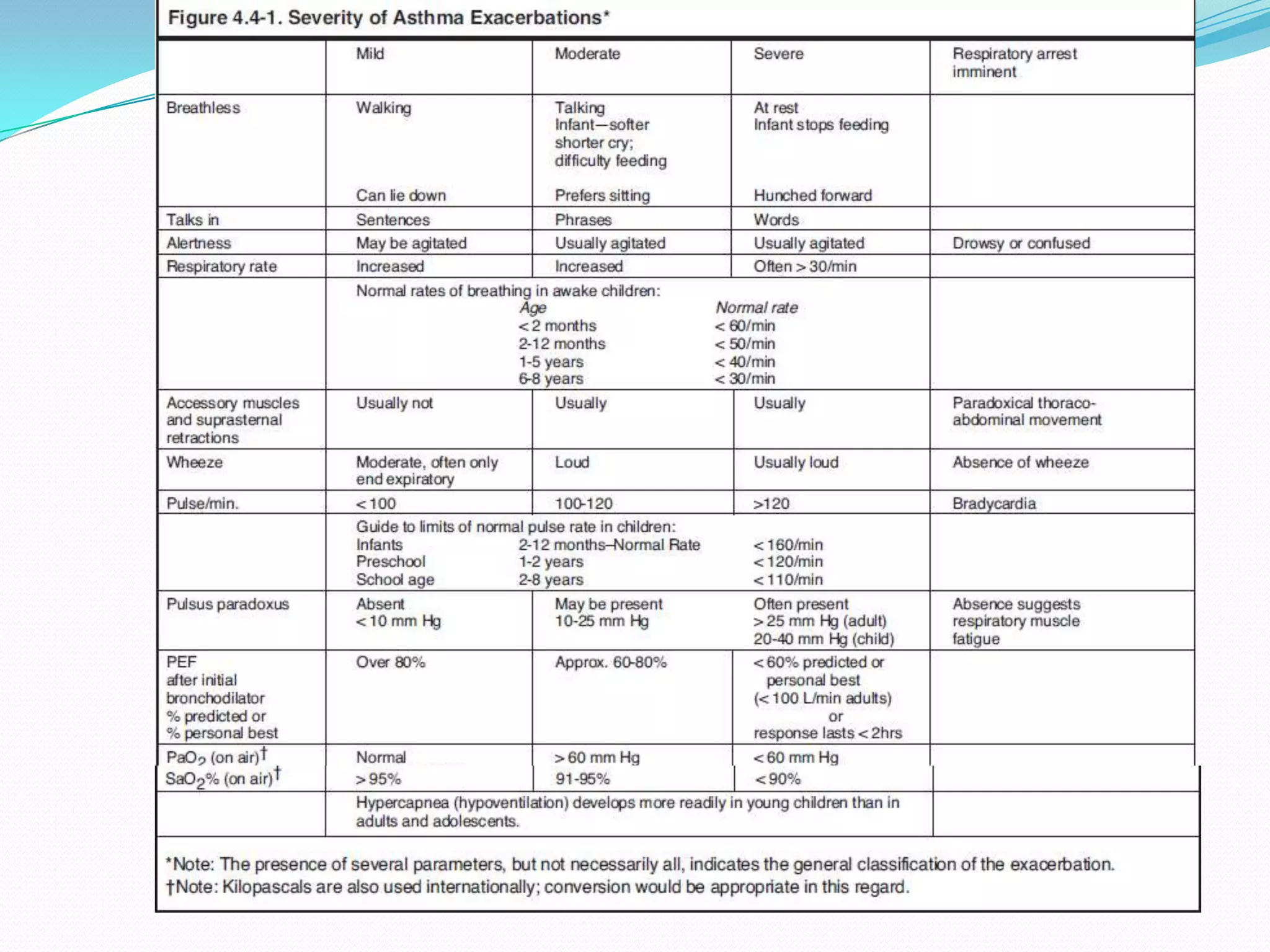
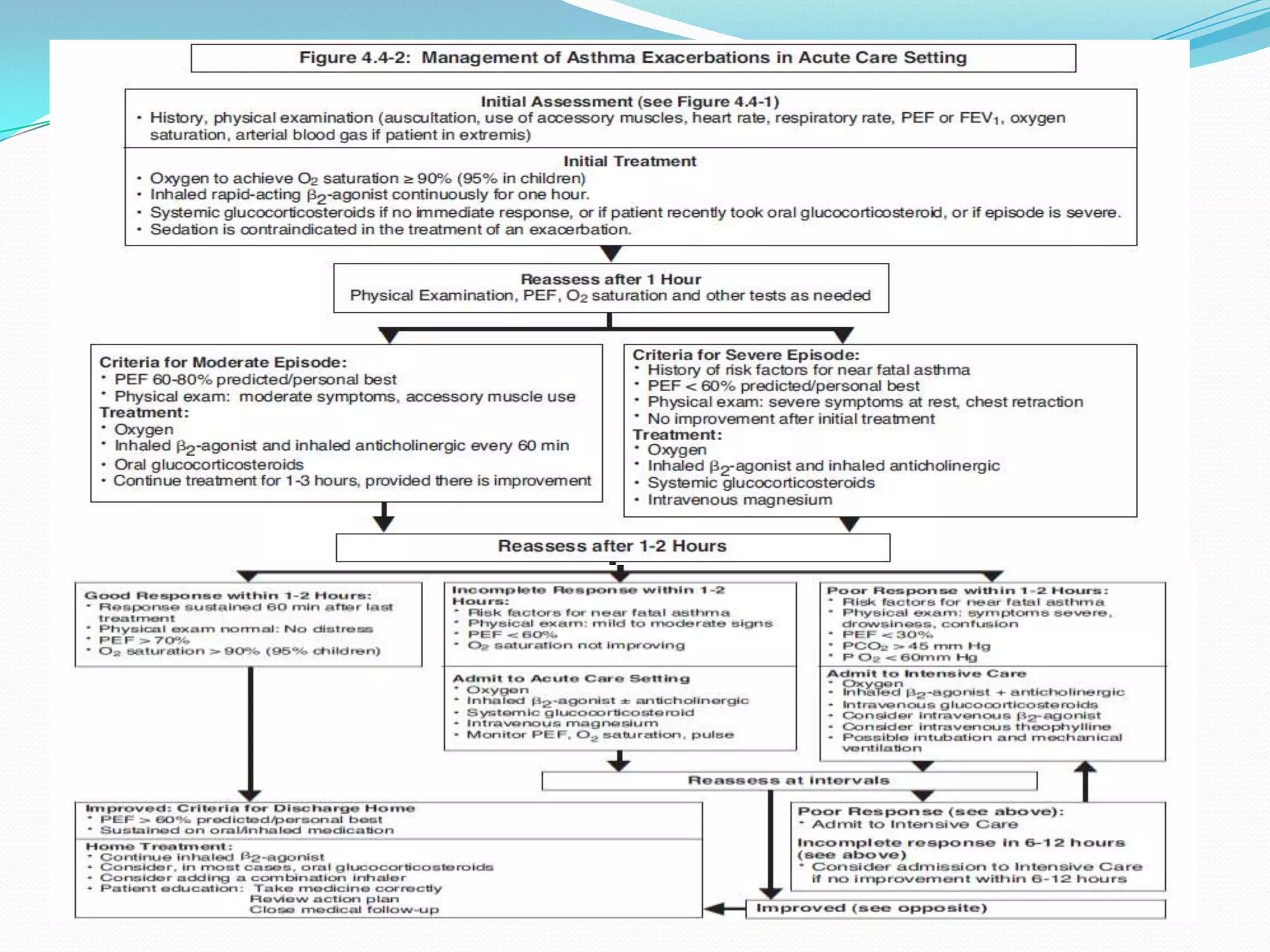
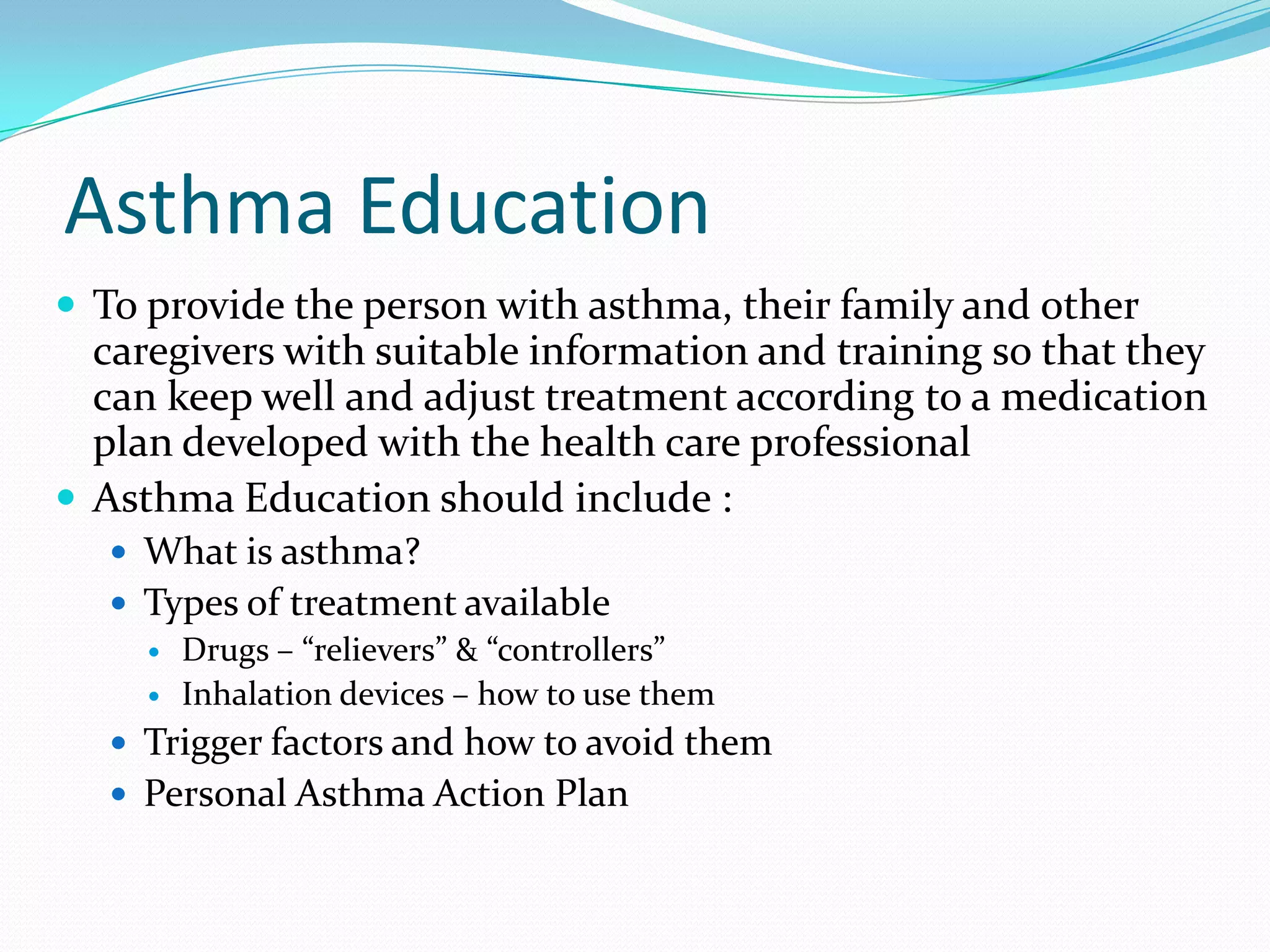
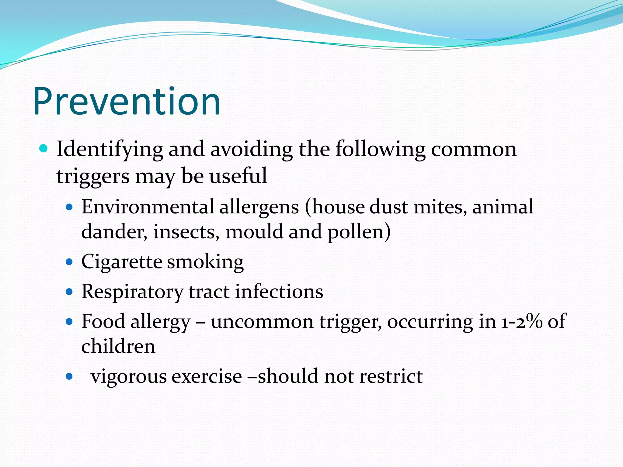
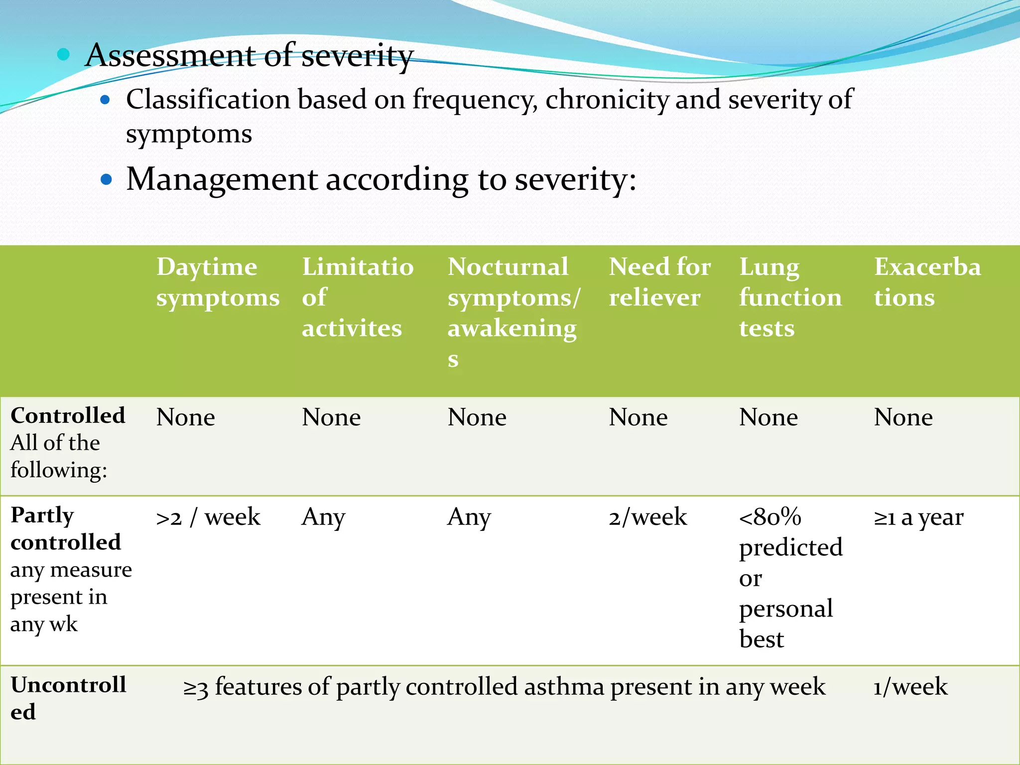
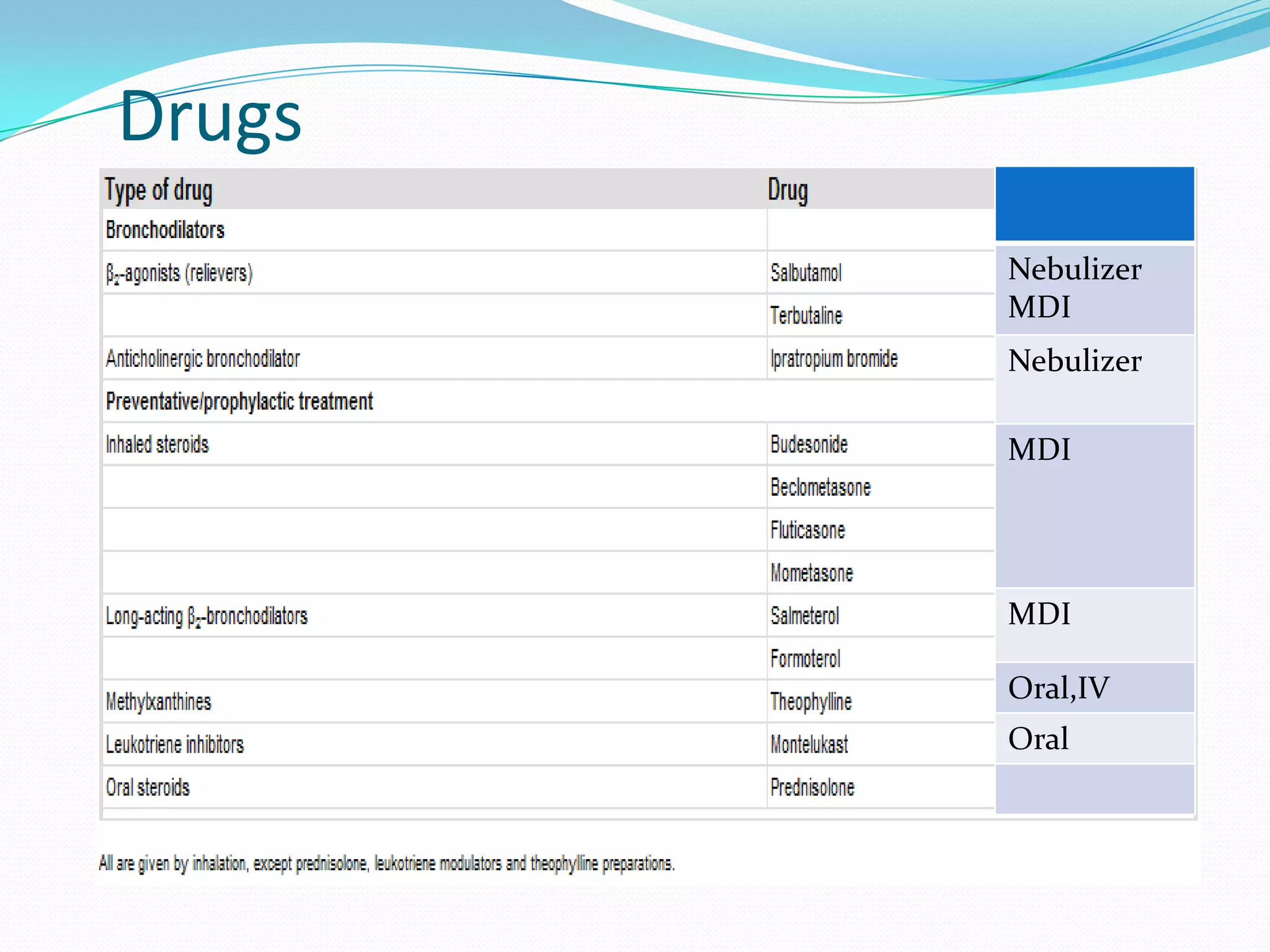
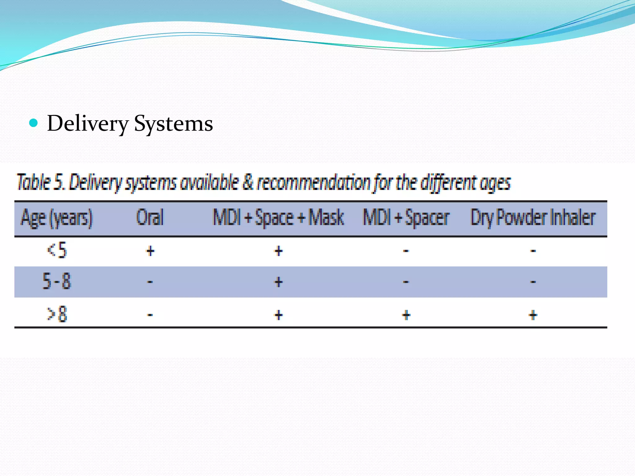
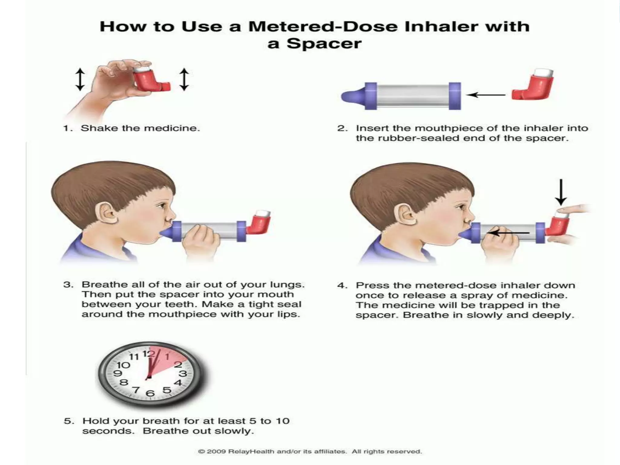
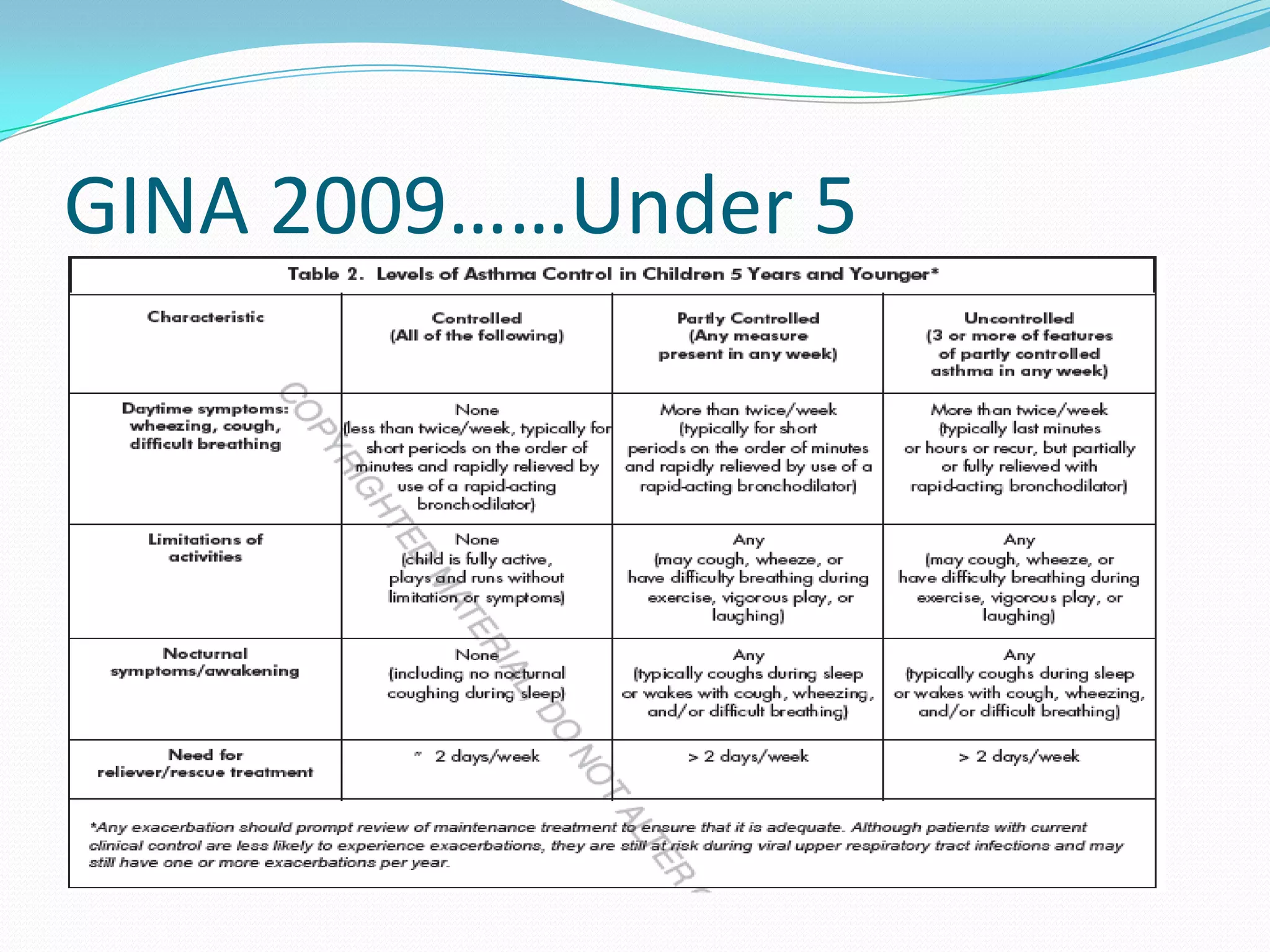
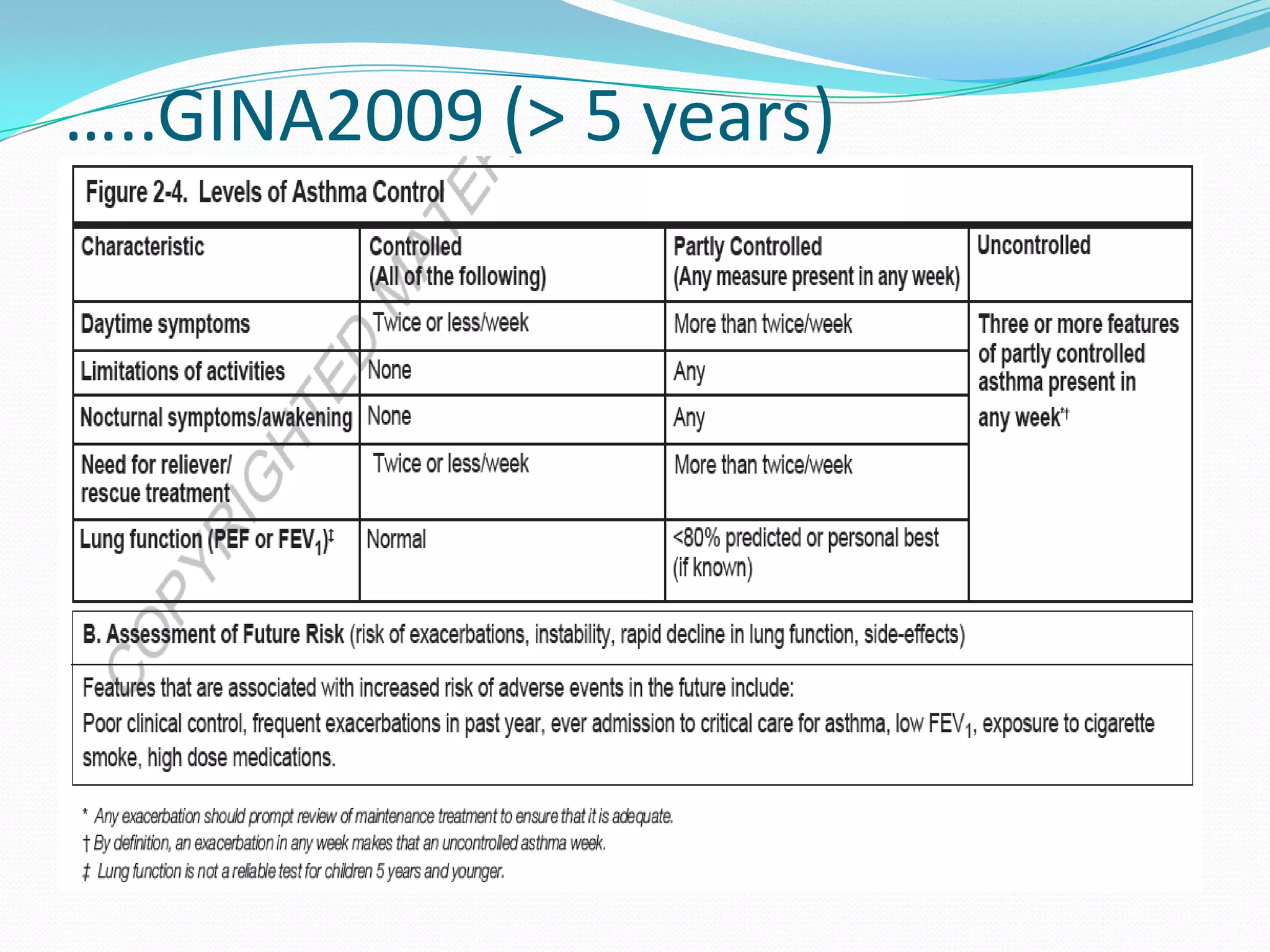
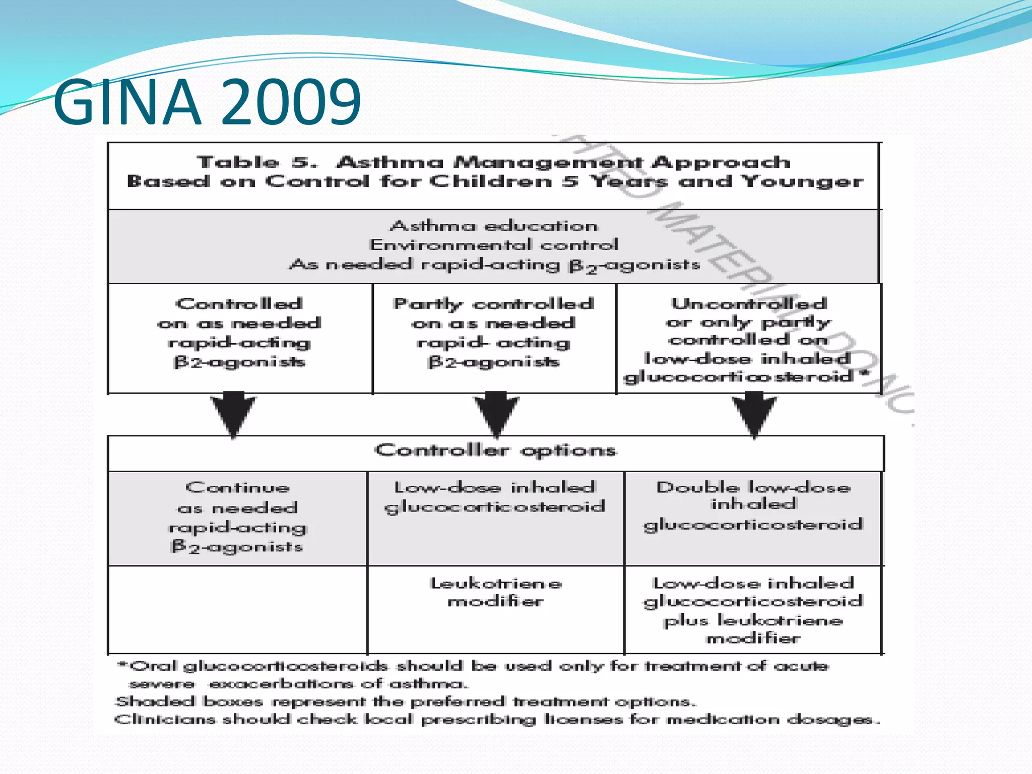
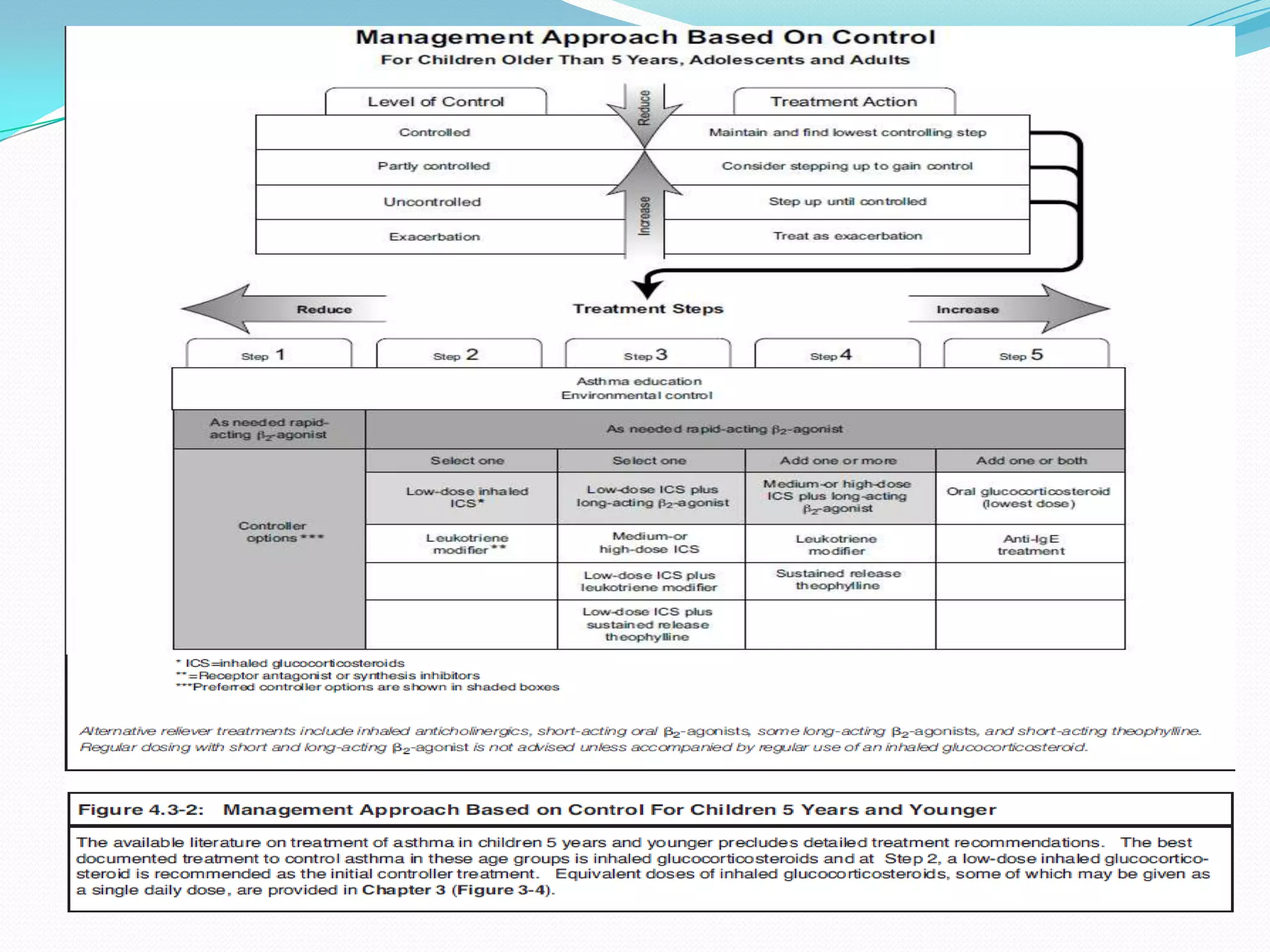
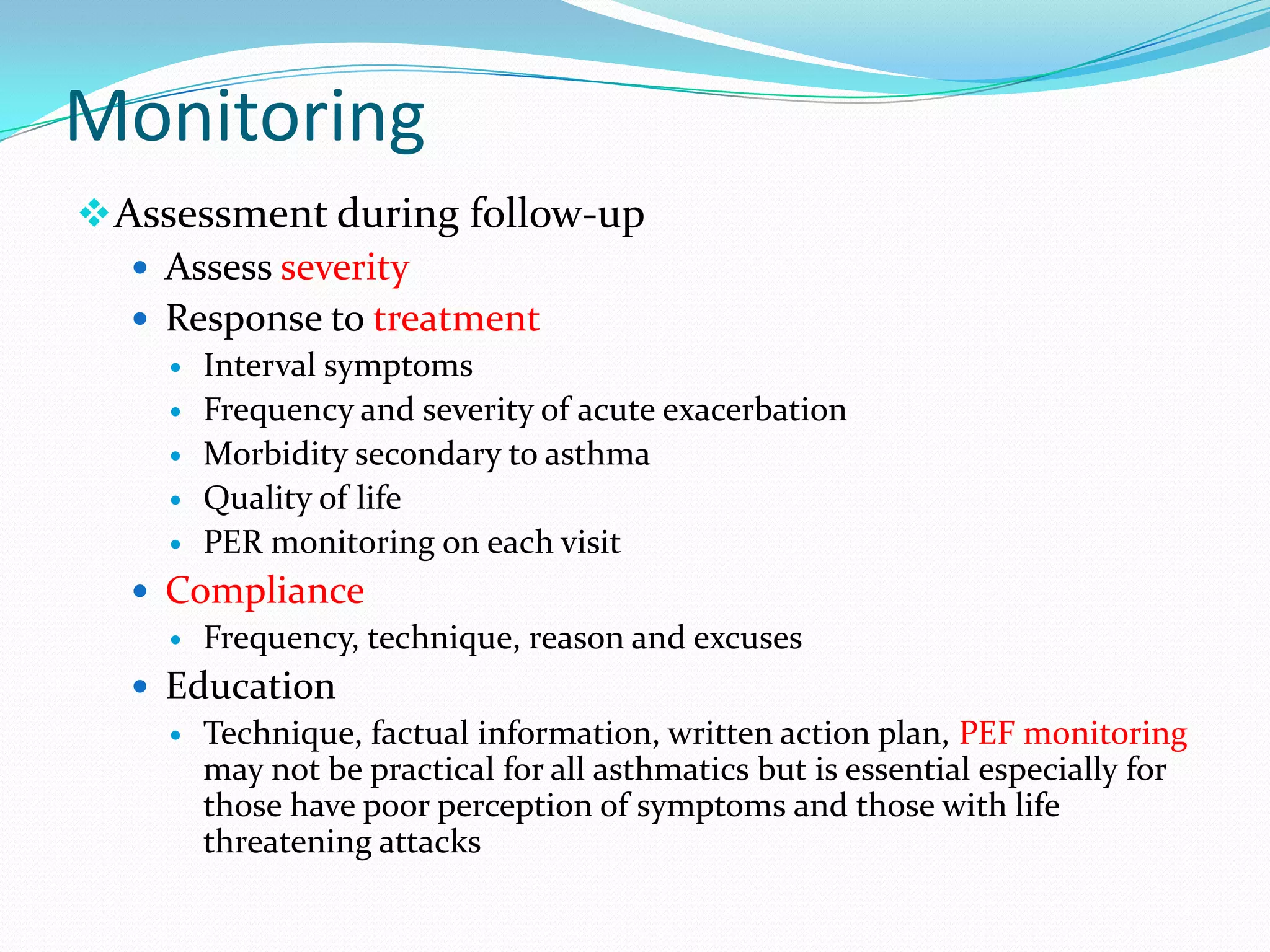
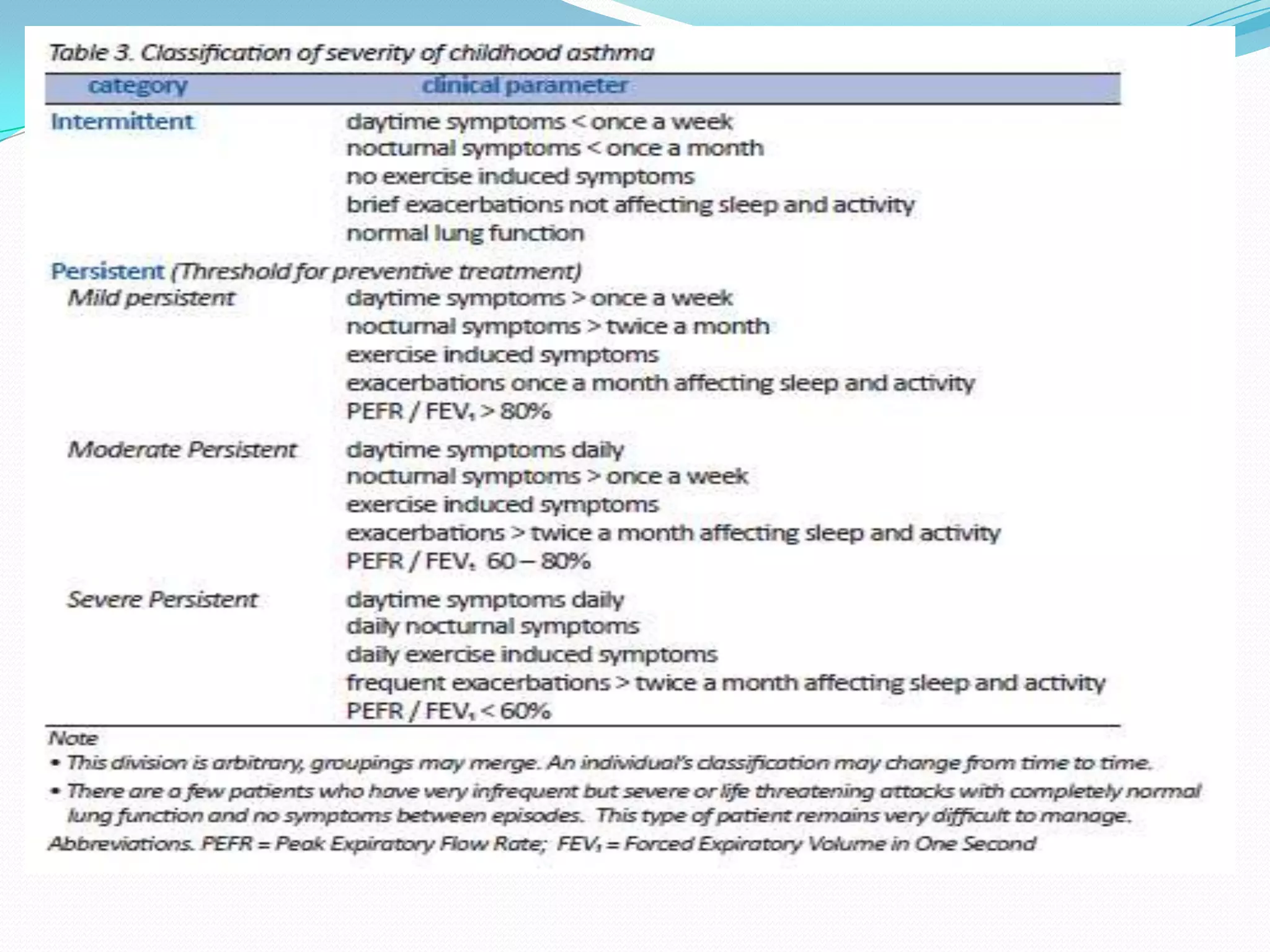
DrSTSA
Wheeze
 A high-pitched whistling/musical sound produced by
    the passage of air through narrowed airways/ bronchi
   Louder during expiration
   A manifestation of lower respiratory tract obstruction
   Site of obstruction may be anywhere from the
    intrathoracic trachea to the small bronchi or large
    bronchioles
   Sound is generated by turbulence in larger airways that
    collapse with forced expiration
Wheezing in Children
 Episodic wheezing and cough are common in children


 Infants and young children (<3 years) are especially
 prone to wheezing
Pathogenesis of Wheezing
Wheeze
 Acute
    Inhaled foreign body
    Acute infection
    Acute allergic reaction
 Recurrent
    Respiratory System
          Asthma
          Other causes*
   •       Non-respiratory system
          Heart failure (left to right shunts)
          GERD (milk inhalation)
Categories of Wheezing in children
<5 years
 Transient early wheezing

 Persistent early-onset wheezing


            Late-onset wheezing
Transient early wheezing
 Result from small airways being obstruct due to
  inflammation secondary to viral infections.
 ↓ lung function from birth
 Risk factors: mother smoking during and/ or
  after pregnancy and prematurity
 Common in ♂
 Resolves by 5 years old
Non atopic wheezing
 Have normal lung function in early life
 Lower respiratory illness due to viral infection
  leads to ↑ wheezing during the 1st 10 years of life.
 cause less severe persistent wheezing
 symptoms improve during adolescence
Ig E-mediated wheezing
             (atopic asthma)
 Lung function – normal at birth
 Recurrent wheeze develops with allergic sensitisation
   ↑ blood Ig E & positive skin prick tests to common
     allergens
 Persistence of symptoms & ↓ lung function later in
  childhood.
 Risk factors:
    Positive family history
    Allergy
    History of eczema
Other causes of recurrent wheeze
in infancy
 Recurrent aspiration of feeds
 Cystic fibrosis
o Inhaled foreign body
 Congenital abnormality of lung, airway or heart
 Idiopathic
 Cow’s milk protein intolerance
History Taking
Wheeze
   Age of onset
   Duration-Acute or recurrent
   Precipitating factors
     URTI symptoms
     Contact with URTI patient
     Triggers for asthma -         ( A,V,C,D,E,F)
     History of atopy
   Associated symptoms
     Rapid breathing
     Cough- dry or productive? Sputum colour?
     Chest tightness
     Nausea or vomiting
     Cyanosis
 Pattern of symptoms
    Daytime/ nocturnal symptoms
    Exercise induced
 Severity
    On any medication? Types?
    Relieved with medication
    Restriction of daily activities
    Sleep disturbances
 Systemic Review
    General condition - LOA, LOW
    Atopy - angioedema, allergic rhinitis, allergic
     conjunctivitis, eczema, urticaria
 Past Medical History
    Number of admission to the hospital
    Number of admission due to similar problem
    Last admission due to similar problem
        Duration of stays
        Medication given and discharge medication
    History of prolonged URTI symptoms
 Birth History
    Antenatal: intrauterine infection
    Intrapartum: Prematurity
    Postnatal: Prolonged labour, NNJ, congenital pulmonary disease
 Immunization History
 Family History
    Asthma
    Atopy
    CHD
    Cystic fibrosis
 Social History
    School performance
    Daily activities
    Social interaction
    Anyone smoking at home
    Location of house
    Environment condition: Pets, flower, dust
 Drug and Allergy History
Physical Examination
 General condition: alert, conscious, drowsiness, irritability
 Signs of respiratory distress
    Sitting propped up
    Shortness of breath
    Use of accessory muscles
    Audible wheeze
    Central cyanosis
 Vital signs
 Anthropometry measurements
 O2 therapy: : nasal prong, face mask, high-flow
  mask, nebulizer
 Speech: sentences, phrases or words
 Hands
    Clubbing of fingernails
    Peripheral cyanosis


 Head
    Ears – inflammation, discharge
    Nose – nasal discharge
    Throat - inflammation, tonsil enlargement
    Tongue – central cyanosis

 Neck
    Lymph nodes
    Tracheal shift
Respiratory System
 Inspection
    Use of accessory muscles : suprasternal, intercostal retraction
    Harrison’s sulcus
    Chest deformities
          pectus carinatum
          Pectus cavum
          Hyperinflacted chest
     Signs of atopy : eczema, dry skin
 Palpation
    Trachea shift ( older child)
    Chest expansion
    Vocal Fremitus
    Apex beat
 Percussion : Hyperresonance

Percussion is usually normal unless there are foreign body, lung collapse, mediasternal
   masses
 Auscultation
    Respiratory
        Air entry
        Prolonged inspiratory or expiratory phase
        Vesicular breath sound
        Added sounds : Rhonchi or crepitations


    Cardiovascular
      S1 and S2
      Additional sounds


 Palpation of abdomen - hepatomegaly
Investigations
1.       Laboratory
            FBC : to look for infection, eosinophilia
            BUSE : hydration status, fluid maintenance
            ABG : to look for respiratory failure if severe condition
            Throat swab or sputum for culture and sensitivity
2. Imaging
   Chest X-Ray
              Foreign body
              Pneumothorax, lobar collapse, mass
              Infection
3. Bronchoscopy
Bronchial Asthma
Asthma (Protocol)
 Chronic airway inflammation leading to ↑ airway
 responsiveness that leads to recurrent episodes of
 wheezing, breathlessness, chest tightness and
 coughing particularly night / early morning.
 often associated with widespread but variable
 airway obstruction that is often reversible either
 spontaneously or with treatment.
Pathophysiology
                                                       Genetic
Environmental factors
                                                    predisposition


                           Bronchial inflammation


                        Bronchial hyperactivity + trigger   Trigger factors:*
                                    factors                 • Common viral infections of
                                                            resp: tract
                                                            •Allergens
                                    Oedema                  •Cig smoking,Coldair,Chemic
                                                            al irritants
                              Bronchoconstriction
                                                            •Dust
                               ↑ mucus production           •Exercise,Emotional upset
                                                            •Food

                               Airways narrowing


                    Symptoms: cough , wheeze , breathlessness
                               & chest tightness
Risk factors

   Allergic rhinitis
   Atopic dermatitis
   Allergy- food
   Bronchiolitis,Pneumonia
   Severe LRTI
   Male
   LBW
   Exposure to tobacco smoke
   Parental Asthma
History taking
 Current symptoms
 Pattern of symptoms
 Precipitating factors
 Prolonged URTI Symptom
 Present Treatment
 Previous hospital admission
 Response to prior Treatment
 Typical exacerbations
 History of atopy
 Home/school environment
 Impact on lifestyle: school, sports, sleep
 Family history
Physical Exam:
Growth & nutrition
                                Signs of acute exacerbation:
                                      • Drowsiness,fatigue
                                      • Tachypnoea
                                      • Tachycardia
                                      • Hyperinflated chest
Signs of chronic illness:             • Accessory muscles
                                      • Cyanosis
   • Harrison sulci                   • Pulsus paradoxus
   • Hyperinflated chest              • Prolonged expiratory
                                        phase, wheeze
   • Eczema/dry skin                  • Silent chest
   • Hypertrophied turbinates         • Rhonchi
Tests for Diagnosis and Monitoring
of Asthma
 Spirometry – FEV1 and FVC
 PEFR
    To confirm the diagnosis of asthma ( improvement of >15%
     after bronchodilator)
    useful for assessing the severity of asthma
    response of the patient to therapy
    Normal value are available & relate to height
    To identify environmental (including occupational) causes of
     asthma symptoms
 Skin prick test with allergens
 Exercise challenge
Peak Flow Meter


http://www.nhs.uk/Pathways/asthm
a/Pages/Diagnosis.aspx
Management of acute asthma
 Assessment of Severity
 Initial (Acute assessment)
  Diagnosis
 - Symptoms e.g. cough, wheezing, breathlessness
  Triggering factors
 - Food, weather, exercise, infection, emotion, drugs, aeroallergens
 • Severity
 - Respiratory rate, colour, respiratory effort, conscious level
Criteria for Admission

 Failure to respond to standard home treatment
 Failure of those with mild or moderate acute asthma to
  respond to nebulised β2-agonists
 Relapse within 4 hrs of nebulised β2-agonists
 Severe acute asthma
Monitor vital s/-
   Pulse,SpO2 ,colour,ABG,PEFR

Definitive Treatment

1. O2 therapy (SpO2 >95%)

2. Bronchodilator therapy
    • β2-agonists
    • Ipratropium bromide
    • Aminophylline

3. Maintenance therapy

Supportive Treatment

1.Hydration & fluid maintenance
2.+/- antibiotics
(ONLY if bacterial infection suspected)
GINA 2009…..Under 5
>5 years
Upon Discharge

 Review asthma medications


 Provide Asthma Action Plan
   How to recognize worsening asthma
   How to treat worsening asthma
   How & when to seek medical attention


 Schedule regular follow-ups to monitor asthma control
Asthma Education
 To provide the person with asthma, their family and other
  caregivers with suitable information and training so that they
  can keep well and adjust treatment according to a medication
  plan developed with the health care professional
 Asthma Education should include :
    What is asthma?
    Types of treatment available
      Drugs – “relievers” & “controllers”
      Inhalation devices – how to use them

    Trigger factors and how to avoid them
    Personal Asthma Action Plan
Prevention
 Identifying and avoiding the following common
 triggers may be useful
   Environmental allergens (house dust mites, animal
      dander, insects, mould and pollen)
     Cigarette smoking
     Respiratory tract infections
     Food allergy – uncommon trigger, occurring in 1-2% of
      children
      vigorous exercise –should not restrict
Assessment of level of control
Management based on control
Drug therapy –types,dosages,delivery
 Assessment of severity
        Classification based on frequency, chronicity and severity of
          symptoms
        Management according to severity:

              Daytime  Limitatio        Nocturnal      Need for    Lung        Exacerba
              symptoms of               symptoms/      reliever    function    tions
                       activites        awakening                  tests
                                        s

Controlled    None        None          None           None        None        None
All of the
following:
Partly        >2 / week   Any           Any            2/week      <80%        ≥1 a year
controlled                                                         predicted
any measure                                                        or
present in
                                                                   personal
any wk
                                                                   best
Uncontroll      ≥3 features of partly controlled asthma present in any week    1/week
ed
Management of Chronic Asthma
 Goal:
    Achieve and maintain control of symptoms
    Maintain normal activity levels, including exercise
    Maintain pulmonary function as close to normal as
     possible
    Prevent asthma exacerbations
    Avoid adverse effects from asthma medications
    Prevent asthma mortality
Treatment of Chronic asthma
Chronic asthma
NOTE:
1. Patients should commence treatment at the step most
   appropriate to the initial severity.
   A short rescue course of prednisolone may help establish
   control promptly.
2. Explain to parents and patient about asthma and all therapy
3. Ensure both compliance and inhaler technique optimal before
   progression to next step.
4. Step-up; assess patient after 1 month of initiation of treatment
   and if control is not adequate, consider step-up after looking
   into factors as in 3
5. Step-down; review treatment every 3 months and if control
   sustained for at least 4-6 months, consider gradual treatment
   reduction.
Drugs
        Nebulizer
        MDI
        Nebulizer

        MDI


        MDI

        Oral,IV
        Oral
 Delivery Systems
Pressurized metered-
            dose inhaler


                        Dry
Nebulizer               powder
                        inhaler
GINA 2009……Under 5
…..GINA2009 (> 5 years)
GINA 2009
Monitoring
 Assessment during follow-up
    Assess severity
    Response to treatment
       Interval symptoms
       Frequency and severity of acute exacerbation
       Morbidity secondary to asthma
       Quality of life
       PER monitoring on each visit
   Compliance
     Frequency, technique, reason and excuses

   Education
     Technique, factual information, written action plan, PEF monitoring
      may not be practical for all asthmatics but is essential especially for
      those have poor perception of symptoms and those with life
      threatening attacks
References
 Pediatric Protocols
 Illustrated Textbook of Pediatric
 GINAReport 2009
 GINA_Under 5 Report 2009
 Nelson Textbook of Pediatric 18th Edition, chapter 381

Asthma Lecture

  • 1.
  • 2.
    Wheeze  A high-pitchedwhistling/musical sound produced by the passage of air through narrowed airways/ bronchi  Louder during expiration  A manifestation of lower respiratory tract obstruction  Site of obstruction may be anywhere from the intrathoracic trachea to the small bronchi or large bronchioles  Sound is generated by turbulence in larger airways that collapse with forced expiration
  • 3.
    Wheezing in Children Episodic wheezing and cough are common in children  Infants and young children (<3 years) are especially prone to wheezing
  • 4.
  • 5.
    Wheeze  Acute  Inhaled foreign body  Acute infection  Acute allergic reaction  Recurrent  Respiratory System  Asthma  Other causes* • Non-respiratory system  Heart failure (left to right shunts)  GERD (milk inhalation)
  • 6.
    Categories of Wheezingin children <5 years  Transient early wheezing  Persistent early-onset wheezing  Late-onset wheezing
  • 7.
    Transient early wheezing Result from small airways being obstruct due to inflammation secondary to viral infections.  ↓ lung function from birth  Risk factors: mother smoking during and/ or after pregnancy and prematurity  Common in ♂  Resolves by 5 years old
  • 8.
    Non atopic wheezing Have normal lung function in early life  Lower respiratory illness due to viral infection leads to ↑ wheezing during the 1st 10 years of life.  cause less severe persistent wheezing  symptoms improve during adolescence
  • 9.
    Ig E-mediated wheezing (atopic asthma)  Lung function – normal at birth  Recurrent wheeze develops with allergic sensitisation  ↑ blood Ig E & positive skin prick tests to common allergens  Persistence of symptoms & ↓ lung function later in childhood.  Risk factors:  Positive family history  Allergy  History of eczema
  • 10.
    Other causes ofrecurrent wheeze in infancy  Recurrent aspiration of feeds  Cystic fibrosis o Inhaled foreign body  Congenital abnormality of lung, airway or heart  Idiopathic  Cow’s milk protein intolerance
  • 11.
    History Taking Wheeze  Age of onset  Duration-Acute or recurrent  Precipitating factors  URTI symptoms  Contact with URTI patient  Triggers for asthma - ( A,V,C,D,E,F)  History of atopy  Associated symptoms  Rapid breathing  Cough- dry or productive? Sputum colour?  Chest tightness  Nausea or vomiting  Cyanosis
  • 12.
     Pattern ofsymptoms  Daytime/ nocturnal symptoms  Exercise induced  Severity  On any medication? Types?  Relieved with medication  Restriction of daily activities  Sleep disturbances
  • 13.
     Systemic Review  General condition - LOA, LOW  Atopy - angioedema, allergic rhinitis, allergic conjunctivitis, eczema, urticaria  Past Medical History  Number of admission to the hospital  Number of admission due to similar problem  Last admission due to similar problem  Duration of stays  Medication given and discharge medication  History of prolonged URTI symptoms
  • 14.
     Birth History  Antenatal: intrauterine infection  Intrapartum: Prematurity  Postnatal: Prolonged labour, NNJ, congenital pulmonary disease  Immunization History  Family History  Asthma  Atopy  CHD  Cystic fibrosis  Social History  School performance  Daily activities  Social interaction  Anyone smoking at home  Location of house  Environment condition: Pets, flower, dust  Drug and Allergy History
  • 15.
    Physical Examination  Generalcondition: alert, conscious, drowsiness, irritability  Signs of respiratory distress  Sitting propped up  Shortness of breath  Use of accessory muscles  Audible wheeze  Central cyanosis  Vital signs  Anthropometry measurements  O2 therapy: : nasal prong, face mask, high-flow mask, nebulizer  Speech: sentences, phrases or words
  • 16.
     Hands  Clubbing of fingernails  Peripheral cyanosis  Head  Ears – inflammation, discharge  Nose – nasal discharge  Throat - inflammation, tonsil enlargement  Tongue – central cyanosis  Neck  Lymph nodes  Tracheal shift
  • 17.
    Respiratory System  Inspection  Use of accessory muscles : suprasternal, intercostal retraction  Harrison’s sulcus  Chest deformities  pectus carinatum  Pectus cavum  Hyperinflacted chest  Signs of atopy : eczema, dry skin  Palpation  Trachea shift ( older child)  Chest expansion  Vocal Fremitus  Apex beat  Percussion : Hyperresonance Percussion is usually normal unless there are foreign body, lung collapse, mediasternal masses
  • 18.
     Auscultation  Respiratory  Air entry  Prolonged inspiratory or expiratory phase  Vesicular breath sound  Added sounds : Rhonchi or crepitations  Cardiovascular  S1 and S2  Additional sounds  Palpation of abdomen - hepatomegaly
  • 19.
    Investigations 1. Laboratory  FBC : to look for infection, eosinophilia  BUSE : hydration status, fluid maintenance  ABG : to look for respiratory failure if severe condition  Throat swab or sputum for culture and sensitivity 2. Imaging  Chest X-Ray  Foreign body  Pneumothorax, lobar collapse, mass  Infection 3. Bronchoscopy
  • 20.
  • 21.
    Asthma (Protocol)  Chronicairway inflammation leading to ↑ airway responsiveness that leads to recurrent episodes of wheezing, breathlessness, chest tightness and coughing particularly night / early morning. often associated with widespread but variable airway obstruction that is often reversible either spontaneously or with treatment.
  • 22.
    Pathophysiology Genetic Environmental factors predisposition Bronchial inflammation Bronchial hyperactivity + trigger Trigger factors:* factors • Common viral infections of resp: tract •Allergens Oedema •Cig smoking,Coldair,Chemic al irritants Bronchoconstriction •Dust ↑ mucus production •Exercise,Emotional upset •Food Airways narrowing Symptoms: cough , wheeze , breathlessness & chest tightness
  • 23.
    Risk factors  Allergic rhinitis  Atopic dermatitis  Allergy- food  Bronchiolitis,Pneumonia  Severe LRTI  Male  LBW  Exposure to tobacco smoke  Parental Asthma
  • 24.
    History taking  Currentsymptoms  Pattern of symptoms  Precipitating factors  Prolonged URTI Symptom  Present Treatment  Previous hospital admission  Response to prior Treatment  Typical exacerbations  History of atopy  Home/school environment  Impact on lifestyle: school, sports, sleep  Family history
  • 25.
    Physical Exam: Growth &nutrition Signs of acute exacerbation: • Drowsiness,fatigue • Tachypnoea • Tachycardia • Hyperinflated chest Signs of chronic illness: • Accessory muscles • Cyanosis • Harrison sulci • Pulsus paradoxus • Hyperinflated chest • Prolonged expiratory phase, wheeze • Eczema/dry skin • Silent chest • Hypertrophied turbinates • Rhonchi
  • 26.
    Tests for Diagnosisand Monitoring of Asthma  Spirometry – FEV1 and FVC  PEFR  To confirm the diagnosis of asthma ( improvement of >15% after bronchodilator)  useful for assessing the severity of asthma  response of the patient to therapy  Normal value are available & relate to height  To identify environmental (including occupational) causes of asthma symptoms  Skin prick test with allergens  Exercise challenge
  • 28.
  • 30.
    Management of acuteasthma Assessment of Severity Initial (Acute assessment)  Diagnosis - Symptoms e.g. cough, wheezing, breathlessness  Triggering factors - Food, weather, exercise, infection, emotion, drugs, aeroallergens • Severity - Respiratory rate, colour, respiratory effort, conscious level
  • 32.
    Criteria for Admission Failure to respond to standard home treatment  Failure of those with mild or moderate acute asthma to respond to nebulised β2-agonists  Relapse within 4 hrs of nebulised β2-agonists  Severe acute asthma
  • 33.
    Monitor vital s/- Pulse,SpO2 ,colour,ABG,PEFR Definitive Treatment 1. O2 therapy (SpO2 >95%) 2. Bronchodilator therapy • β2-agonists • Ipratropium bromide • Aminophylline 3. Maintenance therapy Supportive Treatment 1.Hydration & fluid maintenance 2.+/- antibiotics (ONLY if bacterial infection suspected)
  • 35.
  • 37.
  • 39.
    Upon Discharge  Reviewasthma medications  Provide Asthma Action Plan  How to recognize worsening asthma  How to treat worsening asthma  How & when to seek medical attention  Schedule regular follow-ups to monitor asthma control
  • 40.
    Asthma Education  Toprovide the person with asthma, their family and other caregivers with suitable information and training so that they can keep well and adjust treatment according to a medication plan developed with the health care professional  Asthma Education should include :  What is asthma?  Types of treatment available  Drugs – “relievers” & “controllers”  Inhalation devices – how to use them  Trigger factors and how to avoid them  Personal Asthma Action Plan
  • 41.
    Prevention  Identifying andavoiding the following common triggers may be useful  Environmental allergens (house dust mites, animal dander, insects, mould and pollen)  Cigarette smoking  Respiratory tract infections  Food allergy – uncommon trigger, occurring in 1-2% of children  vigorous exercise –should not restrict
  • 42.
    Assessment of levelof control Management based on control Drug therapy –types,dosages,delivery
  • 43.
     Assessment ofseverity  Classification based on frequency, chronicity and severity of symptoms  Management according to severity: Daytime Limitatio Nocturnal Need for Lung Exacerba symptoms of symptoms/ reliever function tions activites awakening tests s Controlled None None None None None None All of the following: Partly >2 / week Any Any 2/week <80% ≥1 a year controlled predicted any measure or present in personal any wk best Uncontroll ≥3 features of partly controlled asthma present in any week 1/week ed
  • 44.
    Management of ChronicAsthma  Goal:  Achieve and maintain control of symptoms  Maintain normal activity levels, including exercise  Maintain pulmonary function as close to normal as possible  Prevent asthma exacerbations  Avoid adverse effects from asthma medications  Prevent asthma mortality
  • 45.
  • 46.
    Chronic asthma NOTE: 1. Patientsshould commence treatment at the step most appropriate to the initial severity. A short rescue course of prednisolone may help establish control promptly. 2. Explain to parents and patient about asthma and all therapy 3. Ensure both compliance and inhaler technique optimal before progression to next step. 4. Step-up; assess patient after 1 month of initiation of treatment and if control is not adequate, consider step-up after looking into factors as in 3 5. Step-down; review treatment every 3 months and if control sustained for at least 4-6 months, consider gradual treatment reduction.
  • 47.
    Drugs Nebulizer MDI Nebulizer MDI MDI Oral,IV Oral
  • 48.
  • 49.
    Pressurized metered- dose inhaler Dry Nebulizer powder inhaler
  • 51.
  • 52.
  • 53.
  • 55.
    Monitoring  Assessment duringfollow-up  Assess severity  Response to treatment  Interval symptoms  Frequency and severity of acute exacerbation  Morbidity secondary to asthma  Quality of life  PER monitoring on each visit  Compliance  Frequency, technique, reason and excuses  Education  Technique, factual information, written action plan, PEF monitoring may not be practical for all asthmatics but is essential especially for those have poor perception of symptoms and those with life threatening attacks
  • 57.
    References  Pediatric Protocols Illustrated Textbook of Pediatric  GINAReport 2009  GINA_Under 5 Report 2009  Nelson Textbook of Pediatric 18th Edition, chapter 381

Editor's Notes

  • #3 Continuous ossilation,E&gt;I coz airway nlly dilate during I,,absence in severe cond:--therefore poor guide to severity
  • #5 Airway narrowing start fm inflamm;of bronchus/bronchiolesaccumula ;of cells(N,L,E)congestion/oedemanarrowing of wall of bronch,hypertrophy of s/m ,cell produce↑mucous,plug formation
  • #11 Older age- Pul parasitic infestation,Hypersensitivitypneumonitis,T.B,Immunedef,Pciliarydyskinesia
  • #12 Dry&amp; moist =p’ia,Nocturnal = asthma
  • #15 Evidence of poor wt gain suggests Immune def,CF and GOR
  • #23 Chronic inflamm;is precipitated by E and G .Bronchoconstriction is strongly linked to airway hyper-responsiveness to *-irritentexposure,cold/dry air ..etc..Inflamm mediators – E,Cytokines,chemokines, NK ,mast cells(proinflamm cells)inflamm process
  • #27 LFT=spirogram(FEV1,FVC,..)PEF,Flow-volume-loopFEV1/FVC =0.8(80%) useful in determin:ofobst and restrict d/sPEFR morning /evening variation &gt;20% is consistent with asthma,morning dip is s/- of (worsening)uncontrolled asthma
  • #28 Spirometric findings
  • #44 DX,Severityass,Response to Tx, Compliance,PEFR diary
  • #46 Attend school regularly,can participate in sport,sleep well without disturbance
  • #47 Leucotriene synthesis inhibitors-Zilueton,L receptor antagonist- Monteleukast/Zafirleukast
  • #49 Na dichromoglycate=MDI
  • #52 Aerochamber= spacer