COPD
INTRODUCTION
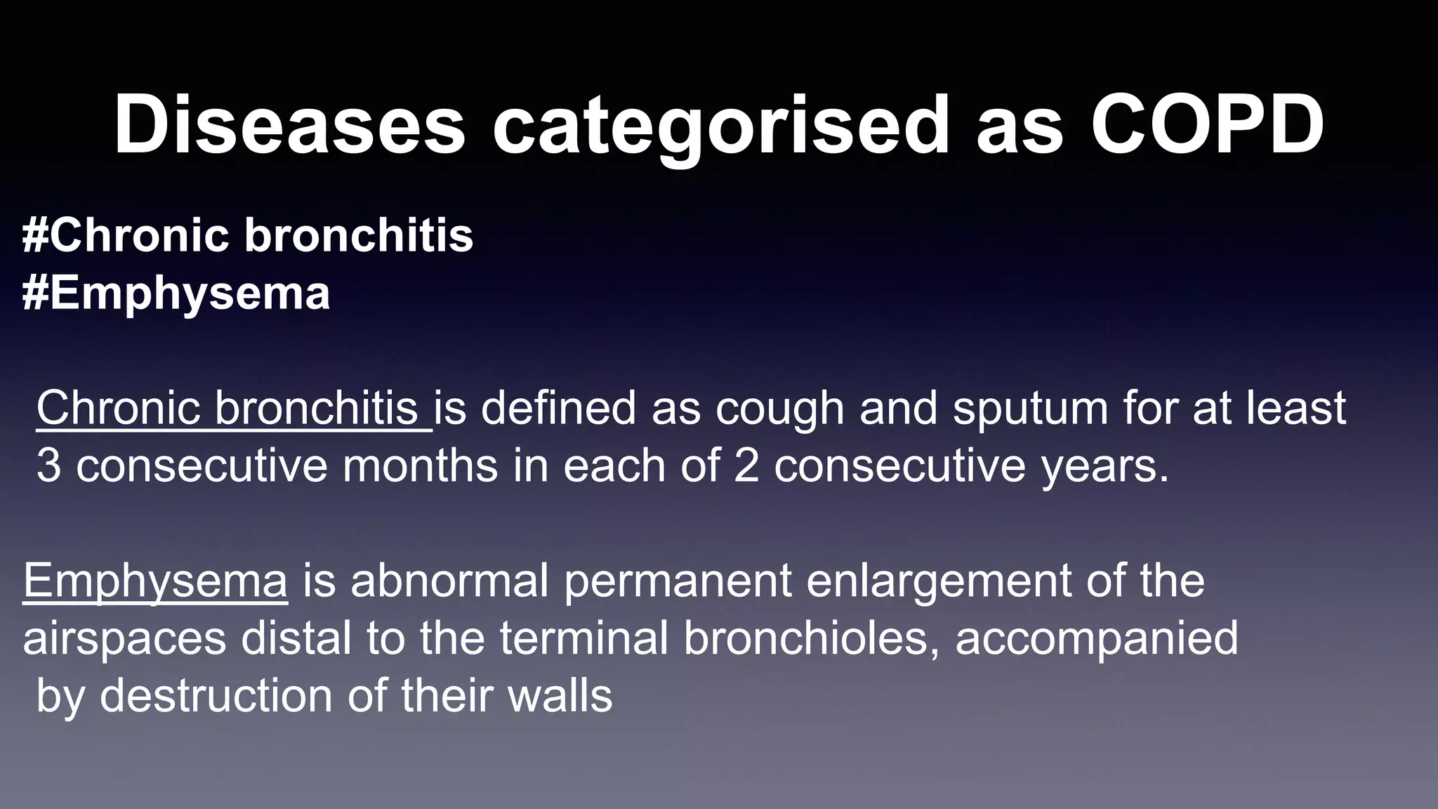
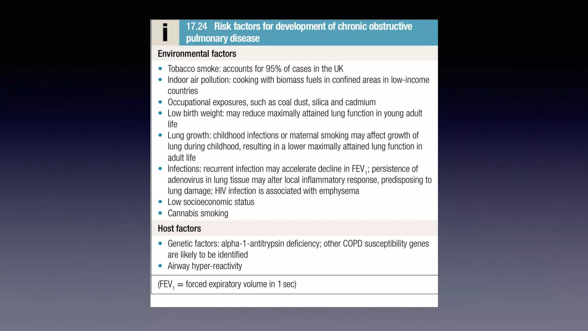
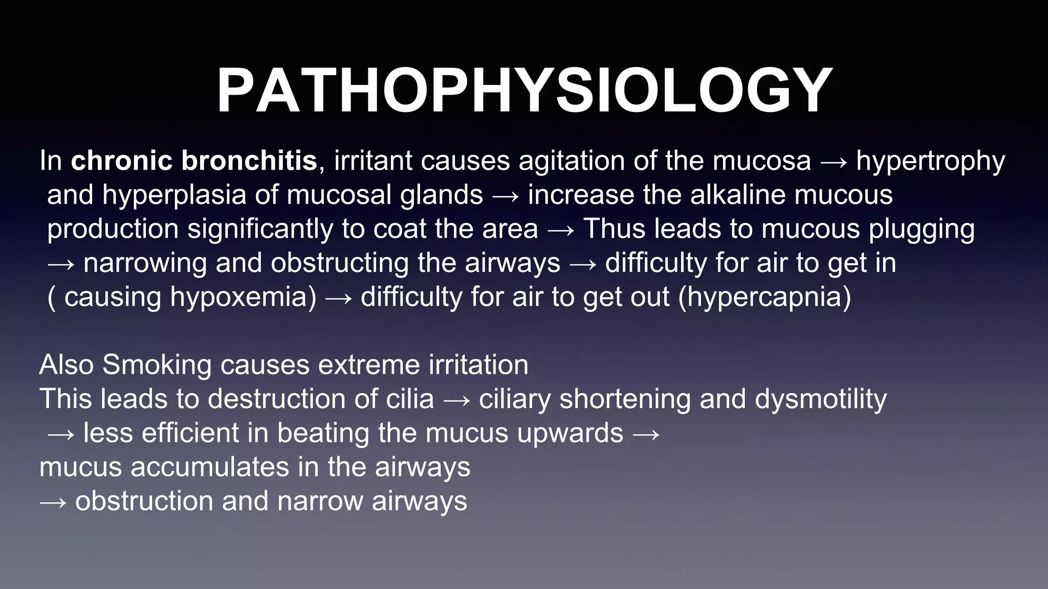
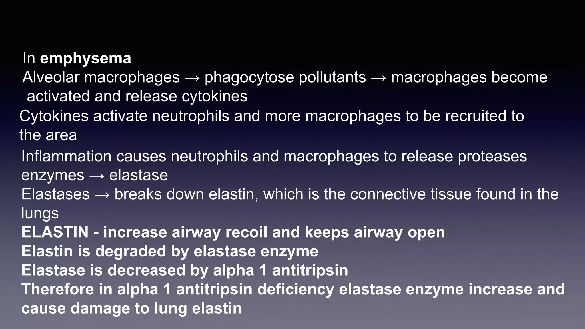
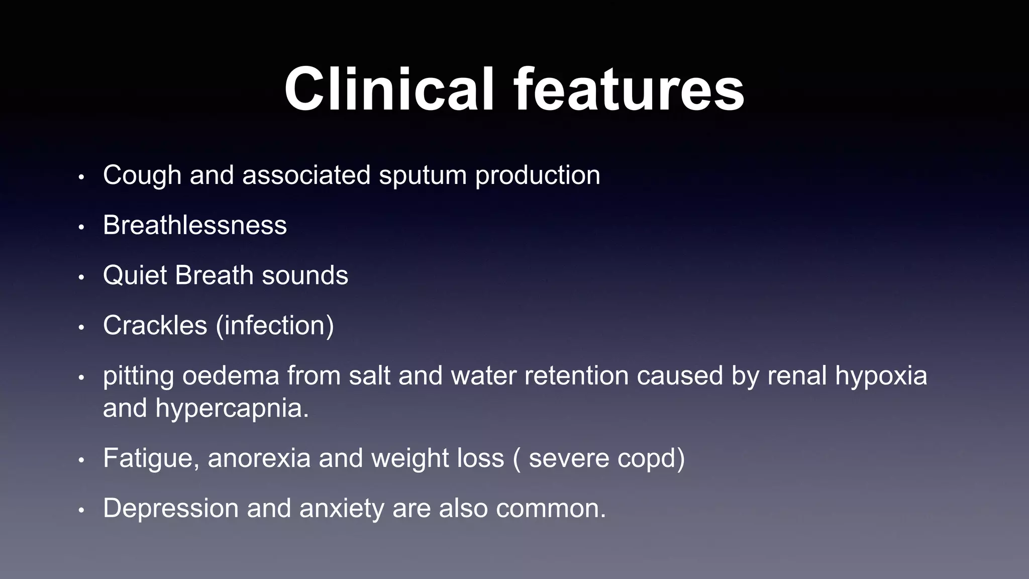
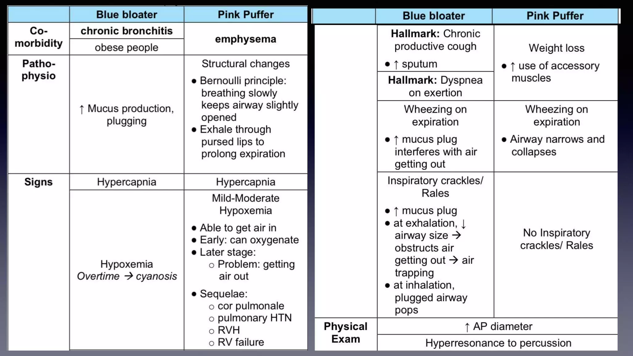
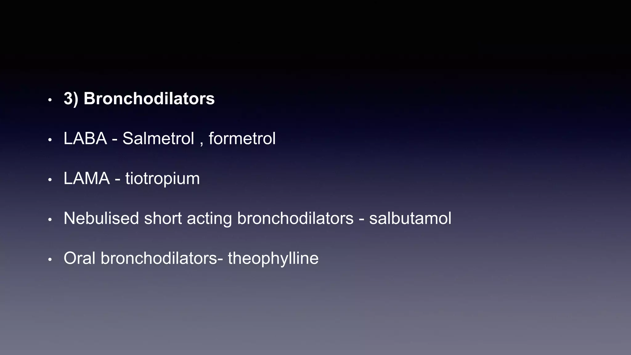
Chronic obstructive pulmonary disease (COPD) is
Defined as a preventable and treatable disease
characterised by persistent respiratory symptoms
and airflow limitation due to airway and/or alveolar
abnormalities,
usually caused by significant exposure to noxious
particles or gases.
Diseases categorised as COPD
#Chronic bronchitis
#Emphysema
Chronic bronchitis is defined as cough and sputum for at least
3 consecutive months in each of 2 consecutive years.
Emphysema is abnormal permanent enlargement of the
airspaces distal to the terminal bronchioles, accompanied
by destruction of their walls
RISK FACTORS
Tobacco smoking
Cigarette smoking represents the most significant risk factor for COPD a
suggesting that individual susceptibility factors are important.
Coal dust exposure
Use of biomass fuels
PATHOPHYSIOLOGY
In chronic bronchitis, irritant causes agitation of the mucosa → hypertrophy
and hyperplasia of mucosal glands → increase the alkaline mucous
production significantly to coat the area → Thus leads to mucous plugging
→ narrowing and obstructing the airways → difficulty for air to get in
( causing hypoxemia) → difficulty for air to get out (hypercapnia)
Also Smoking causes extreme irritation
This leads to destruction of cilia → ciliary shortening and dysmotility
→ less efficient in beating the mucus upwards →
mucus accumulates in the airways
→ obstruction and narrow airways
In emphysema
Alveolar macrophages → phagocytose pollutants → macrophages become
activated and release cytokines
Cytokines activate neutrophils and more macrophages to be recruited to
the area
Inflammation causes neutrophils and macrophages to release proteases
enzymes → elastase
Elastases → breaks down elastin, which is the connective tissue found in the
lungs
ELASTIN - increase airway recoil and keeps airway open
Elastin is degraded by elastase enzyme
Elastase is decreased by alpha 1 antitripsin
Therefore in alpha 1 antitripsin deficiency elastase enzyme increase and
cause damage to lung elastin
Clinical features
• Cough and associated sputum production
• Breathlessness
• Quiet Breath sounds
• Crackles (infection)
• pitting oedema from salt and water retention caused by renal hypoxia
and hypercapnia.
• Fatigue, anorexia and weight loss ( severe copd)
• Depression and anxiety are also common.
Investigations
Chest X-ray is essential to identify alternative diagnoses such as
cardiac failure, lung cancer and the presence of bullae
A blood count is useful to exclude anaemia or document
polycythaemia,
tested for alpha-1-antitrypsin deficiency.
The diagnosis requires objective demonstration of airflow obstruction
by spirometry and is established when the post-bronchodilator
FEV1/ FVC is <70%. The severity of COPD may be defined in relation
to the post-bronchodilator FEV1
• Lung volumes measured - by helium dilution or body
plethysmography - In emphysema decreased gas exchange present
• Exercise tests for assessment of exercise tolerance
• High resolution computed tomography
Management
• 1) Reducing exposure to noxious particles and gases
• Quit smoking
• Non smoking cooking devices and alternate fuels
• 2) Pulmonary rehabilitation
• Encourage exercise
• 3) Bronchodilators
• LABA - Salmetrol , formetrol
• LAMA - tiotropium
• Nebulised short acting bronchodilators - salbutamol
• Oral bronchodilators- theophylline
• 4) combined Inhalational Corticosteroids (ICS) and
bronchodilators
• the form of either a LABA/LAMA/ICS or LABA/ICS combination inhaler.
• 5)Oral anti inflammatories
• Oral glucocorticoids, Roflumilast (phosphodiesterase-4 inhibitor)
• 6) Maintainance measures
• Pneumococcal vaccine , influenza vaccine, mucolytics , treat obesity ,
malnutrition, depression
• 7) Oxygen therapy and home ventilation
• Long-term domiciliary oxygen therapy (LTOT) improves survival in selected
patients with COPD complicated by severe hypoxaemia (arterial PaO2 <7.3 kPa
(55 mmHg)
• 8) Surgical intervention
• Bullectomy - large bullae compress surrounding normal lung tissue.
• Patients with predominantly upper lobe emphysema, preserved gas transfer and
no evidence of pulmonary hypertension may benefit from lung volume reduction
surgery (LVRS). LVRS involves resection of peripheral emphysematous lung
tissue with the aim of reducing hyperinflation and decreasing the work of
breathing.
• Lung transplantation in selected patients
• 9) Palliative
• Morphine
• Benzodiazepines for anxiety
ACUTE EXACERBATION OF COPD
• Acute exacerbations of COPD (AECOPD) are characterised by an
increase in symptoms and deterioration in lung function and health
status. They become more frequent as the disease progresses and
are usually triggered by infection or a change in air quality.
• accompanied by the development of respiratory failure and/or fluid
retention
MANAGEMENT OF AECOPD
• At home with increased bronchodilator therapy
• cyanosis, peripheral oedema or an alteration in consciousness
should prompt referral to hospital
• Oxygen therapy
• In patients with an exacerbation of severe COPD, high
concentrations of oxygen may cause respiratory depression and
worsening acidosis. Controlled oxygen at 24% or 28% should be
used with the aim of main- taining SaO2 of 88%–92% or PaO2 of
more than 8kPa (60mmHg) without worsening acidosis.
• Bronchodilators
• Nebulised short-acting β2-agonists combined with an anticholiner- gic agent (e.g.
salbutamol and ipratropium) are routinely administered. However, the latter is only
appropriate if the patient is unable to take their LAMA inhaler. Nebulisers can be
driven with oxygen, but due to concern regarding oxygen sensitivity, they are more
safely driven by compressed air with supplemental oxygen delivered by nasal
cannula.
• Glucocorticoids
• Oral prednisolone reduces symptoms and improves lung function. Doses of 30 mg
for 5 days are currently recommended but weaning courses are required if the
patient has had a recent course of steroids. Prophylaxis against osteoporosis
should be considered in patients who receive repeated courses of glucocorticoids.
• Antibiotic therapy
• The role of bacteria in exacerbations remains controversial. There is little
evidence for the routine administration of antibiotics, but they are recom-
mended for patients reporting an increase in sputum purulence, sputum
volume or breathlessness.
• Non invasive ventilation
• Invasive ventilation should be considered in patients with deteriorating acidosis
despite optimal NIV settings, those unable to tolerate or wear the inter- face
(e.g. due to facial injury) and those who cannot protect their airway.
• Additional therapy
• Exacerbations may be accompanied by the development of
peripheral oedema; this usually responds to diuretics. Hospital
admission provides a good opportunity to address smoking
cessation, mobility and sputum clear- ance, inhaler technique and
concordance, nutrition and swallowing concerns.
THANK YOU

COPD.pptx

  • 1.
  • 2.
    INTRODUCTION Chronic obstructive pulmonarydisease (COPD) is Defined as a preventable and treatable disease characterised by persistent respiratory symptoms and airflow limitation due to airway and/or alveolar abnormalities, usually caused by significant exposure to noxious particles or gases.
  • 3.
    Diseases categorised asCOPD #Chronic bronchitis #Emphysema Chronic bronchitis is defined as cough and sputum for at least 3 consecutive months in each of 2 consecutive years. Emphysema is abnormal permanent enlargement of the airspaces distal to the terminal bronchioles, accompanied by destruction of their walls
  • 4.
    RISK FACTORS Tobacco smoking Cigarettesmoking represents the most significant risk factor for COPD a suggesting that individual susceptibility factors are important. Coal dust exposure Use of biomass fuels
  • 6.
    PATHOPHYSIOLOGY In chronic bronchitis,irritant causes agitation of the mucosa → hypertrophy and hyperplasia of mucosal glands → increase the alkaline mucous production significantly to coat the area → Thus leads to mucous plugging → narrowing and obstructing the airways → difficulty for air to get in ( causing hypoxemia) → difficulty for air to get out (hypercapnia) Also Smoking causes extreme irritation This leads to destruction of cilia → ciliary shortening and dysmotility → less efficient in beating the mucus upwards → mucus accumulates in the airways → obstruction and narrow airways
  • 7.
    In emphysema Alveolar macrophages→ phagocytose pollutants → macrophages become activated and release cytokines Cytokines activate neutrophils and more macrophages to be recruited to the area Inflammation causes neutrophils and macrophages to release proteases enzymes → elastase Elastases → breaks down elastin, which is the connective tissue found in the lungs ELASTIN - increase airway recoil and keeps airway open Elastin is degraded by elastase enzyme Elastase is decreased by alpha 1 antitripsin Therefore in alpha 1 antitripsin deficiency elastase enzyme increase and cause damage to lung elastin
  • 8.
    Clinical features • Coughand associated sputum production • Breathlessness • Quiet Breath sounds • Crackles (infection) • pitting oedema from salt and water retention caused by renal hypoxia and hypercapnia. • Fatigue, anorexia and weight loss ( severe copd) • Depression and anxiety are also common.
  • 10.
    Investigations Chest X-ray isessential to identify alternative diagnoses such as cardiac failure, lung cancer and the presence of bullae A blood count is useful to exclude anaemia or document polycythaemia, tested for alpha-1-antitrypsin deficiency. The diagnosis requires objective demonstration of airflow obstruction by spirometry and is established when the post-bronchodilator FEV1/ FVC is <70%. The severity of COPD may be defined in relation to the post-bronchodilator FEV1
  • 12.
    • Lung volumesmeasured - by helium dilution or body plethysmography - In emphysema decreased gas exchange present • Exercise tests for assessment of exercise tolerance • High resolution computed tomography
  • 13.
    Management • 1) Reducingexposure to noxious particles and gases • Quit smoking • Non smoking cooking devices and alternate fuels • 2) Pulmonary rehabilitation • Encourage exercise
  • 14.
    • 3) Bronchodilators •LABA - Salmetrol , formetrol • LAMA - tiotropium • Nebulised short acting bronchodilators - salbutamol • Oral bronchodilators- theophylline
  • 15.
    • 4) combinedInhalational Corticosteroids (ICS) and bronchodilators • the form of either a LABA/LAMA/ICS or LABA/ICS combination inhaler. • 5)Oral anti inflammatories • Oral glucocorticoids, Roflumilast (phosphodiesterase-4 inhibitor) • 6) Maintainance measures • Pneumococcal vaccine , influenza vaccine, mucolytics , treat obesity , malnutrition, depression
  • 16.
    • 7) Oxygentherapy and home ventilation • Long-term domiciliary oxygen therapy (LTOT) improves survival in selected patients with COPD complicated by severe hypoxaemia (arterial PaO2 <7.3 kPa (55 mmHg) • 8) Surgical intervention • Bullectomy - large bullae compress surrounding normal lung tissue. • Patients with predominantly upper lobe emphysema, preserved gas transfer and no evidence of pulmonary hypertension may benefit from lung volume reduction surgery (LVRS). LVRS involves resection of peripheral emphysematous lung tissue with the aim of reducing hyperinflation and decreasing the work of breathing. • Lung transplantation in selected patients
  • 17.
    • 9) Palliative •Morphine • Benzodiazepines for anxiety
  • 18.
    ACUTE EXACERBATION OFCOPD • Acute exacerbations of COPD (AECOPD) are characterised by an increase in symptoms and deterioration in lung function and health status. They become more frequent as the disease progresses and are usually triggered by infection or a change in air quality. • accompanied by the development of respiratory failure and/or fluid retention
  • 19.
    MANAGEMENT OF AECOPD •At home with increased bronchodilator therapy • cyanosis, peripheral oedema or an alteration in consciousness should prompt referral to hospital • Oxygen therapy • In patients with an exacerbation of severe COPD, high concentrations of oxygen may cause respiratory depression and worsening acidosis. Controlled oxygen at 24% or 28% should be used with the aim of main- taining SaO2 of 88%–92% or PaO2 of more than 8kPa (60mmHg) without worsening acidosis.
  • 20.
    • Bronchodilators • Nebulisedshort-acting β2-agonists combined with an anticholiner- gic agent (e.g. salbutamol and ipratropium) are routinely administered. However, the latter is only appropriate if the patient is unable to take their LAMA inhaler. Nebulisers can be driven with oxygen, but due to concern regarding oxygen sensitivity, they are more safely driven by compressed air with supplemental oxygen delivered by nasal cannula. • Glucocorticoids • Oral prednisolone reduces symptoms and improves lung function. Doses of 30 mg for 5 days are currently recommended but weaning courses are required if the patient has had a recent course of steroids. Prophylaxis against osteoporosis should be considered in patients who receive repeated courses of glucocorticoids.
  • 21.
    • Antibiotic therapy •The role of bacteria in exacerbations remains controversial. There is little evidence for the routine administration of antibiotics, but they are recom- mended for patients reporting an increase in sputum purulence, sputum volume or breathlessness. • Non invasive ventilation • Invasive ventilation should be considered in patients with deteriorating acidosis despite optimal NIV settings, those unable to tolerate or wear the inter- face (e.g. due to facial injury) and those who cannot protect their airway.
  • 22.
    • Additional therapy •Exacerbations may be accompanied by the development of peripheral oedema; this usually responds to diuretics. Hospital admission provides a good opportunity to address smoking cessation, mobility and sputum clear- ance, inhaler technique and concordance, nutrition and swallowing concerns.
  • 23.