This document describes the technique of dental CT imaging. It discusses how dental CT has become an established method for imaging jaw anatomy prior to dental implant placement. The key aspects of the dental CT technique include acquiring high-resolution axial scans of the jaw and generating curved and orthoradial multiplanar reconstructions. The document outlines the specific scanner protocols used in dental CT, including using a small focal spot, thin slices, and dose reduction strategies. It also describes how the data is reconstructed to generate panoramic and orthogonal views of the jaw anatomy and how the images are presented.
![Received: 2 August 2001
Revised: 17 January 2002
Accepted: 28 January 2002
Published online: 30 April 2002
© Springer-Verlag 2002
Abstract In addition to convention-
al imaging methods, dental CT has
become an established method for
anatomic imaging of the jaws prior
to dental implant placement. More
recently, this high-resolution imag-
ing technique has gained importance
in diagnosing dental-associated dis-
eases of the mandible and maxilla.
Since most radiologists have had lit-
tle experience in these areas, many
of the CT findings remain unde-
scribed. The objective of this review
article is to present the technique of
dental CT, to illustrate the typical ap-
pearance of jaw anatomy and dental-
related diseases of the jaws with
dental CT, and to show where it can
serve as an addition to conventional
imaging methods in dental radiology.
Keywords Computed tomography ·
Jaw · Teeth · Implant
Eur Radiol (2003) 13:366–376
DOI 10.1007/s00330-002-1373-7 H E A D A N D N E C K
André Gahleitner
G. Watzek
H. Imhof
Dental CT: imaging technique, anatomy,
and pathologic conditions of the jaws
Introduction
Dental CT has become an established method for imag-
ing of jaw anatomy prior to dental implant placement [1,
2, 3, 4, 5, 6]. The clinical use of these implants has rap-
idly increased over the past 20 years. It is estimated that
300,000 implants are placed each year with over 50 com-
panies involved in the manufacture, marketing and distri-
bution within the United States [7, 8].
The term “dental CT” does not represent a particular
modality, but rather a specific investigation protocol.
The main features of this protocol include the acquisi-
tion of axial scans of the jaw with the highest possible
resolution together with curved and orthoradial multi-
planar reconstructions (Fig. 1). Dentists commonly di-
agnose and work in the submillimeter scale; hence, a
highly detailed image quality is required and challenges
CT to its technical limits. This article reviews the spe-
cific technique of dental CT and illustrates the typical
appearance of jaw anatomy and dental-related diseases
of the jaw.
Technique
History
The technique of dental CT, also called Dentascan, was
developed by Schwarz et al. in 1987, when these investi-
gators first used curved multiplanar reconstructions of
the jaw [9, 10]. At that time, the number of inserted im-
plants in the jaw had steadily increased and there was a
critical need for accurate imaging of the jaw anatomy,
especially in the bucco-lingual plane. The major disad-
vantage of CT in the jaw region, the metal artifacts from
tooth fillings, was overcome by using the axial plane for
scanning instead of the coronal plane, which kept these
artifacts in the occlusion plane and hence left the jaw
A. Gahleitner (✉) · H. Imhof
Department of Radiology/Osteology,
Medical School, University of Vienna,
Währinger Strasse 25a,
1090 Vienna, Austria
e-mail: andre.gahleitner@univie.ac.at
Tel.: +43-1-427767191
Fax: +43-1-427767039
A. Gahleitner · G. Watzek
Department of Oral Surgery,
Dental School, University of Vienna,
Währinger Strasse 25a,
1090 Vienna, Austria](https://image.slidesharecdn.com/dentalct-130523100307-phpapp02/85/CT-Dental-1-320.jpg)
![367
bone undistorted. This allowed for accurate display of
the vertical as well as the important bucco-lingual di-
mensions of the jaw in actual size, which facilitated the
work of the dentist [11].
Prior to this development, the first useful technique
for pre-implant imaging of jaw anatomy was convention-
al orthoradial tomography, using a complex (circular,
spiral, or hypocycloidal) blurring device, such as the
Scanora or CommCat (Soredex, Marietta, Ga.; Imaging
Sciences International, Roebling, N.J.) [12, 13]. Al-
though this technique is still a valuable procedure, it is
prone to errors, has the known disadvantages of conven-
tional tomography, and does not allow imaging of the
complete jaw within an acceptable time frame. Due to
the higher cost and lesser availability of dental CT, con-
ventional orthoradial tomography is still a standard in-
vestigation in many implanologic centers.
During the past years various studies have been pub-
lished which have validated dental CT as an excellent
tool for diagnosing dental-related pathologies [3, 14, 15,
16, 17, 18, 19, 20, 21]. Since occurring changes may be
very subtle, an optimal image quality with the highest
possible resolution is essential for establishing a correct
diagnosis.
Patient
Prior to imaging, the patient should be informed about
the investigation and instructed not to move or swallow
during the scan. The investigation is performed in the su-
pine position with the cervical spine slightly overextend-
ed backward. The head should be strapped to the head-
rest and positioned as symmetrically as possible. This
can be checked in the scoutview, where both rami and
the angles of the mandible should be perfectly aligned. If
movement of the mandible during the scan is likely to
occur (e.g., edentulous jaws with lack of occlusion), it is
possible to immobilize the jaw by having the patient bite
on a cotton roll or on fast-hardening impression material.
Scanner protocol
Dental CT investigations can be performed either on a
conventional CT, spiral CT, or a multislice CT scanner.
The device should be capable of performing high-resolu-
tion scans with a small focal spot and acquiring thin slic-
es of 1.5 mm or less. A table feed of 1 mm is necessary
to obtain high-quality images with optimal detail in the
scan plane as well as in the multiplanar reconstruction. A
spiral scan technique with 1 s per rotation is sufficient in
most cases where imaging of jaw anatomy is performed
prior to implant placement. If imaging of pathologic con-
ditions is required, small details can be obtained by in-
creasing the scan time to 2 s per rotation. The larger
number of views due to the slower rotation speed of the
tube provides the more detailed information necessary
for imaging of frequently very subtle pathologic fea-
tures. The field of view should be limited to 120 mm or
less to avoid unnecessary imaging of the spine, neck, or
posterior cranial fossa. A well-established universal pro-
tocol is provided as an example in Table 1.
Scan direction is caudocranial beginning with the
mandible base and extends to include the alveolar crest
for the mandible, whereas for the maxilla the scan plane
starts with the alveolar crest and extends upward to in-
clude all root tips. If sinusitis is present, extensions of
the scan are recommended to exclude possible dental-
related causes, such as displaced root remnants or for-
eign bodies.
Dose reduction
Dental CT images are displayed with a very low contrast
setting (bone window) due to the excellent contrast be-
tween bone and soft tissue. Since no contrast medium is
used and displaying soft tissue detail with digital en-
hanced contrast (soft tissue window) is usually not
necessary, dental CT is ideally suited for applying dose-
reduced investigation protocols [4, 22, 23]. This is pri-
marily accomplished by reducing the tube current, which
leads to increased quantum noise noted in the soft tissue
compartment, whereas bone is only marginally affected.
In addition, using 1.5-mm slice thickness instead of 1.0
and/or using a spiral technique with a pitch factor of
more than 1.0 can further reduce dose delivery. The lim-
iting factor here is that the important visualization of the
mandibular canal is degraded when a high pitch factor is
used, although orthoradial reconstructions are displayed
Fig. 1 Dental CT of the maxilla. From axial slices (center) of a
given investigation volume orthoradial reconstructions are calcu-
lated (right). As a single example this reconstruction demonstrates
the alveolar crest and the left maxillary sinus](https://image.slidesharecdn.com/dentalct-130523100307-phpapp02/85/CT-Dental-2-320.jpg)
![370
paralysis or numbness of the chin and edge of the mouth.
Since the neurovascular bundle within the mandibular
canal also supplies the teeth, sudden loss of tooth vitality
within a whole quadrant can occur. Damage to the man-
dibular canal during placement of implants (or extraction
of third molars) therefore represents one of the major
issues for legal steps taken against dentists.
Maxillary sinus. The maxillary sinus can reach far mesi-
ally and between the roots of the molars and premolars.
Due to the close relationship with these other structures,
the maxillary sinus can be easily affected by inflammato-
ry conditions and cystic lesions of the adjacent teeth. As
a frequent variant, a bony septum may be visible in the
sinus floor (Underwood septum), which can complicate
pre-implant augmentative procedures such as the “sinus
lift” (Fig. 4) [24].
Bone volume/resorption/atrophy
After the loss of teeth, atrophy of the alveolar crest is a
typical occurrence. This is due to the loss of chewing
forces in the jaw and can lead to a complete loss of the
alveolar crest in edentulous patients. Since the height of
the alveolar crest in both jaws can be up to 4 cm, a third
of the patient’s face is lost and a typical surplus of soft
tissue is present. Cawood and Howell have described
and classified the bone volume loss in anatomic studies
that can be applied to dental CT due to the perfect visu-
alization of the alveolar crest in the orthoradial plane
[25]. These six resorption classes represent typical ap-
pearances of jaw atrophy after tooth loss (Fig. 5). In gen-
eral, after extraction of a tooth (class 2), a continuous re-
duction of bone occurs until the alveolar crest demon-
strates a “knife-edge” appearance (class 4). If atrophy
continues, further bone height is lost until only the jaw
base remains as a thin bone layer (class 6). Although at-
rophy is a continuous process, single classes can be
skipped. For example, class 2 can directly transform into
class 4 by loss of the buccal cortical bone (juga alveol-
aria), which results in a knife-edge alveolar crest. The re-
sorption classes are an important consideration when im-
plantation is planned, and have an influence on the selec-
tion of implant dimensions and type as well as for the
choice of a possible augmentation procedure such as si-
nus lift, onlay graft, or lateral augmentation. These oper-
ative procedures are used to artificially enhance the
available bone volume by deposition of autologous bone
or artificial bone replacement material.
Bone quality
Bone quality, as described by Lekholm and Zarb, is of
major importance for the success of an implant place-
ment [26]. For preoperative planning bone quality has
been categorized into four classes that basically describe
the relation of cortical and cancellous bone in a specified
region of the jaw (Fig. 6). The amount of cortical bone is
Fig. 3 Axial slice through the alveolar crest of the maxilla. The
numbers of each tooth are given according to international nomen-
clature (the first digit representing the quadrant and the second
digit the tooth counted from the midline). In addition, the incisive
canal (double arrowhead) and a small part of the maxillary sinus
(single arrowhead) is visible
Fig. 4 Axial slice through the maxillary sinus. Prominent Under-
wood septum within both sinuses (arrows). Two root tips of tooth
27 are visible within the left septum (arrowheads)](https://image.slidesharecdn.com/dentalct-130523100307-phpapp02/85/CT-Dental-5-320.jpg)
![371
responsible for the primary stability of the implant,
whereas cancellous bone is responsible for long-term
stability. Although class 1 indicates optimum stability of
the implant, studies have revealed classes 2 and 3 to
have the best long-term results, with class 4 having the
most frequent premature implant loss [5, 27].
Measurements/implants
Evaluation of bone quantity is performed by measuring
the height and width of the alveolar crest for a specified
region. These values serve as an overview of the avail-
able bone quantity and do not serve as a suggested im-
plant size. This is because the oral surgeon has multiple
choices of implant sites and implantation directions us-
ing different angulations and different diameters. The
choice is not solely based on the available amount of
bone (bone-demanded implantation), but must take pro-
sthodontic and cosmetic factors into account. Moreover,
immediately prior to implant placement a canal is
drilled, which usually is 1–2 mm longer than the final in-
serted implant. Thus, injury of anatomic structures can
occur even if the final implant does not reach these
structures; hence, the radiology report concerning mea-
surements cannot serve as the sole factor for implant
choice.
Jaw pathology
In addition to imaging of jaw anatomy and its variants,
dental CT has proved to be an excellent tool for diagnos-
ing pathologic conditions of the jaw. Most lesions in
this region are visible only in the millimeter or even sub-
millimeter scale and therefore visualization of these al-
terations was not possible with typical CT protocols.
This changed with the advent of dental CT where it was
possible to visualize objects on the submillimeter scale.
Still, conventional imaging (dental film, panoramic radi-
ography) is the basic screening investigation for diagnos-
ing pathologic conditions, but CT can aid in revealing
additional features and in localization of a lesion or even
help to exclude the presence of a pathologic condition
more easily. In the following section, the most frequent
pathologies found in the jaw are summarized.
Chronic apical periodontitis
Chronic apical periodontitis (CAP) is a frequent finding
in patients with pulpitis and in patients after dental treat-
ment by root canal filling. It is characterized by an en-
largement of the periodontal space at the periapical re-
gion of the tooth. Dental CT can demonstrate the root tip
within a small osteolytic region (the enlarged periodontal
space) [16]. If bacteria spreads into the surrounding can-
cellous bone in the chronic stage, a reactive enlargement
of trabeculae occurs. This entity is described as “scleros-
ing ostitis,” a form of chronic osteomyelitis. When the
CAP reaches cortical bone, a periosteal reaction (Fig. 7)
Fig. 5 Classification of bone
atrophy according to Cawood
and Howell [25]. Schematic
representation of atrophic
changes in the anterior midline
of the maxilla (upper part) and
mandible (lower part)
Fig. 6 Classification of bone quality according to Lekholm and
Zarb [26]](https://image.slidesharecdn.com/dentalct-130523100307-phpapp02/85/CT-Dental-6-320.jpg)
![372
or reactive sinusitis can be visible (Fig. 8). After perfora-
tion of the apical periodontitis through the cortical bone
into the surrounding tissue, an infiltrate with soft tissue
edema can be seen in this compartment.
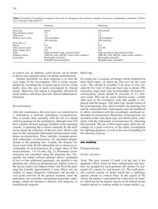
Radicular cysts
When a CAP remains untreated, it tends to grow until
the granulation tissue around the root apex transforms
and becomes a cystic, epithelial-lined lesion. These ra-
dicular cysts are the most common type of cyst in the
jaw and can reach a substantial size with the risk of frac-
ture [17, 28, 29]. They are characterized by a large, well-
defined radiolucency (>1 cm) with the apex of a non-
vital root in the epicenter of the lesion (Fig. 9). The size
at which the transformation from CAP occurs is approxi-
mately 1 cm with a large overlap. Although a clear dif-
ferentiation between CAP and radicular cyst based on
criteria other than size is impossible, the choice of treat-
ment is often different (operative vs endodontic treat-
ment).
Dentigerous cysts
The second most common cyst in the jaw, the dentiger-
ous cyst, also called follicular cyst, is located around
the crown of an impacted tooth and attaches to the ce-
mento-enamel junction, which helps in the differentia-
tion from radicular cysts [30, 31]. Dentigerous cysts are
sharply delineated and frequently demonstrate a local
Fig. 7 Axial slices demonstrat-
ing Chronic apical periodontitis
(CAP) of tooth 35 (arrow) with
surrounding sclerosing ostitis
and lingual-sided perforation
and periosteal reaction (arrow-
heads)
Fig. 8 Panoramic slices of the
maxilla. A CAP of tooth 27
with perforation into the left
maxillary sinus (arrows) and
reactive dentogen sinusitis
(arrowheads)](https://image.slidesharecdn.com/dentalct-130523100307-phpapp02/85/CT-Dental-7-320.jpg)
![374
expansion of the buccal or lingual cortical plate. Adja-
cent roots of neighboring teeth can be easily reached,
but usually are not surrounded by the cyst completely
(Fig. 10).
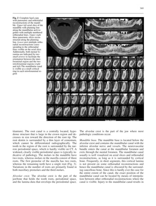
Root fractures
Horizontal root fractures, which usually occur after trau-
ma, are easily diagnosed by clinical examination and
conventional radiographic techniques, whereas vertical
root fractures are visualized with dental film only when
the fracture line is oriented at least partially within the
direction of the X-ray beam. Studies have demonstrated
that dental CT is superior to dental film in diagnosing
vertical root fractures because CT is not sensitive to
beam orientation [19]. These fractures usually occur as a
result of conservative restorations of a tooth with a post
or in endodontically treated teeth.
The limitations of dental CT in the diagnosis of den-
tal fractures, resulting in false-negative readings, in-
clude small fissures below the resolution capability of
CT and superimposed metal artifacts from root posts.
In addition to obscuring a root, these artifacts can also
mimic fracture lines, but these limitations can be over-
come if the fracture extends below the root post and is
visible there. Although horizontally oriented fractures
lying in the scanning plane are difficult to visualize
with CT, oblique fractures remain easily detectable
(Fig. 11).
Fig. 12 Orthoradial reconstruc-
tion of region 17: extraction
socket (arrowheads) demon-
strating an oro-antral fistula
from the trifurcation (arrow) to
the right maxillary sinus with
reactive sinusitis
Fig. 13 Foreign body in the lingual-sided soft tissue of the mandi-
ble (arrows). Panoramic radiography revealed the impression of
the foreign body located within the visible extraction socket (arrow-
heads)](https://image.slidesharecdn.com/dentalct-130523100307-phpapp02/85/CT-Dental-9-320.jpg)
![375
Sinus fistula
Sinus fistula frequently occurs as a complication after
tooth extraction or after root resection in the maxillary
molar region and must be treated to prevent maxillary si-
nus inflammation. Dental CT can clearly localize the
corresponding osseous defect in the alveolar ridge and
frequently orthoradial reconstructions offer optimal visu-
alization for preoperative planning (Fig. 12) [14].
Foreign bodies
Dental CT can help in localizing foreign bodies that can
be found after or during dental treatment, which other-
wise would be hard to detect with conventional radiolog-
ic methods. Most dental instruments and materials are
radio-opaque, which helps in identifying the source and
exact location. Materials usually found include root or
crown fillings, gutta-percha, endodontic instruments, and
root posts. These materials are typically located in the
maxillary sinus, the alveolar ridge, or in the adjacent soft
tissue and can be a source of chronic infection and pain
(Fig. 13).
Implant placement “periimplantitis”
Correct implant placement is crucial to prevent early im-
plant loss or clinical complications, which is especially
important if implant perforation into the maxillary sinus
or nasal cavity occurs [32, 33, 34, 35, 36]. Another ma-
jor complication is perforation of the implant into the
mandibular canal, which can lead to paresthesia of the
mental region and loss of vitality of the more mesially
located teeth. The term “periimplantitis” describes an os-
teolytic layer around implants due to chronic infection
and/or malocclusion and it is the major radiologic indi-
cator for imminent implant loss (Fig. 14).
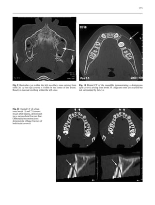
Impacted teeth
Dental CT offers superb visualization of impacted teeth
and can help the clinician to plan his treatment preoper-
atively or prior to orthodontic therapy [37, 38, 39]. The
position of the tooth within the alveolar crest as well as
the relation to surrounding structures is clearly dis-
closed. Resorption of adjacent roots and hooks, in par-
ticular, are easily detected and quantified by dental CT
(Fig. 15).
Conclusion
This review summarizes the capabilities of dental CT as
an imaging method for dentistry. Anatomic features as
well as the appearance of frequent dental pathologies are
described with their typical findings, which the radiolo-
gist should communicate to the referring clinician.
Fig. 14 Axial slice demonstrating extensive “periimplantitis” in
region 26 (arrow) and misplacement of implants in the anterior
maxilla (arrowheads)
Fig. 15 Axial slice of the maxilla. Horizontally positioned and
impacted tooth 23 with a hook (arrow) at the apex within the left
maxillary sinus complicating a planned extraction](https://image.slidesharecdn.com/dentalct-130523100307-phpapp02/85/CT-Dental-10-320.jpg)
![376
References
1. Abrahams JJ (2001) Dental CT imag-
ing: a look at the jaw. Radiology
219:334–345
2. Homolka P, Gahleitner A, Kudler H,
Nowotny R (2001) A simple method
for estimating effective dose in dental
CT. Conversion factors and calculation
examples for a clinical low dose proto-
col. Rofo Fortschr Geb Rontgenstr
Neuen Bildgeb Verfahr 173:558–562
3. Solar P, Gahleitner A (1999) Dental CT
in the planning of surgical procedures.
Its significance in the oro-maxillofacial
region from the viewpoint of the den-
tist. Radiologe 39:1051–1063
4. Schorn C, Visser H, Hermann KP,
Alamo L, Funke M, Grabbe E (1999)
Dental CT: image quality and radiation
exposure in relation to scan parame-
ters. Rofo Fortschr Geb Rontgenstr
Neuen Bildgeb Verfahr 170:137–144
5. Norton MR, Gamble C (2001) Bone
classification an objective scale of
bone density using the computerized
tomography scan. Clin Oral Impl Res
12:79–84
6. Lenglinger FX, Muhr T, Krennmair G
(1999) Dental CT: examination method,
radiation dosage and anatomy. Radio-
loge 39:1027–1034
7. Brunski JB (1999) In vivo bone re-
sponse to biomechanical loading at the
bone/dental-implant interface. Adv Dent
Res 13:99–119
8. Dunlap J (1988) Implants: implications
for general dentists. Dent Econ 78:101–
112
9. Schwarz MS, Rothman SL, Rhodes
ML, Chafetz N (1987) Computed to-
mography. I. Preoperative assessment
of the mandible for endosseous implant
surgery. Int J Oral Maxillofac Implants
2:137–141
10. Schwarz MS, Rothman SL, Rhodes
ML, Chafetz N (1987) Computed to-
mography. II. Preoperative assessment
of the maxilla for endosseous implant
surgery. Int J Oral Maxillofac Implants
2:143–148
11. Rothman SL, Chaftez N, Rhodes ML,
Schwarz MS, Schwartz MS (1988) CT
in the preoperative assessment of the
mandible and maxilla for endosseous
implant surgery. Work in progress
[published erratum appears in Radiol-
ogy 169:581]. Radiology 168:171–175
12. Hanssens JF (1996) Pre-implantation
evaluation using medical imagery:
scanner or Scanora? Rev Belge Med
Dent 51:101–110
13. Ekestubbe A (1999) Conventional spi-
ral and low-dose computed mandibular
tomography for dental implant plan-
ning. Swed Dent J (Suppl) 138:1–82
14. Abrahams JJ, Berger SB (1995) Oral-
maxillary sinus fistula (oroantral fistu-
la): clinical features and findings on
multiplanar CT. Am J Roentgenol
165:1273–1276
15. Abrahams JJ, Hayt MW (1999) Dental
CT in pathologic changes of the max-
illo-mandibular region. Radiologe
39:1035–1043
16. Abrahams JJ, Berger SB (1998) In-
flammatory disease of the jaw: appear-
ance on reformatted CT scans. Am J
Roentgenol 170:1085–1091
17. Bodner L, Bar-Ziv J, Kaffe I (1994) CT
of cystic jaw lesions. J Comput Assist
Tomogr 18:22–26
18. Fuhrmann RA, Bucker A, Diedrich PR
(1997) Furcation involvement: compar-
ison of dental radiographs and HR-CT
slices in human specimens. J Periodon-
tal Res 32:409–418
19. Youssefzadeh S, Gahleitner A,
Dorffner R, Bernhart T, Kainberger FM
(1999) Dental vertical root fractures:
value of CT in detection. Radiology
210:545–549
20. Royal SA, Hedlund GL, Wiatrak BJ
(1999) Single central maxillary incisor
with nasal pyriform aperture stenosis:
CT diagnosis prior to tooth eruption.
Pediatr Radiol 29:357–359
21. Lenglinger FX, Krennmair G, Muhr T,
Zisch RJ (1995) Improved imaging of
mandibular cysts using dental-CT.
Aktuelle Radiol 5:315–318
22. Hassfeld S, Streib S, Sahl H, Strat-
mann U, Fehrentz D, Zoller J (1998)
Low-dose computerized tomography of
the jaw bone in pre-implantation diag-
nosis. Limits of dose reduction and ac-
curacy of distance measurements.
Mund Kiefer Gesichtschir 2:188–193
23. Rustemeyer P, Streubuhr U, Hohn
HP, Rustemeyer R, Eich HT, John-
Mikolajewski V, Muller RD (1999)
Low-dosage dental CT. Rofo Fortschr
Geb Rontgenstr Neuen Bildgeb Verfahr
171:130–135
24. Abrahams JJ, Hayt MW, Rock R (2000)
Sinus lift procedure of the maxilla in
patients with inadequate bone for den-
tal implants: radiographic appearance.
Am J Roentgenol 174:1289–1292
25. Cawood JI, Howell RA (1988) A clas-
sification of the edentulous jaws.
Int J Oral Maxillofac Surg 17:232–
236
26. Lekholm U, Zarb GA (1985) Patient
selection and preparation. In: Bråne-
mark PI, Zarb GA, Albrektsson T (eds)
Osseointegration in clinical dentistry.
Quintessence, Chicago, pp 199–209
27. Jaffin RA, Berman CL (1991) The ex-
cesssive loss of Brånemark implants
in type IV bone: a 5 year analysis.
J Periodontol 62:2–4
28. Krennmair G, Lenglinger F (1995) Im-
aging of mandibular cysts with a dental
computed tomography software pro-
gram. Int J Oral Maxillofac Surg 24:48–
52
29. Abrahams JJ, Oliverio PJ (1993) Odon-
togenic cysts: improved imaging with
a dental CT software program. Am J
Neuroradiol 14:367–374
30. Som PM, Shangold LM, Biller HF
(1992) A palatal dentigerous cyst aris-
ing from a mesiodente. Am J Neuro-
radiol 13:212–214
31. Lehrman BJ, Mayer DP, Tidwell OF,
Brooks ML (1991) Computed tomo-
graphy of odontogenic keratocysts.
Comput Med Imaging Graph 15:365–
368
32. Widlitzek H, Konig S, Golin U (1996)
Value of dental CT for the implant spe-
cialty in mouth, jaw and facial surgery.
Radiologe 36:229–235
33. Schuller H (1996) Computerized to-
mography of the alveolar process.
Radiologe 36:221–225
34. Dula K, Buser D (1996) Computed to-
mography/oral implantology. Dental-
CT: a program for the computed tomo-
graphic imaging of the jaws. The indi-
cations for preimplantological clarifi-
cation. Schweiz Monatsschr Zahnmed
106:550–563
35. Wicht L, Moegelin A, Schedel H,
Pentzold C, Bier J, Langer R, Felix R
(1994) A dental CT study for preopera-
tive assessment of maxillary atrophy.
Aktuelle Radiol 4:64–69
36. Abrahams JJ, Kalyanpur A (1995)
Dental implants and dental CT soft-
ware programs. Semin Ultrasound CT
MR 16:468–486
37. Hirschfelder U (1994) Radiological
survey imaging of the dentition: dental
CT versus orthopantomography.
Fortschr Kieferorthop 55:14–20
38. Bodner L, Sarnat H, Bar-Ziv J, Kaffe I
(1994) Computed tomography in the
management of impacted teeth in chil-
dren. ASDC J Dent Child 61:370–377
39. Krennmair G, Lenglinger FX, Traxler
M (1995) Imaging of unerupted and
displaced teeth by cross-sectional CT
scans. Int J Oral Maxillofac Surg
24:413–416](https://image.slidesharecdn.com/dentalct-130523100307-phpapp02/85/CT-Dental-11-320.jpg)


