SlideShare a Scribd company logo
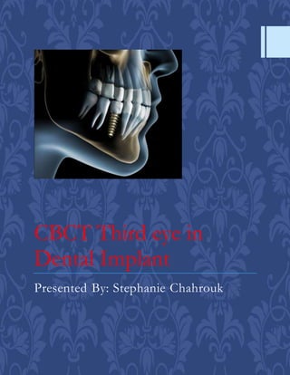
CBCT Third eye in
Dental Implant
Presented By: Stephanie Chahrouk
Introduction
"Imaging is a critically important aspect of diagnostics and
treatment planning when placing dental implants."
"High-quality, accurate images collected at the presurgical stage can
yield dividends both in implant outcomes and patient satisfaction."
Does Traditionaland CT Radiographic Methods Ideal?
"Traditional radiographic methods are not ideal for planning
implant placement "
"because you are visualizing a three dimensional (3D) object in two
dimensions and missing important information along the way"
"The development of computed tomography (CT) allowed clinicians to take
radiographic cross-sections, paving the way for 3D reconstructions of
maxillofacial features, though this improvement came at the cost of higher
radiation exposure"
Advantages ofCBCT
 CBCT technology overcomes limitations.
 It is possible to view all aspects of the insertion site on the computer screen, virtually,
noninvasively, as though you are dissecting your actual patient.
 Modern software packages generally provide various perspectivesthat usually are
customizable and adjustable based on the clinician’s preference.
 It is possible to view a 3D model (volumetric view) of the entire scanned object or only parts
of it or to create tomographic slices in all three planes of space and navigate through the
volume in increments of designated thicknesses, which also are customizable (Kau et al.,
2005).
 CBCT technology allows an exact visual identification of the location, shape, and divergence
of the mesial and distal dental roots, the floor of the maxillary sinus, and the buccal and
lingual wall of the alveolar process.
 Qualitative assessments, therefore, can be made with much greater accuracy than on a
regular 2D radiograph.
 CBCT data can be quantified using the appropriate software packages.
 Thus distances between two points such as interradicular distance or buccal bone thickness
can be measured with considerable accuracy (Timock et al., 2011);
 angulation between three points can be calculated (e.g., to determine root divergence);
 The density of objects including bone can be assessed (Marquezan et al., 2012).
 More detailed approach to planning and placing of the miniscrew implants.
Disadvantages
 the only major disadvantage is that a large amount of information provided by the CBCT
can lead to
o confusion or, even worse, may provide a false sense of security and override the
clinical chairside assessment for inexperienced clinicians.
 The transfer of the information obtained on the virtual model to the real patient also can be
difficult.
B: Procedure of Image acquisition in CBCT
“Principle of Action Of CBCT”
“The principle of CBCT is based on a:”
• “fixed x-ray source and detectorwith a rotating gantry.”
“The x-ray source emits a cone-shapedbeam of ionizing radiation that passes throughthe centre of the
scan region of interest (ROI) in the patient’s head to the x-ray detectoron the otherside.”
“The gantry bearing the x-ray source and detectorrotates around the patient’s head in 360 degree arcs.”
A: Difference between Cone –Beam and Fan-Beam Geometry
 “While rotating,the x-ray source emits radiation in a continuous or pulsed
mode allowing projection radiographs or“basis images”.”
 “These are similar to lateral cephalometric radiographic images, each slightly
offset from one another.”
 “This series of basis projection images is referred to as the projection data.”
 “Software programs incorporatingsophisticatedalgorithms including back-
filtered projection are applied to these image data to generate a 3D volumetric
data set,which can be used to provide primary reconstruction images in 3
orthogonalplanes (axial, sagittaland coronal).”
"A multiplanar display panel of
CBCT showing axial, coronal,
sagittal and 3D –reconstructed
images"
I-IMAGING FOR PREOPERATIVE IMPLANT TREATMENT PLANNING
"Recent Ideal Method In RadiographicImaging Diagnosis?"
• "*CBCT is an advancement of the CT technology that uses a
cone-shaped X-ray beam and a two-dimensional image receptor"
• "*to generate high-quality 3D reconstructions with significantly
lower radiation exposure"
"Diagnostic treatment Imaging Of Implant Dentistry Requires:"
• "1) Imaging for preoperativeimplant treatmentplanning."
• "2) Imaging for postoperative implant care."
"A-A comprehensive evaluation of the oral cavity begins with"
"A detailed medicaland dentalhistory"
"Dentalhard and soft tissue clinical examination"
"Followed by conventional imaging such as two dimensional imaging
(intraoral periapical and panoramic radiography)"
"Followed by cross-sectionalimaging using MDCT or CBCT"
"Preoperative radiographs should reveal:"
" 1. Position and size of relevant normal anatomic structures, including the:"
• "a. Inferior alveolar canals"
• "b. Mental foramina"
• "c. Incisive or nasopalatine foramen and canal"
• "d. Nasal floor"
" 2. Shape and size of the antra, including the position of the antral floor
and its relation to adjacent teeth"
"3. Presence of any underlying disease that could compromise the
outcome of treatment e.g. osteoscleorosis"
"4. Presence of any buried teeth or retained roots"
"5. Quantity of alveolar crest/basal bone, allowing direct measuremtns of the height,
width and shape"
"6. Quality (density) of bone"
"Characteristics of Ideal Imaging Techniques in Implants:"
"1. Ability to visualize the site of implant site in the mesiodistal,
faciolingual, and superoinferior dimensions"
"2. ability to determine axial orientation of implants"
"3. ability to allow reliable, accurate measurements"
"4. capacity to evaluate trabecular bone density and cortical thickness"
"5. capacity to correlate the imaged site with the clinical site"
"6. reasonable access and cost to the patient"
"7. minimal radiation risk Preoperative Radiographic Assessment"
Right and left volumetric views with full course of inferior
alveolar nerve illustrated (green line)
Prominent nasopalatine canal.
A: Axial CBCT section reveals an enlarged nasopalatine canal that measures
approximately 6 mm in diameter (arrow). Correlation with the clinical exam will
help determine the need for biopsy in this borderline case.
B: Coronal CBCT section of the same patient displaying the enlarged
nasopalatine canal (arrow)
Implant planningusing cross-sectional imaging.
"Diagnosis depends on:"
• "the amount of residual alveolar ridge atrophy"
• "*the bone quality"
• "*bone quantity"
• "*remaining bone height"
• "*bone width"
• "*available bone volume"
Highly atrophied mandible and visualisation of the mandibular canalrunning
overthe alveolar ridge with prominant corticalcone overall.the mentalspines
can be seen above the level of the alveolar ridge, particularly in the 3D rendering
Pronounced atrophy in region 45 to 46. As the bone structure is markedly
reduced horizontally and there are hardly any cancellous structures
between the corticallamellea, lateral bone augmentationin indicated.
CBCT is Ideal to detect the important landmarks in the
maxillofacial region:
Remarques!!
• "*The residual alveolar ridge topography needs to be understood and addressed rather carefully, as
the positioning of an implant is crucial for prosthetic treatment planning."
• "*The identification of vital anatomic landmarks and their relation or vicinity to the future implant
site/s is a crucial factor."
• "*Potential implant sites need to be safely identified"
In the maxilla:
• "Nasal floor",
• "naso-palatine canal",
• "anterior superior alveolar canal",
• "maxillary sinus and related structures,"
• "posterior superior alveolar canal,"
• "maxillary tuberosity, pterygoid plates."
In the mandible:
• "Lingual foramen",
• "incisive canal",
• "genial tubercles",
• "inferior alveolar nerve canal,"
• "mental foramina,"
• "retromolar foramen,"
• "sublingual fossa (lingual undercut),"
• "mylohyoid undercut,"
• "lingula of ascending ramus."
In the zygomatic region:
• "Orbital floor,"
• "infraorbital foramen,"
• "zygomatic bone."
"The decision for use of CBCT must be based on:"
"Patient history"
"Examination "
"Individualized need"
"The benefit must outweigh the potential risk."
"It is recommendedthat CBCT be used as an imaging alternative for computer-aided
implant planning."
The planning may include:
"Implant placement
in an esthetic zone"
"Pre- and post-
grafting/augmentat
ion procedures"
"Post-implant
complications"
"History or suspected trauma
to the jaw"
Planing augmentation in the case of multiple agenesis. As well as the vertical bone
defect, pronounced retraction of the alveolar process and an absence of cancellous
bone in the crestalpart of the alveolar ridge can be seen.
Planing Of Implant insertion in the maxilla in a case of agenesis of tooth
25.the Bone structure in the region 25 shows a pronounced, thin cortical
layer vestibularly, eith slightly mineralized fine-mesh cancellous tissue. As
an incidental finding, teeth 18, 38, and 48 appear impacted, and round
mucosal proliferation is seen in the maxillary sinus.
"Planning and surgery of primary reconstruction immediately after tumor
ablative surgery"
"Reconstruction of large maxillofacial defects with free vascularized grafts
directly after tumor ablation has become a standard treatment modality and is
widely accepted and used (Taylor et al., 1975)."
"Direct reconstruction provides jaw stability and tissue support for favorable
esthetic reconstruction of the face and adequate filling of the defect."
"The resection of a bone tumor or bone invading tumor can be planned
virtually from CBCT data."
"The shape of the graft can also be planned virtually."
"Virtual shaping of the graft at the donor site helps to adequately fill the
defect created by tumor resection."
Planing Of implant insertion in the maxilla. To avoid a sinus floor
elevation procedure, an angulated implant position is chosen.
3D virtual planning of the bone graft and the implants
• "*Once the setup of the missing dentition is determined, the planning continues with
the selection of the type of donorgraft."
The choice of the graft usually has severalaspects.
• "*First, the graft has to anatomically fill the defect and provide sufficient support to
the implant-supported dentalstructure."
• "*Next, the blood supply of the graft has to be sufficient, with sufficient vessel length
for recirculation attachment."
• "*The distance of the graft to the acceptorvessels of the neck can be large, especially
when the reconstruction concerns a defect in the maxilla."
Preparation of the recipient jaw area
•"*In most large maxillofacial defects the bone needs to be shaped to fit the graft properly
without compromising the blood supply of the graft."
•"*This includes the shaping of the bony borders of the defect and the local soft tissue."
There are generally two ways to prepare the defect.
•"*One possibility is to design a cutting guide, either bone or dentition supported,"
•"*to perform the shaping of the defect."
•"*The planned graft will fit into the planned resection."
•"*Another possibility is to print the 3D planned suprastructure and the connected bone graft
in a 3D stereolithographic model."
•"*This 3D model resembles the transplant exactly and can be used intraorally in the defect to
prepare the defect"
Once the modelfits the defect, the transplant will fit as well
Axial tomographic slice of
maxillary arch demonstrating
anticipated miniscrew
position relative to incisive
canal.
Safety distance around
miniscrew outlined using
Dolphin 3D (Dolphin
Imaging software).
Bone depth assessment on a
sagittal tomographic slice.
II-IMAGING FOR POSTOPERATIVE IMPLANT TREATMENT PLANNING
Evaluation of the surgery CBCT scans provides the possibility of
postoperative analysis for evaluation of the outcome of the surgery.
The CBCT scan shows all dimensions of the reconstruction outcome
The DICOM files from the scan can be imported into ProPlan; these
can then be superimposed on the original reconstruction plan
Postoperative CBCT scans can also be used to evaluate
consolidation of the graft bone segments to the defect edges
Reasons for a post-op insertion outcome evaluation
The misinterpretation of the images can be attributed to scatter radiation and alteration of
the screw dimensions on the scan.
This is a useful example of how findings from diagnostic imaging should be placed in the
perspective of clinical observations.
Clinician self-assessment.
The orthodontist placing miniscrews should evaluate if the implemented clinical protocol led
to the desired outcome or at least be aware of how close the final result came to the planned
“ideal insertion.”
This self-assessment is the primary and important approach to improve future TAD
insertions.
While a review of the final miniscrew position can be interesting, it is more meaningful if
compared with the virtually placed implant (Figure 18.12).
Figure 18.12
Treatment planning for the miniscrew
A: Coronal view of virtual insertion
B: Axial view of virtual insertion.
3D superimposition of pre-treatment
and post-expansion time points using
the Hybrid-Hyrax advancer. Tan, pre-
expansion; blue, post-expansion.
References:
 Cone beam computed tomography - oral and maxillofacial diagnosis and
applications
 Cone Beam Computed Tomography in Orthodontics Indications,Insights, and
Innovations, 1ed (2014)

More Related Content

What's hot

Implant dentistry in esthetic zone
Implant dentistry in esthetic zone Implant dentistry in esthetic zone
Implant dentistry in esthetic zone
Private Office
 
IMPLANT IMAGING TECHNIQUE
IMPLANT IMAGING TECHNIQUEIMPLANT IMAGING TECHNIQUE
IMPLANT IMAGING TECHNIQUE
Dr Ripunjay Tripathi
 
Osseointegration in Dental Implants
Osseointegration in Dental ImplantsOsseointegration in Dental Implants
Osseointegration in Dental Implants
Naveed AnJum
 
Diagnosis and treatment planning in implant dentistry
Diagnosis and treatment planning in implant dentistryDiagnosis and treatment planning in implant dentistry
Diagnosis and treatment planning in implant dentistry
Laju Mahesh
 
Implant prosthetic dentistry
Implant prosthetic dentistryImplant prosthetic dentistry
Implant prosthetic dentistry
Ruhi Kashmiri
 
Impression techniques in implants
Impression techniques in implantsImpression techniques in implants
Impression techniques in implants
Mohammad Algraisi
 
Clinical evaluation of the implant patient
Clinical evaluation of the implant patientClinical evaluation of the implant patient
Clinical evaluation of the implant patient
Dr.SANDIP Bhattacharyya
 
Loading protocols in implant
Loading protocols in implantLoading protocols in implant
Loading protocols in implant
PiyaliBhattacharya10
 
PLATFORM SWITCHING
PLATFORM SWITCHINGPLATFORM SWITCHING
PLATFORM SWITCHING
shari kurup
 
Cone beam
Cone beamCone beam
Periodontal regeneration
Periodontal regeneration Periodontal regeneration
Periodontal regeneration
Navneet Randhawa
 
Imaging in implants
Imaging in implantsImaging in implants
Imaging in implants
Dr Arpita Dutta
 
"GUIDED TISSUE REGENERATION"
"GUIDED TISSUE REGENERATION""GUIDED TISSUE REGENERATION"
"GUIDED TISSUE REGENERATION"
Dr.Pradnya Wagh
 
Flapless implant surgery
Flapless implant surgeryFlapless implant surgery
Flapless implant surgery
Dr Ripunjay Tripathi
 
Guidelines for selecting the implant diameter
Guidelines for selecting the implant diameterGuidelines for selecting the implant diameter
Guidelines for selecting the implant diameter
Dr Ripunjay Tripathi
 
Implant placement protocol
Implant placement protocolImplant placement protocol
Implant placement protocol
marwan mohamed
 
Dental implant in esthetic zone
Dental implant in esthetic zoneDental implant in esthetic zone
Dental implant in esthetic zoneIsraa Awadh
 
Periimplantitis
PeriimplantitisPeriimplantitis
Periimplantitis
Shilpa Shiv
 
Prosthetic considerations for implant patients
Prosthetic considerations for implant patientsProsthetic considerations for implant patients
Prosthetic considerations for implant patients
DR. OINAM MONICA DEVI
 

What's hot (20)

Implant dentistry in esthetic zone
Implant dentistry in esthetic zone Implant dentistry in esthetic zone
Implant dentistry in esthetic zone
 
IMPLANT IMAGING TECHNIQUE
IMPLANT IMAGING TECHNIQUEIMPLANT IMAGING TECHNIQUE
IMPLANT IMAGING TECHNIQUE
 
Osseointegration in Dental Implants
Osseointegration in Dental ImplantsOsseointegration in Dental Implants
Osseointegration in Dental Implants
 
Diagnosis and treatment planning in implant dentistry
Diagnosis and treatment planning in implant dentistryDiagnosis and treatment planning in implant dentistry
Diagnosis and treatment planning in implant dentistry
 
Implant prosthetic dentistry
Implant prosthetic dentistryImplant prosthetic dentistry
Implant prosthetic dentistry
 
Impression techniques in implants
Impression techniques in implantsImpression techniques in implants
Impression techniques in implants
 
Clinical evaluation of the implant patient
Clinical evaluation of the implant patientClinical evaluation of the implant patient
Clinical evaluation of the implant patient
 
Loading protocols in implant
Loading protocols in implantLoading protocols in implant
Loading protocols in implant
 
PLATFORM SWITCHING
PLATFORM SWITCHINGPLATFORM SWITCHING
PLATFORM SWITCHING
 
Cone beam
Cone beamCone beam
Cone beam
 
Periodontal regeneration
Periodontal regeneration Periodontal regeneration
Periodontal regeneration
 
Imaging in implants
Imaging in implantsImaging in implants
Imaging in implants
 
"GUIDED TISSUE REGENERATION"
"GUIDED TISSUE REGENERATION""GUIDED TISSUE REGENERATION"
"GUIDED TISSUE REGENERATION"
 
Flapless implant surgery
Flapless implant surgeryFlapless implant surgery
Flapless implant surgery
 
Guidelines for selecting the implant diameter
Guidelines for selecting the implant diameterGuidelines for selecting the implant diameter
Guidelines for selecting the implant diameter
 
Implant placement protocol
Implant placement protocolImplant placement protocol
Implant placement protocol
 
Dental implant in esthetic zone
Dental implant in esthetic zoneDental implant in esthetic zone
Dental implant in esthetic zone
 
Periimplantitis
PeriimplantitisPeriimplantitis
Periimplantitis
 
CBCT
CBCTCBCT
CBCT
 
Prosthetic considerations for implant patients
Prosthetic considerations for implant patientsProsthetic considerations for implant patients
Prosthetic considerations for implant patients
 

Similar to use of Cbct in dental implant

Comparison of ct and cbct
Comparison of ct and cbct Comparison of ct and cbct
Comparison of ct and cbct
Amritha James
 
Cbct in endodontics ppt
Cbct in endodontics pptCbct in endodontics ppt
Cbct in endodontics ppt
Dr kausar banu
 
CBCT anatomical structures
CBCT anatomical structuresCBCT anatomical structures
CBCT anatomical structures
sarahelgammal2
 
CBCT in dental practice
CBCT in dental practiceCBCT in dental practice
CBCT in dental practiceZana Hussein
 
CBCT; In Clinical Orthodontic Practice
CBCT; In Clinical Orthodontic PracticeCBCT; In Clinical Orthodontic Practice
CBCT; In Clinical Orthodontic PracticeAbu-Hussein Muhamad
 
CBCT in Periodontology
CBCT in PeriodontologyCBCT in Periodontology
CBCT in Periodontology
Rinisha Sinha
 
Cone Beam Computed Tomography.pptx
Cone Beam Computed Tomography.pptxCone Beam Computed Tomography.pptx
Cone Beam Computed Tomography.pptx
DrAnand22
 
Implications of cbct in orthodontics
Implications of cbct in orthodonticsImplications of cbct in orthodontics
Implications of cbct in orthodontics
Ali Waqar Hasan
 
Three-dimensional imaging techniques: A literature review By; Orhan Hakki Ka...
Three-dimensional imaging techniques: A literature review  By; Orhan Hakki Ka...Three-dimensional imaging techniques: A literature review  By; Orhan Hakki Ka...
Three-dimensional imaging techniques: A literature review By; Orhan Hakki Ka...
Dr. Yahya Alogaibi
 
Diagnostic imaging in head and neck pathology
Diagnostic imaging in head and neck pathologyDiagnostic imaging in head and neck pathology
Diagnostic imaging in head and neck pathologyHayat Youssef
 
CBCT.pptx
CBCT.pptxCBCT.pptx
CBCT.pptx
AyushiGautam41
 
CBCT IN ORTHODONTICS
CBCT IN ORTHODONTICSCBCT IN ORTHODONTICS
CBCT IN ORTHODONTICS
Roni Kuttickal
 
Advanced radiographic techniques in pedodontics
Advanced radiographic techniques in pedodonticsAdvanced radiographic techniques in pedodontics
Advanced radiographic techniques in pedodontics
Dr. Deepashree Paul
 
CT Dental
CT DentalCT Dental
CT Dental
mujahidjojo
 
Cbct
CbctCbct
CBCT in Endodontics, Uses, Advanatges, Disadvantages, and Recent Advancements...
CBCT in Endodontics, Uses, Advanatges, Disadvantages, and Recent Advancements...CBCT in Endodontics, Uses, Advanatges, Disadvantages, and Recent Advancements...
CBCT in Endodontics, Uses, Advanatges, Disadvantages, and Recent Advancements...
ShahdHIbrahim
 
ASSESSING THE EFFECT OF UNICONDYLAR KNEE ARTHROPLASTY ON PROXIMAL TIBIA BONE ...
ASSESSING THE EFFECT OF UNICONDYLAR KNEE ARTHROPLASTY ON PROXIMAL TIBIA BONE ...ASSESSING THE EFFECT OF UNICONDYLAR KNEE ARTHROPLASTY ON PROXIMAL TIBIA BONE ...
ASSESSING THE EFFECT OF UNICONDYLAR KNEE ARTHROPLASTY ON PROXIMAL TIBIA BONE ...
ijbesjournal
 
Diagnostic imaging /certified fixed orthodontic courses by Indian dental acad...
Diagnostic imaging /certified fixed orthodontic courses by Indian dental acad...Diagnostic imaging /certified fixed orthodontic courses by Indian dental acad...
Diagnostic imaging /certified fixed orthodontic courses by Indian dental acad...
Indian dental academy
 
cbct.pptx
cbct.pptxcbct.pptx
Diagnostic imaging./ cosmetic dentistry training
Diagnostic imaging./ cosmetic dentistry trainingDiagnostic imaging./ cosmetic dentistry training
Diagnostic imaging./ cosmetic dentistry training
Indian dental academy
 

Similar to use of Cbct in dental implant (20)

Comparison of ct and cbct
Comparison of ct and cbct Comparison of ct and cbct
Comparison of ct and cbct
 
Cbct in endodontics ppt
Cbct in endodontics pptCbct in endodontics ppt
Cbct in endodontics ppt
 
CBCT anatomical structures
CBCT anatomical structuresCBCT anatomical structures
CBCT anatomical structures
 
CBCT in dental practice
CBCT in dental practiceCBCT in dental practice
CBCT in dental practice
 
CBCT; In Clinical Orthodontic Practice
CBCT; In Clinical Orthodontic PracticeCBCT; In Clinical Orthodontic Practice
CBCT; In Clinical Orthodontic Practice
 
CBCT in Periodontology
CBCT in PeriodontologyCBCT in Periodontology
CBCT in Periodontology
 
Cone Beam Computed Tomography.pptx
Cone Beam Computed Tomography.pptxCone Beam Computed Tomography.pptx
Cone Beam Computed Tomography.pptx
 
Implications of cbct in orthodontics
Implications of cbct in orthodonticsImplications of cbct in orthodontics
Implications of cbct in orthodontics
 
Three-dimensional imaging techniques: A literature review By; Orhan Hakki Ka...
Three-dimensional imaging techniques: A literature review  By; Orhan Hakki Ka...Three-dimensional imaging techniques: A literature review  By; Orhan Hakki Ka...
Three-dimensional imaging techniques: A literature review By; Orhan Hakki Ka...
 
Diagnostic imaging in head and neck pathology
Diagnostic imaging in head and neck pathologyDiagnostic imaging in head and neck pathology
Diagnostic imaging in head and neck pathology
 
CBCT.pptx
CBCT.pptxCBCT.pptx
CBCT.pptx
 
CBCT IN ORTHODONTICS
CBCT IN ORTHODONTICSCBCT IN ORTHODONTICS
CBCT IN ORTHODONTICS
 
Advanced radiographic techniques in pedodontics
Advanced radiographic techniques in pedodonticsAdvanced radiographic techniques in pedodontics
Advanced radiographic techniques in pedodontics
 
CT Dental
CT DentalCT Dental
CT Dental
 
Cbct
CbctCbct
Cbct
 
CBCT in Endodontics, Uses, Advanatges, Disadvantages, and Recent Advancements...
CBCT in Endodontics, Uses, Advanatges, Disadvantages, and Recent Advancements...CBCT in Endodontics, Uses, Advanatges, Disadvantages, and Recent Advancements...
CBCT in Endodontics, Uses, Advanatges, Disadvantages, and Recent Advancements...
 
ASSESSING THE EFFECT OF UNICONDYLAR KNEE ARTHROPLASTY ON PROXIMAL TIBIA BONE ...
ASSESSING THE EFFECT OF UNICONDYLAR KNEE ARTHROPLASTY ON PROXIMAL TIBIA BONE ...ASSESSING THE EFFECT OF UNICONDYLAR KNEE ARTHROPLASTY ON PROXIMAL TIBIA BONE ...
ASSESSING THE EFFECT OF UNICONDYLAR KNEE ARTHROPLASTY ON PROXIMAL TIBIA BONE ...
 
Diagnostic imaging /certified fixed orthodontic courses by Indian dental acad...
Diagnostic imaging /certified fixed orthodontic courses by Indian dental acad...Diagnostic imaging /certified fixed orthodontic courses by Indian dental acad...
Diagnostic imaging /certified fixed orthodontic courses by Indian dental acad...
 
cbct.pptx
cbct.pptxcbct.pptx
cbct.pptx
 
Diagnostic imaging./ cosmetic dentistry training
Diagnostic imaging./ cosmetic dentistry trainingDiagnostic imaging./ cosmetic dentistry training
Diagnostic imaging./ cosmetic dentistry training
 

More from Stephanie Chahrouk

Deep Spaces infection of the Head and Neck
Deep Spaces infection of the Head and NeckDeep Spaces infection of the Head and Neck
Deep Spaces infection of the Head and Neck
Stephanie Chahrouk
 
Guidelines for dental radiographs for pediatric and adolescent
Guidelines for dental radiographs for pediatric and adolescent Guidelines for dental radiographs for pediatric and adolescent
Guidelines for dental radiographs for pediatric and adolescent
Stephanie Chahrouk
 
Dental Clinical Comprehensive Case (CCC)
Dental Clinical Comprehensive Case (CCC)Dental Clinical Comprehensive Case (CCC)
Dental Clinical Comprehensive Case (CCC)
Stephanie Chahrouk
 
The neutral zone concept in complete denture final
The neutral zone concept in complete denture finalThe neutral zone concept in complete denture final
The neutral zone concept in complete denture final
Stephanie Chahrouk
 
Laboratory investigations for different systemic diseases
Laboratory investigations for different systemic diseasesLaboratory investigations for different systemic diseases
Laboratory investigations for different systemic diseases
Stephanie Chahrouk
 
Advanced periodontal regenerations
Advanced periodontal regenerations Advanced periodontal regenerations
Advanced periodontal regenerations
Stephanie Chahrouk
 
BIOLOGICALLY ORIENTED PREPARATION TECHNIQUE (BOPT)
BIOLOGICALLY ORIENTED PREPARATION TECHNIQUE (BOPT)BIOLOGICALLY ORIENTED PREPARATION TECHNIQUE (BOPT)
BIOLOGICALLY ORIENTED PREPARATION TECHNIQUE (BOPT)
Stephanie Chahrouk
 
mixed radiolucent radiopaque lesions of oral cavity
mixed radiolucent radiopaque lesions of oral cavitymixed radiolucent radiopaque lesions of oral cavity
mixed radiolucent radiopaque lesions of oral cavity
Stephanie Chahrouk
 
Hard tissue changes within the pulp
Hard tissue changes within the pulpHard tissue changes within the pulp
Hard tissue changes within the pulp
Stephanie Chahrouk
 
Malignant melanoma
Malignant melanomaMalignant melanoma
Malignant melanoma
Stephanie Chahrouk
 
Effect of Nutrition and Diet on Periodontal health
Effect of Nutrition and Diet on Periodontal healthEffect of Nutrition and Diet on Periodontal health
Effect of Nutrition and Diet on Periodontal health
Stephanie Chahrouk
 
Steps For fixed Provisionals
Steps For fixed ProvisionalsSteps For fixed Provisionals
Steps For fixed Provisionals
Stephanie Chahrouk
 
Antibiotic prophylaxis for infective endocartitis
Antibiotic prophylaxis for infective endocartitisAntibiotic prophylaxis for infective endocartitis
Antibiotic prophylaxis for infective endocartitis
Stephanie Chahrouk
 
Tissue damage resulting from removable partial denture
Tissue damage resulting from removable partial dentureTissue damage resulting from removable partial denture
Tissue damage resulting from removable partial denture
Stephanie Chahrouk
 
Periodontal therapy in the female patient
Periodontal therapy in the female patientPeriodontal therapy in the female patient
Periodontal therapy in the female patient
Stephanie Chahrouk
 
Case presentation in Oral Diagnosis and Periodontology
Case presentation in Oral Diagnosis and PeriodontologyCase presentation in Oral Diagnosis and Periodontology
Case presentation in Oral Diagnosis and Periodontology
Stephanie Chahrouk
 
Effect of different smoking habits on periodontiti1
Effect of different smoking habits on periodontiti1Effect of different smoking habits on periodontiti1
Effect of different smoking habits on periodontiti1
Stephanie Chahrouk
 
Future Trends In Pain Control
Future Trends In Pain ControlFuture Trends In Pain Control
Future Trends In Pain Control
Stephanie Chahrouk
 
Bonding to Enamel and Dentin Bonding to Enamel and Dentin
Bonding to Enamel and Dentin Bonding to Enamel and DentinBonding to Enamel and Dentin Bonding to Enamel and Dentin
Bonding to Enamel and Dentin Bonding to Enamel and Dentin
Stephanie Chahrouk
 
dental Management of Systemic Lupus Erythematous & Scleroderma
dental Management of Systemic Lupus Erythematous & Sclerodermadental Management of Systemic Lupus Erythematous & Scleroderma
dental Management of Systemic Lupus Erythematous & Scleroderma
Stephanie Chahrouk
 

More from Stephanie Chahrouk (20)

Deep Spaces infection of the Head and Neck
Deep Spaces infection of the Head and NeckDeep Spaces infection of the Head and Neck
Deep Spaces infection of the Head and Neck
 
Guidelines for dental radiographs for pediatric and adolescent
Guidelines for dental radiographs for pediatric and adolescent Guidelines for dental radiographs for pediatric and adolescent
Guidelines for dental radiographs for pediatric and adolescent
 
Dental Clinical Comprehensive Case (CCC)
Dental Clinical Comprehensive Case (CCC)Dental Clinical Comprehensive Case (CCC)
Dental Clinical Comprehensive Case (CCC)
 
The neutral zone concept in complete denture final
The neutral zone concept in complete denture finalThe neutral zone concept in complete denture final
The neutral zone concept in complete denture final
 
Laboratory investigations for different systemic diseases
Laboratory investigations for different systemic diseasesLaboratory investigations for different systemic diseases
Laboratory investigations for different systemic diseases
 
Advanced periodontal regenerations
Advanced periodontal regenerations Advanced periodontal regenerations
Advanced periodontal regenerations
 
BIOLOGICALLY ORIENTED PREPARATION TECHNIQUE (BOPT)
BIOLOGICALLY ORIENTED PREPARATION TECHNIQUE (BOPT)BIOLOGICALLY ORIENTED PREPARATION TECHNIQUE (BOPT)
BIOLOGICALLY ORIENTED PREPARATION TECHNIQUE (BOPT)
 
mixed radiolucent radiopaque lesions of oral cavity
mixed radiolucent radiopaque lesions of oral cavitymixed radiolucent radiopaque lesions of oral cavity
mixed radiolucent radiopaque lesions of oral cavity
 
Hard tissue changes within the pulp
Hard tissue changes within the pulpHard tissue changes within the pulp
Hard tissue changes within the pulp
 
Malignant melanoma
Malignant melanomaMalignant melanoma
Malignant melanoma
 
Effect of Nutrition and Diet on Periodontal health
Effect of Nutrition and Diet on Periodontal healthEffect of Nutrition and Diet on Periodontal health
Effect of Nutrition and Diet on Periodontal health
 
Steps For fixed Provisionals
Steps For fixed ProvisionalsSteps For fixed Provisionals
Steps For fixed Provisionals
 
Antibiotic prophylaxis for infective endocartitis
Antibiotic prophylaxis for infective endocartitisAntibiotic prophylaxis for infective endocartitis
Antibiotic prophylaxis for infective endocartitis
 
Tissue damage resulting from removable partial denture
Tissue damage resulting from removable partial dentureTissue damage resulting from removable partial denture
Tissue damage resulting from removable partial denture
 
Periodontal therapy in the female patient
Periodontal therapy in the female patientPeriodontal therapy in the female patient
Periodontal therapy in the female patient
 
Case presentation in Oral Diagnosis and Periodontology
Case presentation in Oral Diagnosis and PeriodontologyCase presentation in Oral Diagnosis and Periodontology
Case presentation in Oral Diagnosis and Periodontology
 
Effect of different smoking habits on periodontiti1
Effect of different smoking habits on periodontiti1Effect of different smoking habits on periodontiti1
Effect of different smoking habits on periodontiti1
 
Future Trends In Pain Control
Future Trends In Pain ControlFuture Trends In Pain Control
Future Trends In Pain Control
 
Bonding to Enamel and Dentin Bonding to Enamel and Dentin
Bonding to Enamel and Dentin Bonding to Enamel and DentinBonding to Enamel and Dentin Bonding to Enamel and Dentin
Bonding to Enamel and Dentin Bonding to Enamel and Dentin
 
dental Management of Systemic Lupus Erythematous & Scleroderma
dental Management of Systemic Lupus Erythematous & Sclerodermadental Management of Systemic Lupus Erythematous & Scleroderma
dental Management of Systemic Lupus Erythematous & Scleroderma
 

Recently uploaded

Superficial & Deep Fascia of the NECK.pptx
Superficial & Deep Fascia of the NECK.pptxSuperficial & Deep Fascia of the NECK.pptx
Superficial & Deep Fascia of the NECK.pptx
Dr. Rabia Inam Gandapore
 
Physiology of Special Chemical Sensation of Taste
Physiology of Special Chemical Sensation of TastePhysiology of Special Chemical Sensation of Taste
Physiology of Special Chemical Sensation of Taste
MedicoseAcademics
 
Evaluation of antidepressant activity of clitoris ternatea in animals
Evaluation of antidepressant activity of clitoris ternatea in animalsEvaluation of antidepressant activity of clitoris ternatea in animals
Evaluation of antidepressant activity of clitoris ternatea in animals
Shweta
 
For Better Surat #ℂall #Girl Service ❤85270-49040❤ Surat #ℂall #Girls
For Better Surat #ℂall #Girl Service ❤85270-49040❤ Surat #ℂall #GirlsFor Better Surat #ℂall #Girl Service ❤85270-49040❤ Surat #ℂall #Girls
For Better Surat #ℂall #Girl Service ❤85270-49040❤ Surat #ℂall #Girls
Savita Shen $i11
 
Ophthalmology Clinical Tests for OSCE exam
Ophthalmology Clinical Tests for OSCE examOphthalmology Clinical Tests for OSCE exam
Ophthalmology Clinical Tests for OSCE exam
KafrELShiekh University
 
Couples presenting to the infertility clinic- Do they really have infertility...
Couples presenting to the infertility clinic- Do they really have infertility...Couples presenting to the infertility clinic- Do they really have infertility...
Couples presenting to the infertility clinic- Do they really have infertility...
Sujoy Dasgupta
 
Tom Selleck Health: A Comprehensive Look at the Iconic Actor’s Wellness Journey
Tom Selleck Health: A Comprehensive Look at the Iconic Actor’s Wellness JourneyTom Selleck Health: A Comprehensive Look at the Iconic Actor’s Wellness Journey
Tom Selleck Health: A Comprehensive Look at the Iconic Actor’s Wellness Journey
greendigital
 
Ocular injury ppt Upendra pal optometrist upums saifai etawah
Ocular injury  ppt  Upendra pal  optometrist upums saifai etawahOcular injury  ppt  Upendra pal  optometrist upums saifai etawah
Ocular injury ppt Upendra pal optometrist upums saifai etawah
pal078100
 
MANAGEMENT OF ATRIOVENTRICULAR CONDUCTION BLOCK.pdf
MANAGEMENT OF ATRIOVENTRICULAR CONDUCTION BLOCK.pdfMANAGEMENT OF ATRIOVENTRICULAR CONDUCTION BLOCK.pdf
MANAGEMENT OF ATRIOVENTRICULAR CONDUCTION BLOCK.pdf
Jim Jacob Roy
 
Are There Any Natural Remedies To Treat Syphilis.pdf
Are There Any Natural Remedies To Treat Syphilis.pdfAre There Any Natural Remedies To Treat Syphilis.pdf
Are There Any Natural Remedies To Treat Syphilis.pdf
Little Cross Family Clinic
 
Antiulcer drugs Advance Pharmacology .pptx
Antiulcer drugs Advance Pharmacology .pptxAntiulcer drugs Advance Pharmacology .pptx
Antiulcer drugs Advance Pharmacology .pptx
Rohit chaurpagar
 
ARTHROLOGY PPT NCISM SYLLABUS AYURVEDA STUDENTS
ARTHROLOGY PPT NCISM SYLLABUS AYURVEDA STUDENTSARTHROLOGY PPT NCISM SYLLABUS AYURVEDA STUDENTS
ARTHROLOGY PPT NCISM SYLLABUS AYURVEDA STUDENTS
Dr. Vinay Pareek
 
Pulmonary Thromboembolism - etilogy, types, medical- Surgical and nursing man...
Pulmonary Thromboembolism - etilogy, types, medical- Surgical and nursing man...Pulmonary Thromboembolism - etilogy, types, medical- Surgical and nursing man...
Pulmonary Thromboembolism - etilogy, types, medical- Surgical and nursing man...
VarunMahajani
 
The hemodynamic and autonomic determinants of elevated blood pressure in obes...
The hemodynamic and autonomic determinants of elevated blood pressure in obes...The hemodynamic and autonomic determinants of elevated blood pressure in obes...
The hemodynamic and autonomic determinants of elevated blood pressure in obes...
Catherine Liao
 
Maxilla, Mandible & Hyoid Bone & Clinical Correlations by Dr. RIG.pptx
Maxilla, Mandible & Hyoid Bone & Clinical Correlations by Dr. RIG.pptxMaxilla, Mandible & Hyoid Bone & Clinical Correlations by Dr. RIG.pptx
Maxilla, Mandible & Hyoid Bone & Clinical Correlations by Dr. RIG.pptx
Dr. Rabia Inam Gandapore
 
24 Upakrama.pptx class ppt useful in all
24 Upakrama.pptx class ppt useful in all24 Upakrama.pptx class ppt useful in all
24 Upakrama.pptx class ppt useful in all
DrSathishMS1
 
micro teaching on communication m.sc nursing.pdf
micro teaching on communication m.sc nursing.pdfmicro teaching on communication m.sc nursing.pdf
micro teaching on communication m.sc nursing.pdf
Anurag Sharma
 
Surat @ℂall @Girls ꧁❤8527049040❤꧂@ℂall @Girls Service Vip Top Model Safe
Surat @ℂall @Girls ꧁❤8527049040❤꧂@ℂall @Girls Service Vip Top Model SafeSurat @ℂall @Girls ꧁❤8527049040❤꧂@ℂall @Girls Service Vip Top Model Safe
Surat @ℂall @Girls ꧁❤8527049040❤꧂@ℂall @Girls Service Vip Top Model Safe
Savita Shen $i11
 
Charaka Samhita Sutra sthana Chapter 15 Upakalpaniyaadhyaya
Charaka Samhita Sutra sthana Chapter 15 UpakalpaniyaadhyayaCharaka Samhita Sutra sthana Chapter 15 Upakalpaniyaadhyaya
Charaka Samhita Sutra sthana Chapter 15 Upakalpaniyaadhyaya
Dr KHALID B.M
 
TEST BANK for Operations Management, 14th Edition by William J. Stevenson, Ve...
TEST BANK for Operations Management, 14th Edition by William J. Stevenson, Ve...TEST BANK for Operations Management, 14th Edition by William J. Stevenson, Ve...
TEST BANK for Operations Management, 14th Edition by William J. Stevenson, Ve...
kevinkariuki227
 

Recently uploaded (20)

Superficial & Deep Fascia of the NECK.pptx
Superficial & Deep Fascia of the NECK.pptxSuperficial & Deep Fascia of the NECK.pptx
Superficial & Deep Fascia of the NECK.pptx
 
Physiology of Special Chemical Sensation of Taste
Physiology of Special Chemical Sensation of TastePhysiology of Special Chemical Sensation of Taste
Physiology of Special Chemical Sensation of Taste
 
Evaluation of antidepressant activity of clitoris ternatea in animals
Evaluation of antidepressant activity of clitoris ternatea in animalsEvaluation of antidepressant activity of clitoris ternatea in animals
Evaluation of antidepressant activity of clitoris ternatea in animals
 
For Better Surat #ℂall #Girl Service ❤85270-49040❤ Surat #ℂall #Girls
For Better Surat #ℂall #Girl Service ❤85270-49040❤ Surat #ℂall #GirlsFor Better Surat #ℂall #Girl Service ❤85270-49040❤ Surat #ℂall #Girls
For Better Surat #ℂall #Girl Service ❤85270-49040❤ Surat #ℂall #Girls
 
Ophthalmology Clinical Tests for OSCE exam
Ophthalmology Clinical Tests for OSCE examOphthalmology Clinical Tests for OSCE exam
Ophthalmology Clinical Tests for OSCE exam
 
Couples presenting to the infertility clinic- Do they really have infertility...
Couples presenting to the infertility clinic- Do they really have infertility...Couples presenting to the infertility clinic- Do they really have infertility...
Couples presenting to the infertility clinic- Do they really have infertility...
 
Tom Selleck Health: A Comprehensive Look at the Iconic Actor’s Wellness Journey
Tom Selleck Health: A Comprehensive Look at the Iconic Actor’s Wellness JourneyTom Selleck Health: A Comprehensive Look at the Iconic Actor’s Wellness Journey
Tom Selleck Health: A Comprehensive Look at the Iconic Actor’s Wellness Journey
 
Ocular injury ppt Upendra pal optometrist upums saifai etawah
Ocular injury  ppt  Upendra pal  optometrist upums saifai etawahOcular injury  ppt  Upendra pal  optometrist upums saifai etawah
Ocular injury ppt Upendra pal optometrist upums saifai etawah
 
MANAGEMENT OF ATRIOVENTRICULAR CONDUCTION BLOCK.pdf
MANAGEMENT OF ATRIOVENTRICULAR CONDUCTION BLOCK.pdfMANAGEMENT OF ATRIOVENTRICULAR CONDUCTION BLOCK.pdf
MANAGEMENT OF ATRIOVENTRICULAR CONDUCTION BLOCK.pdf
 
Are There Any Natural Remedies To Treat Syphilis.pdf
Are There Any Natural Remedies To Treat Syphilis.pdfAre There Any Natural Remedies To Treat Syphilis.pdf
Are There Any Natural Remedies To Treat Syphilis.pdf
 
Antiulcer drugs Advance Pharmacology .pptx
Antiulcer drugs Advance Pharmacology .pptxAntiulcer drugs Advance Pharmacology .pptx
Antiulcer drugs Advance Pharmacology .pptx
 
ARTHROLOGY PPT NCISM SYLLABUS AYURVEDA STUDENTS
ARTHROLOGY PPT NCISM SYLLABUS AYURVEDA STUDENTSARTHROLOGY PPT NCISM SYLLABUS AYURVEDA STUDENTS
ARTHROLOGY PPT NCISM SYLLABUS AYURVEDA STUDENTS
 
Pulmonary Thromboembolism - etilogy, types, medical- Surgical and nursing man...
Pulmonary Thromboembolism - etilogy, types, medical- Surgical and nursing man...Pulmonary Thromboembolism - etilogy, types, medical- Surgical and nursing man...
Pulmonary Thromboembolism - etilogy, types, medical- Surgical and nursing man...
 
The hemodynamic and autonomic determinants of elevated blood pressure in obes...
The hemodynamic and autonomic determinants of elevated blood pressure in obes...The hemodynamic and autonomic determinants of elevated blood pressure in obes...
The hemodynamic and autonomic determinants of elevated blood pressure in obes...
 
Maxilla, Mandible & Hyoid Bone & Clinical Correlations by Dr. RIG.pptx
Maxilla, Mandible & Hyoid Bone & Clinical Correlations by Dr. RIG.pptxMaxilla, Mandible & Hyoid Bone & Clinical Correlations by Dr. RIG.pptx
Maxilla, Mandible & Hyoid Bone & Clinical Correlations by Dr. RIG.pptx
 
24 Upakrama.pptx class ppt useful in all
24 Upakrama.pptx class ppt useful in all24 Upakrama.pptx class ppt useful in all
24 Upakrama.pptx class ppt useful in all
 
micro teaching on communication m.sc nursing.pdf
micro teaching on communication m.sc nursing.pdfmicro teaching on communication m.sc nursing.pdf
micro teaching on communication m.sc nursing.pdf
 
Surat @ℂall @Girls ꧁❤8527049040❤꧂@ℂall @Girls Service Vip Top Model Safe
Surat @ℂall @Girls ꧁❤8527049040❤꧂@ℂall @Girls Service Vip Top Model SafeSurat @ℂall @Girls ꧁❤8527049040❤꧂@ℂall @Girls Service Vip Top Model Safe
Surat @ℂall @Girls ꧁❤8527049040❤꧂@ℂall @Girls Service Vip Top Model Safe
 
Charaka Samhita Sutra sthana Chapter 15 Upakalpaniyaadhyaya
Charaka Samhita Sutra sthana Chapter 15 UpakalpaniyaadhyayaCharaka Samhita Sutra sthana Chapter 15 Upakalpaniyaadhyaya
Charaka Samhita Sutra sthana Chapter 15 Upakalpaniyaadhyaya
 
TEST BANK for Operations Management, 14th Edition by William J. Stevenson, Ve...
TEST BANK for Operations Management, 14th Edition by William J. Stevenson, Ve...TEST BANK for Operations Management, 14th Edition by William J. Stevenson, Ve...
TEST BANK for Operations Management, 14th Edition by William J. Stevenson, Ve...
 

use of Cbct in dental implant

  • 1. CBCT Third eye in Dental Implant Presented By: Stephanie Chahrouk
  • 2. Introduction "Imaging is a critically important aspect of diagnostics and treatment planning when placing dental implants." "High-quality, accurate images collected at the presurgical stage can yield dividends both in implant outcomes and patient satisfaction." Does Traditionaland CT Radiographic Methods Ideal? "Traditional radiographic methods are not ideal for planning implant placement " "because you are visualizing a three dimensional (3D) object in two dimensions and missing important information along the way" "The development of computed tomography (CT) allowed clinicians to take radiographic cross-sections, paving the way for 3D reconstructions of maxillofacial features, though this improvement came at the cost of higher radiation exposure"
  • 3. Advantages ofCBCT  CBCT technology overcomes limitations.  It is possible to view all aspects of the insertion site on the computer screen, virtually, noninvasively, as though you are dissecting your actual patient.  Modern software packages generally provide various perspectivesthat usually are customizable and adjustable based on the clinician’s preference.  It is possible to view a 3D model (volumetric view) of the entire scanned object or only parts of it or to create tomographic slices in all three planes of space and navigate through the volume in increments of designated thicknesses, which also are customizable (Kau et al., 2005).  CBCT technology allows an exact visual identification of the location, shape, and divergence of the mesial and distal dental roots, the floor of the maxillary sinus, and the buccal and lingual wall of the alveolar process.  Qualitative assessments, therefore, can be made with much greater accuracy than on a regular 2D radiograph.  CBCT data can be quantified using the appropriate software packages.  Thus distances between two points such as interradicular distance or buccal bone thickness can be measured with considerable accuracy (Timock et al., 2011);  angulation between three points can be calculated (e.g., to determine root divergence);  The density of objects including bone can be assessed (Marquezan et al., 2012).  More detailed approach to planning and placing of the miniscrew implants. Disadvantages  the only major disadvantage is that a large amount of information provided by the CBCT can lead to o confusion or, even worse, may provide a false sense of security and override the clinical chairside assessment for inexperienced clinicians.  The transfer of the information obtained on the virtual model to the real patient also can be difficult.
  • 4. B: Procedure of Image acquisition in CBCT “Principle of Action Of CBCT” “The principle of CBCT is based on a:” • “fixed x-ray source and detectorwith a rotating gantry.” “The x-ray source emits a cone-shapedbeam of ionizing radiation that passes throughthe centre of the scan region of interest (ROI) in the patient’s head to the x-ray detectoron the otherside.” “The gantry bearing the x-ray source and detectorrotates around the patient’s head in 360 degree arcs.” A: Difference between Cone –Beam and Fan-Beam Geometry
  • 5.  “While rotating,the x-ray source emits radiation in a continuous or pulsed mode allowing projection radiographs or“basis images”.”  “These are similar to lateral cephalometric radiographic images, each slightly offset from one another.”  “This series of basis projection images is referred to as the projection data.”  “Software programs incorporatingsophisticatedalgorithms including back- filtered projection are applied to these image data to generate a 3D volumetric data set,which can be used to provide primary reconstruction images in 3 orthogonalplanes (axial, sagittaland coronal).” "A multiplanar display panel of CBCT showing axial, coronal, sagittal and 3D –reconstructed images"
  • 6. I-IMAGING FOR PREOPERATIVE IMPLANT TREATMENT PLANNING "Recent Ideal Method In RadiographicImaging Diagnosis?" • "*CBCT is an advancement of the CT technology that uses a cone-shaped X-ray beam and a two-dimensional image receptor" • "*to generate high-quality 3D reconstructions with significantly lower radiation exposure" "Diagnostic treatment Imaging Of Implant Dentistry Requires:" • "1) Imaging for preoperativeimplant treatmentplanning." • "2) Imaging for postoperative implant care." "A-A comprehensive evaluation of the oral cavity begins with" "A detailed medicaland dentalhistory" "Dentalhard and soft tissue clinical examination" "Followed by conventional imaging such as two dimensional imaging (intraoral periapical and panoramic radiography)" "Followed by cross-sectionalimaging using MDCT or CBCT"
  • 7. "Preoperative radiographs should reveal:" " 1. Position and size of relevant normal anatomic structures, including the:" • "a. Inferior alveolar canals" • "b. Mental foramina" • "c. Incisive or nasopalatine foramen and canal" • "d. Nasal floor" " 2. Shape and size of the antra, including the position of the antral floor and its relation to adjacent teeth" "3. Presence of any underlying disease that could compromise the outcome of treatment e.g. osteoscleorosis" "4. Presence of any buried teeth or retained roots" "5. Quantity of alveolar crest/basal bone, allowing direct measuremtns of the height, width and shape" "6. Quality (density) of bone" "Characteristics of Ideal Imaging Techniques in Implants:" "1. Ability to visualize the site of implant site in the mesiodistal, faciolingual, and superoinferior dimensions" "2. ability to determine axial orientation of implants" "3. ability to allow reliable, accurate measurements" "4. capacity to evaluate trabecular bone density and cortical thickness" "5. capacity to correlate the imaged site with the clinical site" "6. reasonable access and cost to the patient" "7. minimal radiation risk Preoperative Radiographic Assessment"
  • 8. Right and left volumetric views with full course of inferior alveolar nerve illustrated (green line) Prominent nasopalatine canal. A: Axial CBCT section reveals an enlarged nasopalatine canal that measures approximately 6 mm in diameter (arrow). Correlation with the clinical exam will help determine the need for biopsy in this borderline case. B: Coronal CBCT section of the same patient displaying the enlarged nasopalatine canal (arrow) Implant planningusing cross-sectional imaging.
  • 9. "Diagnosis depends on:" • "the amount of residual alveolar ridge atrophy" • "*the bone quality" • "*bone quantity" • "*remaining bone height" • "*bone width" • "*available bone volume"
  • 10. Highly atrophied mandible and visualisation of the mandibular canalrunning overthe alveolar ridge with prominant corticalcone overall.the mentalspines can be seen above the level of the alveolar ridge, particularly in the 3D rendering Pronounced atrophy in region 45 to 46. As the bone structure is markedly reduced horizontally and there are hardly any cancellous structures between the corticallamellea, lateral bone augmentationin indicated.
  • 11. CBCT is Ideal to detect the important landmarks in the maxillofacial region: Remarques!! • "*The residual alveolar ridge topography needs to be understood and addressed rather carefully, as the positioning of an implant is crucial for prosthetic treatment planning." • "*The identification of vital anatomic landmarks and their relation or vicinity to the future implant site/s is a crucial factor." • "*Potential implant sites need to be safely identified" In the maxilla: • "Nasal floor", • "naso-palatine canal", • "anterior superior alveolar canal", • "maxillary sinus and related structures," • "posterior superior alveolar canal," • "maxillary tuberosity, pterygoid plates." In the mandible: • "Lingual foramen", • "incisive canal", • "genial tubercles", • "inferior alveolar nerve canal," • "mental foramina," • "retromolar foramen," • "sublingual fossa (lingual undercut)," • "mylohyoid undercut," • "lingula of ascending ramus." In the zygomatic region: • "Orbital floor," • "infraorbital foramen," • "zygomatic bone."
  • 12. "The decision for use of CBCT must be based on:" "Patient history" "Examination " "Individualized need" "The benefit must outweigh the potential risk."
  • 13. "It is recommendedthat CBCT be used as an imaging alternative for computer-aided implant planning." The planning may include: "Implant placement in an esthetic zone" "Pre- and post- grafting/augmentat ion procedures" "Post-implant complications" "History or suspected trauma to the jaw"
  • 14. Planing augmentation in the case of multiple agenesis. As well as the vertical bone defect, pronounced retraction of the alveolar process and an absence of cancellous bone in the crestalpart of the alveolar ridge can be seen. Planing Of Implant insertion in the maxilla in a case of agenesis of tooth 25.the Bone structure in the region 25 shows a pronounced, thin cortical layer vestibularly, eith slightly mineralized fine-mesh cancellous tissue. As an incidental finding, teeth 18, 38, and 48 appear impacted, and round mucosal proliferation is seen in the maxillary sinus.
  • 15. "Planning and surgery of primary reconstruction immediately after tumor ablative surgery" "Reconstruction of large maxillofacial defects with free vascularized grafts directly after tumor ablation has become a standard treatment modality and is widely accepted and used (Taylor et al., 1975)." "Direct reconstruction provides jaw stability and tissue support for favorable esthetic reconstruction of the face and adequate filling of the defect." "The resection of a bone tumor or bone invading tumor can be planned virtually from CBCT data." "The shape of the graft can also be planned virtually." "Virtual shaping of the graft at the donor site helps to adequately fill the defect created by tumor resection."
  • 16.
  • 17. Planing Of implant insertion in the maxilla. To avoid a sinus floor elevation procedure, an angulated implant position is chosen.
  • 18. 3D virtual planning of the bone graft and the implants • "*Once the setup of the missing dentition is determined, the planning continues with the selection of the type of donorgraft." The choice of the graft usually has severalaspects. • "*First, the graft has to anatomically fill the defect and provide sufficient support to the implant-supported dentalstructure." • "*Next, the blood supply of the graft has to be sufficient, with sufficient vessel length for recirculation attachment." • "*The distance of the graft to the acceptorvessels of the neck can be large, especially when the reconstruction concerns a defect in the maxilla."
  • 19. Preparation of the recipient jaw area •"*In most large maxillofacial defects the bone needs to be shaped to fit the graft properly without compromising the blood supply of the graft." •"*This includes the shaping of the bony borders of the defect and the local soft tissue." There are generally two ways to prepare the defect. •"*One possibility is to design a cutting guide, either bone or dentition supported," •"*to perform the shaping of the defect." •"*The planned graft will fit into the planned resection." •"*Another possibility is to print the 3D planned suprastructure and the connected bone graft in a 3D stereolithographic model." •"*This 3D model resembles the transplant exactly and can be used intraorally in the defect to prepare the defect" Once the modelfits the defect, the transplant will fit as well
  • 20. Axial tomographic slice of maxillary arch demonstrating anticipated miniscrew position relative to incisive canal. Safety distance around miniscrew outlined using Dolphin 3D (Dolphin Imaging software). Bone depth assessment on a sagittal tomographic slice.
  • 21. II-IMAGING FOR POSTOPERATIVE IMPLANT TREATMENT PLANNING Evaluation of the surgery CBCT scans provides the possibility of postoperative analysis for evaluation of the outcome of the surgery. The CBCT scan shows all dimensions of the reconstruction outcome The DICOM files from the scan can be imported into ProPlan; these can then be superimposed on the original reconstruction plan
  • 22. Postoperative CBCT scans can also be used to evaluate consolidation of the graft bone segments to the defect edges
  • 23. Reasons for a post-op insertion outcome evaluation The misinterpretation of the images can be attributed to scatter radiation and alteration of the screw dimensions on the scan. This is a useful example of how findings from diagnostic imaging should be placed in the perspective of clinical observations. Clinician self-assessment. The orthodontist placing miniscrews should evaluate if the implemented clinical protocol led to the desired outcome or at least be aware of how close the final result came to the planned “ideal insertion.” This self-assessment is the primary and important approach to improve future TAD insertions. While a review of the final miniscrew position can be interesting, it is more meaningful if compared with the virtually placed implant (Figure 18.12). Figure 18.12 Treatment planning for the miniscrew A: Coronal view of virtual insertion B: Axial view of virtual insertion. 3D superimposition of pre-treatment and post-expansion time points using the Hybrid-Hyrax advancer. Tan, pre- expansion; blue, post-expansion.
  • 24. References:  Cone beam computed tomography - oral and maxillofacial diagnosis and applications  Cone Beam Computed Tomography in Orthodontics Indications,Insights, and Innovations, 1ed (2014)