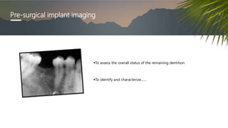
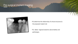
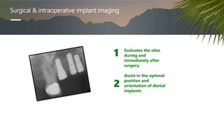
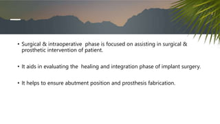
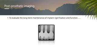
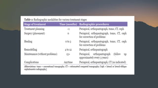
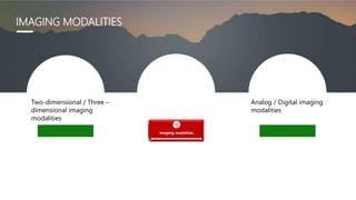
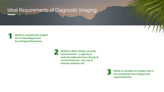
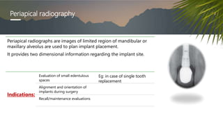
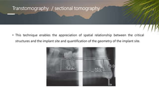
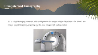
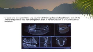
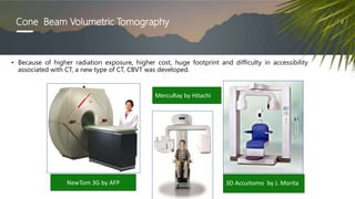
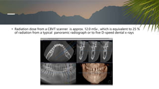
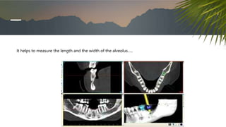
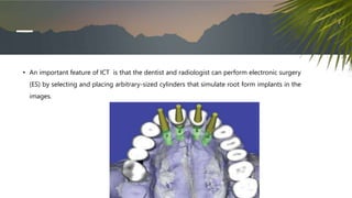
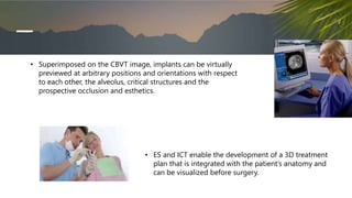
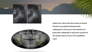
SlideShare a Scribd company logo
1 of 81
RADIOGRAPHIC AIDS IN DENTAL IMPLANTS
THASLIM FATHIMA N.
SECOND YEAR POSTGRADUATE
DEPARTMENT OF
PERIODONTOLOGY
CONTENTS
• Introduction
• Phases of imaging
• Imaging modalities
• Periapical radiography
• Digital radiography
• Panoramic radiography
• Tomography & computed tomography
• Cone beam volumetric tomography
• Interactive computed tomography
• Imaging of vital structures
• Diagnostic templates
• Conclusion
• References
INTRODUCTION
 Diagnostic imaging techniques must always be interpreted
in conjunction with a good clinical examination.
 The decision is a balance between these factors and the
desire to minimize risk of complications to the patient.
Multiple factors influence the selection of radiographic technique(s) for a particular case,
including cost, availability, radiation exposure, and case type.
Some of the key parameters in implant planning where appropriate
imaging can contribute are:
• Bone height (craniocaudal dimension)
• Faciolingual/faciopalatal width
• Mesiodistal dimension
• Bone morphology
• Presence and prominence of anatomic features:
Sublingual and submandibular fossae
Incisive and canine fossae
• Neurovascular canals and foramina, including:
Mandibular canal and the mental foramen
Incisive canal and foramen
Mandibular lingual canals and foramina (Laboda 1990; Mason et al. 1990; Katakami et al. 2009;
Tagaya et al. 2009; Givol et al. 2000)
Greater palatine canal and foramen
• Cortical thickness and density
• Extent and morphology of the alveolar recesses of the maxillary sinus bases.
PHASES OF IMAGING
1
.
2 Phase II Surgical and intraoperative implant imaging
Phase I Pre-surgical implant imaging
Phase III Post-prosthetic imaging
Pre-surgical implant imaging
To assess the overall status of the remaining dentition
To identify and characterize……
Pre-surgical implant imaging
To determine the relationship of critical structures to
the proposed implant site
To detect regional anatomic abnormalities and
pathologies.
Surgical & intraoperative implant imaging
Evaluates the sites
during and
immediately after
surgery
Assist in the optimal
position and
orientation of dental
implants
1
2
• Surgical & intraoperative phase is focused on assisting in surgical &
prosthetic intervention of patient.
• It aids in evaluating the healing and integration phase of implant surgery.
• It helps to ensure abutment position and prosthesis fabrication.
Post-prosthetic imaging
• To evaluate the long-term maintenance of implant rigid fixation and function……
Two-dimensional / Three –
dimensional imaging
modalities
Imaging modalities
Analog / Digital imaging
modalities
IMAGING MODALITIES
Ideal Requirements of Diagnostic Imaging
Ability to visualize the implant
site in mesiolingual and
buccolingual dimensions.
1
Ability to allow reliable, accurate
measurements ; a capacity to
evaluate trabecular bone density &
cortical thickness; less cost &
minimal radiation risk.
2
Ability to visualize the implant site in
the mesiodistal, buccolingual and
superioinferiorly.
3
DIAGNOSTIC IMAGING TECHNIQUES
Periapical radiography
Periapical radiographs are images of limited region of mandibular or
maxillary alveolus are used to plan implant placement.
It provides two dimensional information regarding the implant site.
Indications:
Evaluation of small edentulous
spaces
Eg: in case of single tooth
replacement
Alignment and orientation of
implants during surgery
Recall/maintenance evaluations
ADVANTAGES
 Amount of bone loss and peri- implantitis can be visualized.
 Subtle variations in bone activity is clearly seen.
 Minimal magnification with high resolution.
 Provides an assessment of the quantity and quality of the
edentulous alveolar ridge & adjacent teeth.
 They are easy to obtain in dental office, Inexpensive.
 Delivers low radiation to the patient.
DISADVANTAGES
• Distortion is particularly accentuated in edentulous areas, where missing
teeth and resorption of the alveolus necessitate film placement at
significant angulation in relation to the long axis of the teeth and alveolar
bone.
• Often, it is not possible to image the entire height of the remaining alveolar
ridge, and when extensive mesial-distal areas need to be evaluated,
multiple periapical films are required
OCCLUSAL RADIOGRAPHY
• Occlusal radiograph is a planer radiograph produced by placing film intraorally parallel to the occlusal
plane with the central x-ray beam perpendicular to the film for the mandibular image and oblique (usually
45 degrees ) to the film for maxillary image.
ADVANTAGES
High resolution planer image
DISADVANTAGES
• The degree of mineralization of
trabecular bone is not
determined by this projection
& spatial relationship between
critical structures such as
mandibular canal & mental
foramen.
• The proposed implant site is
lost with this projection
BITEWING RADIOGRAPHY
• Bitewing radiographs are used as post prosthetic imaging.
• The x-ray beam should be angled to be perpendicular to the crestal bone region of the implant
or abutment to implant connection
• Quality bitewing radiographs placed parallel to the implant body with central ray of
source oriented perpendicular to the film enable sequential radiograph for crestal and
perimplant bone loss.
• Used as postprosthetic imaging.
PANORAMIC RADIOGRAPH
INDICATIONS
• Indicated when multiple implant placements are planned.
• Initial assessment of vertical height of bone
• Evaluation of gross anatomy of the jaws and any related pathologic
findings
• They display image slices through jaws by producing a single image of maxilla & mandible their supporting
structures in a frontal plane.
• The image receptor is either radiograph film or can also be a digital storage phosphor plate.
Advantages:
o They display anatomic structures like nasal cavity, maxillary sinus, inferior alveolar canal and mental
foramen.
Disadvantages
 The resolution is lesser when compared to intraoral radiographs.
 When compared to intraoral radiograph Cross sectional view is not demonstrated and is of little use
in depicting the spatial relationship between the structures. 10-20% image magnification occurs,
which is non uniform.
Tomography
• Tomographic units produce cross-sectional slices of the jaws that can be as thin as 1 mm
and are suitable for pre- and post-implant assessment.
TOMO- slice
GRAPH- picture
• The basic principle of tomography is that the x-ray tube and film are connected by a rigid bar called the
fulcrum bar, which pivots on a point called fulcrum.
• When the system is energized, the x-ray tube moves in one direction with the film plane moving in the
opposite direction and the system pivoting about the fulcrum
• Linear tomography is the simplest form , which has a one dimensional motion and produces
blurring of adjacent sections and results in linear steak artifacts in the resulting image, which
may obfuscate the section of interest.
• Circular, spiral and hypocycloidal are two- dimensional motions
• Hypocycloidal is generally accepted as the most effective blurring motion
• This technique is difficult to use in cases of multiple implant sites.
Transtomography / sectional tomography
• This technique enables the appreciation of spatial relationship between the critical
structures and the implant site and quantification of the geometry of the implant site.
• The tomographic layers are thick and have adjacent structures that are
blurred and superimposed on image, limiting usefulness of this technique
for individual sites,especially in the anterior regions where geometry of the
alveolar changes rapidly.
Advantages
• Cross- sectional views
• Constant magnification Disadvantages:
• Technique- sensitive
• Blurred images
• High radiation dose
• Multiple images needed
• Expensive
Computerised Tomography
CT is a digital imaging technique, which can generate 3D images using a very narrow “fan beam” that
rotates around the patient, acquiring one thin slice (image) with each revolution
• CT scans have been shown to be very accurate with the magnification effect, the same for both the
anterior and posterior area, from a range of 0% to 6% in horizontal as well as 0-4% in the vertical
dimension.
•
• Advantages
• Negligible magnification
• High contrast image
• 3 dimensional bone models
• Interactive treatment planning
Disadvantages
High dose of radiation
Technique – sensitive
Expensive
Recent advances in CT
CONE BEAM VOLUMETRIC TOMOGRAPHY
MICROTOMOGRAPH- acquiring serial sections
of bone implant interface
MULTI SLICE HELICAL CT-higher accuracy of
images
• CBCT is a three dimensional imaging technique.
• Results in optimal implant placement & improved clinical outcome.
• It helps in improved visualization and comprehension of the anatomy in
the areas in which implants are being planned for placement,
• Helps to visualize the implant site in axial, reconstructed panoramic and
cross sectional views of the jaws.
• CBCT generates cone shaped beams and the images are acquired in one
rotation by an image intensifier of flat panel detector, resulting in
reasonably low levels of radiation dosage.
Dentascan
DentaScan is a computed tomography (CT) software
program that allows the mandible and maxilla to be
imaged in three planes: axial, panoramic and cross-
sectional.
Cone Beam Volumetric Tomography
• Because of higher radiation exposure, higher cost, huge footprint and difficulty in accessibility
associated with CT, a new type of CT, CBVT was developed.
NewTom 3G by AFP
MercuRay by Hitachi
3D Accuitomo by J. Morita
• The x- ray tube on these scanners rotates around 360 degrees and will capture
images of the maxilla and mandible in 36 seconds , in which only 5.6 seconds is
needed for exposure
“ Cone beam” geometry
• The images recorded are placed onto a charge- coupled device chip and is
then converted into axial, sagittal and coronal slices and permit reformatting
to view traditional radiographic images as well as 3- dimensional soft tissue
or osseous images.
Indications:
• Assess the positions…….
Inferior alveolar canal
Location of neurovascular bundle
The incisive and mental foramina
Pneumatization of the maxillary sinus
Floor of the maxillary sinus
Nasal fossa
• Radiation dose from a CBVT scanner is approx. 12.0 mSv , which is equivalent to 25 %
of radiation from a typical panoramic radiograph or to five D-speed dental x-rays
Advantages
Almost 0% magnification.
No superimposition or
overlapping of images,
Minimal distortion
Lower cost
More feasible compared to the
CT
• Evaluation of bone resorption and root retention, as well as lesions of the facial skeleton.
INDICATIONS:
CONTRAINDICATIONS
Interactive computed tomography
• This technique enables the radiologist to transfer the imaging study to the practitioner as a
computer file and enables the practitioner to view and interact with the imaging study on their
own computer
It helps to measure the length and the width of the alveolus…..
• An important feature of ICT is that the dentist and radiologist can perform electronic surgery
(ES) by selecting and placing arbitrary-sized cylinders that simulate root form implants in the
images.
• Superimposed on the CBVT image, implants can be virtually
previewed at arbitrary positions and orientations with respect
to each other, the alveolus, critical structures and the
prospective occlusion and esthetics.
• ES and ICT enable the development of a 3D treatment
plan that is integrated with the patient’s anatomy and
can be visualized before surgery.
MAGNETIC RESONANCE IMAGING
• It is a 3 dimensional imaging technique. MR images can be varied to obtain fat, water, or balanced
imaging of the patient’s anatomy.
• Oriented MR imaging of the posterior mandible is dimensionally quantitative between critical
structures and the proposed implant site
Surgical guides
• Computer generated drilling guides that are fabricated through the process of
stereolithography using SimPlant software for ideal implant positioning
• These successive diameter surgical osteotomy drill guides may be either bone, teeth or mucosa-borne.
• Surgiguides have metal cylindrical tubes that correspond to the number of desired osteotomy
preparations and specific drill diameters.
• Process wherein the film is replaced by a sensor that collects the data.
DIGITAL RADIOGRAPHY
 The resultant image can be modified in terms of gray scale,
brightness, contrast, inversion and color enhancement
 Computerised software programs like Dexisimplant are
available that allowing for calibration of magnified images ,
ensuring accurate measurements
ADVANTAGES
• Less radiation
• Superior resolution
• Instantaneous speed of image
formation is highly useful during
surgical placement of implants and
the prosthetic verification of
component placement
DISADVANTAGES
• Size and thickness of the film and
position of the connecting cord
sometimes makes film placement
difficult in some sites , such as
those adjacent to tori or in case of
tapered arch form in the region of
canines
IMAGING OF VITAL STRUCTURES IN ORAL
IMPLANTOLOGY
Mental foramen and mandibular canal
While using the two-dimensional radiographs for imaging Mental foramen and
mandibular canal ,it is mainly dependent on the positioning.
• If the image is taken from the mesio-oblique orientation, measurements will be
fore-shortened
Studies have shown that the location of mental
foramen on periapical and panoramic
radiograph are inaccurate and sometimes on
panoramic radiograph it represents a portion of
the mental canal as it leaves the mandibular
canal
Several studies have shown CT to be the most accurate
and highly recommended when measurements are
needed for the inferior alveolar canal and mental foramen
Mandibular lingual concavities
• When there is advanced atrophy of posterior
mandible, lingual concavities may be present .
• Within these concavities branches of facial artery
may be present
Mandibular ramus
Single Onlay Cortical Bone Grafting
The term onlay bone grafting was coined by
Campbell. The technique involves a bone
grafting in which the transplanted tissue is laid
directly onto the surface of the recipient bone.
The technique is more of historical importance.
The technique is rarely used.
• Donor site for autogenous onlay bone grafting and is extremely variable in the amount of
bone present .
• Hence adequate assessment of host bone present is not possible
• Recommended : CT
Mandibular symphysis
• Donor site for autogenous graft
• Panoramic radiographs mostly overestimate the
height of the available bone in the anterior
region
• Recommended: conventional CT
Maxillary sinus
• For implant placement detailed
information is needed regarding the
position of septa, maxillary sinus
anatomy, sinus pathologies.
• CT- the gold standard for evaluation
of sinuses
• To verify the positioning and location of an osteotomy site or for identification of a vital structure,
processing of standard radiograph film can take upto 6 minutes and hence is time- ineffcient .
• Current day digital imaging system gives instant images that can be manipulated, and allows accurate
measurements and maintains aseptic protocol
INTRA OPERATIVE
IMAGING
Immediate post operative imaging
• A periapical or panoramic radiograph should be taken post surgically so that a baseline image can be
used to evaluate against future images.
Abutment and prosthetic component imaging
• When evaluating for transfer of impressions along with the two- piece implant abutment component
placement , radiographs should be taken to verify secure adaptation.
• Intraoral radiographs are recommended because of their high geometric resolution to evaluate for any fit
discrepancy.
Post prosthetic imaging
• When investigating complications after implant placement, a panoramic radiograph is the most
ideal technique.
• Whenever Single implant image or a detailed information of implant viewed on panoramic image
is needed, a periapical radiograph is taken.
Recall and maintenance imaging
• Follow up or recall radiographs are to be taken 1 year of functional loading and yearly for the first 3
years to assess the marginal bone levels.
Radiographic sequence for dental implants imaging
• Pretreatment
• Immediate post surgical (baseline)
• Healing period ( if necessary)
• Second stage surgery
• Post prosthetic surgery ( baseline)
• 1 year postoperatively
• After 1st year, every 2 years
Fabrication of diagnostic templates
• The surfaces of the proposed restorations and the exact position and orientation
of each dental implant should be incorporated into the diagnostic CT templates.
• The design may vary from a vaccuformed reproduction of the wax-up - to one
processed from acrylic reproduction of the diagnostic wax-up – to a sophisticated
specially designed radiopaque denture teeth.
• The processed acrylic template may be modified by coating the proposed restorations with a
thin film of barium sulfate and filling a hole drilled through the occlusal surface of the
restoration with a gutta-percha.
The vaccuform templates involve either coating of
the proposed restorations with a thin film of barium
sulfate. This does not depict the ideal position and
orientation of the proposed implant
Another method is to blend 10% barium sulfate
and 90% cold cure acrylic , which makes the
proposed restoration radiopaque and evident but
again does not give idea about the position and
orientation
• Recently radiographic teeth specifically designed for the fabrication of diagnostic
templates have been introduced .
• These are time saving, placed easily, provide consistently high radiopacity, have moulds
corresponding to prosthetic teeth used in the final restoration and are bonded easily with
the template-based material
CONCLUSION
References
• Misch CE. Contemporary implant dentistry. 3rd ed. St. Louis: Mosby Elsevier;
2008.Diagnostic imaging and techniques; p.38-67.
• Caranza 11th edition
• Ganz SD. Presurgical planning with CT-derived fabrication of surgical guides. J
Oral Maxillofac Surg 2005; 63: 59-71.
• Vercruyssen M, Jacobs R, Van Assche N, van Steenberghe D. The use of CT scan
based planning for oral rehabilitation by means of implants and its transfer to
the surgical field: A critical review on accuracy. J Oral Rehabil 2008; 35: 454-74.
• Mi les DA. The future of dental and maxillofacial imaging. Dent Clin North Am 2008; 52: 917-28.
• Frederiksen NL. Diagnostic imaging in dental implantology. Oral Surgery Oral Medicine Oral
Pathology 1995; 80: 540-54.
• Spector L. Computer-aided dental implant planning. Dent Clin North Am 2008; 52: 761-75.
• K Niranjani et al. Radiographic Aids in Implantology: A Review. RESEARCH AND REVIEWS: JOURNAL
OF DENTAL SCIENCES 2014
Radiographic aids in dental implants

More Related Content

What's hot

Digital Radiography dentistry
Digital Radiography dentistryDigital Radiography dentistry
Digital Radiography dentistryNishtha Singhal
 
Benefits of cbct in implant planning
Benefits of cbct in implant planningBenefits of cbct in implant planning
Benefits of cbct in implant planningUE
 
CBCT in Implants- Summary
CBCT in Implants- SummaryCBCT in Implants- Summary
CBCT in Implants- Summaryghidalawand
 
Dental CBCT Evidence Based Guideline 2012 European Commission
Dental CBCT Evidence Based Guideline 2012 European CommissionDental CBCT Evidence Based Guideline 2012 European Commission
Dental CBCT Evidence Based Guideline 2012 European CommissionNeil Pande
 
implant placement protocol where and when
implant placement protocol where and when implant placement protocol where and when
implant placement protocol where and when marwan mohamed
 
Bone density for dental implant.
Bone density for dental implant.Bone density for dental implant.
Bone density for dental implant.Riad Mahmud
 
Computer guided implant surgery
Computer guided implant surgeryComputer guided implant surgery
Computer guided implant surgeryUS Dental
 
PROJECTION GEOMETRY/ dental implant courses
PROJECTION GEOMETRY/ dental implant coursesPROJECTION GEOMETRY/ dental implant courses
PROJECTION GEOMETRY/ dental implant coursesIndian dental academy
 
Basics Of Implantology - Dr Harshavardhan Patwal
Basics Of Implantology - Dr Harshavardhan PatwalBasics Of Implantology - Dr Harshavardhan Patwal
Basics Of Implantology - Dr Harshavardhan PatwalDr Harshavardhan Patwal
 
Emdogain by dr. maryam salman
Emdogain by dr. maryam salmanEmdogain by dr. maryam salman
Emdogain by dr. maryam salmanDr.Maryam Salman
 

What's hot (20)

Digital Radiography dentistry
Digital Radiography dentistryDigital Radiography dentistry
Digital Radiography dentistry
 
Benefits of cbct in implant planning
Benefits of cbct in implant planningBenefits of cbct in implant planning
Benefits of cbct in implant planning
 
Immediate implant lecture
Immediate implant lectureImmediate implant lecture
Immediate implant lecture
 
CBCT in Implants- Summary
CBCT in Implants- SummaryCBCT in Implants- Summary
CBCT in Implants- Summary
 
Dental CBCT Evidence Based Guideline 2012 European Commission
Dental CBCT Evidence Based Guideline 2012 European CommissionDental CBCT Evidence Based Guideline 2012 European Commission
Dental CBCT Evidence Based Guideline 2012 European Commission
 
use of Cbct in dental implant
use of Cbct  in dental implantuse of Cbct  in dental implant
use of Cbct in dental implant
 
CBCT Basic.pptx
CBCT Basic.pptxCBCT Basic.pptx
CBCT Basic.pptx
 
implant placement protocol where and when
implant placement protocol where and when implant placement protocol where and when
implant placement protocol where and when
 
CBCT in Dentistry
CBCT in DentistryCBCT in Dentistry
CBCT in Dentistry
 
Bone density for dental implant.
Bone density for dental implant.Bone density for dental implant.
Bone density for dental implant.
 
IMPLANT IMAGING TECHNIQUE
IMPLANT IMAGING TECHNIQUEIMPLANT IMAGING TECHNIQUE
IMPLANT IMAGING TECHNIQUE
 
Cone beam
Cone beamCone beam
Cone beam
 
Computer guided implant surgery
Computer guided implant surgeryComputer guided implant surgery
Computer guided implant surgery
 
TMJ IMAGING.pptx
TMJ IMAGING.pptxTMJ IMAGING.pptx
TMJ IMAGING.pptx
 
Digital imaging IN DENTISTRY
Digital imaging IN DENTISTRYDigital imaging IN DENTISTRY
Digital imaging IN DENTISTRY
 
PROJECTION GEOMETRY/ dental implant courses
PROJECTION GEOMETRY/ dental implant coursesPROJECTION GEOMETRY/ dental implant courses
PROJECTION GEOMETRY/ dental implant courses
 
Basics Of Implantology - Dr Harshavardhan Patwal
Basics Of Implantology - Dr Harshavardhan PatwalBasics Of Implantology - Dr Harshavardhan Patwal
Basics Of Implantology - Dr Harshavardhan Patwal
 
Available bone
Available boneAvailable bone
Available bone
 
Dental Diagnostic imaging
Dental Diagnostic imagingDental Diagnostic imaging
Dental Diagnostic imaging
 
Emdogain by dr. maryam salman
Emdogain by dr. maryam salmanEmdogain by dr. maryam salman
Emdogain by dr. maryam salman
 

Similar to Radiographic aids in dental implants

Diagnostic imaging for the implant patient
Diagnostic imaging for the implant patientDiagnostic imaging for the implant patient
Diagnostic imaging for the implant patientDR. OINAM MONICA DEVI
 
Radiographic Considerations in Implants
Radiographic Considerations in ImplantsRadiographic Considerations in Implants
Radiographic Considerations in ImplantsDr.Richa Sahai
 
Imaging Modalities in Dental Implant
Imaging Modalities in Dental ImplantImaging Modalities in Dental Implant
Imaging Modalities in Dental Implantassem awad
 
Orofacial implant
Orofacial implantOrofacial implant
Orofacial implantIAU Dent
 
Diagnostic aids in implant seminar in detail
Diagnostic aids in implant seminar in detailDiagnostic aids in implant seminar in detail
Diagnostic aids in implant seminar in detailNikitaChhabariya
 
Diagnostic imaging in implants /certified fixed orthodontic courses by Indian...
Diagnostic imaging in implants /certified fixed orthodontic courses by Indian...Diagnostic imaging in implants /certified fixed orthodontic courses by Indian...
Diagnostic imaging in implants /certified fixed orthodontic courses by Indian...Indian dental academy
 
Diagnostic imaging./ cosmetic dentistry training
Diagnostic imaging./ cosmetic dentistry trainingDiagnostic imaging./ cosmetic dentistry training
Diagnostic imaging./ cosmetic dentistry trainingIndian dental academy
 
Diagnostic imaging / dental implant courses by Indian dental academy
Diagnostic imaging / dental implant courses by Indian dental academy Diagnostic imaging / dental implant courses by Indian dental academy
Diagnostic imaging / dental implant courses by Indian dental academy Indian dental academy
 
Diagnostic imaging / dental crown & bridge courses
Diagnostic imaging / dental crown & bridge coursesDiagnostic imaging / dental crown & bridge courses
Diagnostic imaging / dental crown & bridge coursesIndian dental academy
 
Diagnostic imaging /certified fixed orthodontic courses by Indian dental acad...
Diagnostic imaging /certified fixed orthodontic courses by Indian dental acad...Diagnostic imaging /certified fixed orthodontic courses by Indian dental acad...
Diagnostic imaging /certified fixed orthodontic courses by Indian dental acad...Indian dental academy
 
Diagnostic imaging/ oral surgery courses  
Diagnostic imaging/ oral surgery courses  Diagnostic imaging/ oral surgery courses  
Diagnostic imaging/ oral surgery courses  Indian dental academy
 
Role of radiology in orthodontics
Role of radiology in orthodonticsRole of radiology in orthodontics
Role of radiology in orthodonticsMahmoud Ghareib
 
Specialised Techniques in Oral Radiology
Specialised Techniques in Oral RadiologySpecialised Techniques in Oral Radiology
Specialised Techniques in Oral RadiologyIAU Dent
 
Recent advances in radiographic technique in orthodontics
Recent advances in radiographic technique in orthodonticsRecent advances in radiographic technique in orthodontics
Recent advances in radiographic technique in orthodonticsIndian dental academy
 
Recent advances in radiographic technique in orthodontics
Recent advances in radiographic technique in orthodonticsRecent advances in radiographic technique in orthodontics
Recent advances in radiographic technique in orthodonticsIndian dental academy
 
SURGICAL TREATMENT OBJECTIVES
SURGICAL TREATMENT OBJECTIVESSURGICAL TREATMENT OBJECTIVES
SURGICAL TREATMENT OBJECTIVESShehnaz Jahangir
 
Role of Image Guidance in Radiotherapy
Role of Image Guidance in RadiotherapyRole of Image Guidance in Radiotherapy
Role of Image Guidance in RadiotherapySusan Rochelle
 
Limitations of cephalometric radiographs
Limitations of cephalometric radiographsLimitations of cephalometric radiographs
Limitations of cephalometric radiographsIndian dental academy
 

Similar to Radiographic aids in dental implants (20)

Diagnostic imaging for the implant patient
Diagnostic imaging for the implant patientDiagnostic imaging for the implant patient
Diagnostic imaging for the implant patient
 
Radiographic Considerations in Implants
Radiographic Considerations in ImplantsRadiographic Considerations in Implants
Radiographic Considerations in Implants
 
Imaging Modalities in Dental Implant
Imaging Modalities in Dental ImplantImaging Modalities in Dental Implant
Imaging Modalities in Dental Implant
 
Orofacial implant
Orofacial implantOrofacial implant
Orofacial implant
 
Diagnostic aids in implant seminar in detail
Diagnostic aids in implant seminar in detailDiagnostic aids in implant seminar in detail
Diagnostic aids in implant seminar in detail
 
Diagnostic imaging in implants /certified fixed orthodontic courses by Indian...
Diagnostic imaging in implants /certified fixed orthodontic courses by Indian...Diagnostic imaging in implants /certified fixed orthodontic courses by Indian...
Diagnostic imaging in implants /certified fixed orthodontic courses by Indian...
 
Diagnostic imaging./ cosmetic dentistry training
Diagnostic imaging./ cosmetic dentistry trainingDiagnostic imaging./ cosmetic dentistry training
Diagnostic imaging./ cosmetic dentistry training
 
Diagnostic imaging / dental implant courses by Indian dental academy
Diagnostic imaging / dental implant courses by Indian dental academy Diagnostic imaging / dental implant courses by Indian dental academy
Diagnostic imaging / dental implant courses by Indian dental academy
 
Diagnostic imaging / dental crown & bridge courses
Diagnostic imaging / dental crown & bridge coursesDiagnostic imaging / dental crown & bridge courses
Diagnostic imaging / dental crown & bridge courses
 
Diagnostic imaging /certified fixed orthodontic courses by Indian dental acad...
Diagnostic imaging /certified fixed orthodontic courses by Indian dental acad...Diagnostic imaging /certified fixed orthodontic courses by Indian dental acad...
Diagnostic imaging /certified fixed orthodontic courses by Indian dental acad...
 
Diagnostic imaging/ oral surgery courses  
Diagnostic imaging/ oral surgery courses  Diagnostic imaging/ oral surgery courses  
Diagnostic imaging/ oral surgery courses  
 
Role of radiology in orthodontics
Role of radiology in orthodonticsRole of radiology in orthodontics
Role of radiology in orthodontics
 
Imaging in implants
Imaging in implantsImaging in implants
Imaging in implants
 
Orthopantomogram.ppt
Orthopantomogram.pptOrthopantomogram.ppt
Orthopantomogram.ppt
 
Specialised Techniques in Oral Radiology
Specialised Techniques in Oral RadiologySpecialised Techniques in Oral Radiology
Specialised Techniques in Oral Radiology
 
Recent advances in radiographic technique in orthodontics
Recent advances in radiographic technique in orthodonticsRecent advances in radiographic technique in orthodontics
Recent advances in radiographic technique in orthodontics
 
Recent advances in radiographic technique in orthodontics
Recent advances in radiographic technique in orthodonticsRecent advances in radiographic technique in orthodontics
Recent advances in radiographic technique in orthodontics
 
SURGICAL TREATMENT OBJECTIVES
SURGICAL TREATMENT OBJECTIVESSURGICAL TREATMENT OBJECTIVES
SURGICAL TREATMENT OBJECTIVES
 
Role of Image Guidance in Radiotherapy
Role of Image Guidance in RadiotherapyRole of Image Guidance in Radiotherapy
Role of Image Guidance in Radiotherapy
 
Limitations of cephalometric radiographs
Limitations of cephalometric radiographsLimitations of cephalometric radiographs
Limitations of cephalometric radiographs
 

More from Thaslim Fathima

More from Thaslim Fathima (14)

Osseointegration
OsseointegrationOsseointegration
Osseointegration
 
Minimally invasive periodontal surgery
Minimally invasive periodontal surgeryMinimally invasive periodontal surgery
Minimally invasive periodontal surgery
 
Tissue engineering
Tissue engineeringTissue engineering
Tissue engineering
 
Stress and periodontium
Stress and periodontiumStress and periodontium
Stress and periodontium
 
Periodontal splinting
Periodontal splintingPeriodontal splinting
Periodontal splinting
 
Periodontal dressing
Periodontal dressingPeriodontal dressing
Periodontal dressing
 
Aids and periodontium
Aids and periodontiumAids and periodontium
Aids and periodontium
 
Local drug delivery
Local drug deliveryLocal drug delivery
Local drug delivery
 
Facial artery
Facial arteryFacial artery
Facial artery
 
Gingival surgical techniques/Gingivectomy
Gingival surgical techniques/GingivectomyGingival surgical techniques/Gingivectomy
Gingival surgical techniques/Gingivectomy
 
Chronic periodontitis
Chronic periodontitisChronic periodontitis
Chronic periodontitis
 
Sterilization and infection control
Sterilization and infection controlSterilization and infection control
Sterilization and infection control
 
Blood
BloodBlood
Blood
 
Mandibular nerve
Mandibular nerveMandibular nerve
Mandibular nerve
 

Recently uploaded

Speech, hearing, noise, intelligibility.pptx
Speech, hearing, noise, intelligibility.pptxSpeech, hearing, noise, intelligibility.pptx
Speech, hearing, noise, intelligibility.pptxpriyankatabhane
 
Is RISC-V ready for HPC workload? Maybe?
Is RISC-V ready for HPC workload? Maybe?Is RISC-V ready for HPC workload? Maybe?
Is RISC-V ready for HPC workload? Maybe?Patrick Diehl
 
Temporomandibular joint Muscles of Mastication
Temporomandibular joint Muscles of MasticationTemporomandibular joint Muscles of Mastication
Temporomandibular joint Muscles of Masticationvidulajaib
 
Twin's paradox experiment is a meassurement of the extra dimensions.pptx
Twin's paradox experiment is a meassurement of the extra dimensions.pptxTwin's paradox experiment is a meassurement of the extra dimensions.pptx
Twin's paradox experiment is a meassurement of the extra dimensions.pptxEran Akiva Sinbar
 
TOTAL CHOLESTEROL (lipid profile test).pptx
TOTAL CHOLESTEROL (lipid profile test).pptxTOTAL CHOLESTEROL (lipid profile test).pptx
TOTAL CHOLESTEROL (lipid profile test).pptxdharshini369nike
 
insect anatomy and insect body wall and their physiology
insect anatomy and insect body wall and their  physiologyinsect anatomy and insect body wall and their  physiology
insect anatomy and insect body wall and their physiologyDrAnita Sharma
 
Vision and reflection on Mining Software Repositories research in 2024
Vision and reflection on Mining Software Repositories research in 2024Vision and reflection on Mining Software Repositories research in 2024
Vision and reflection on Mining Software Repositories research in 2024AyushiRastogi48
 
LIGHT-PHENOMENA-BY-CABUALDIONALDOPANOGANCADIENTE-CONDEZA (1).pptx
LIGHT-PHENOMENA-BY-CABUALDIONALDOPANOGANCADIENTE-CONDEZA (1).pptxLIGHT-PHENOMENA-BY-CABUALDIONALDOPANOGANCADIENTE-CONDEZA (1).pptx
LIGHT-PHENOMENA-BY-CABUALDIONALDOPANOGANCADIENTE-CONDEZA (1).pptxmalonesandreagweneth
 
Harmful and Useful Microorganisms Presentation
Harmful and Useful Microorganisms PresentationHarmful and Useful Microorganisms Presentation
Harmful and Useful Microorganisms Presentationtahreemzahra82
 
Call Us ≽ 9953322196 ≼ Call Girls In Mukherjee Nagar(Delhi) |
Call Us ≽ 9953322196 ≼ Call Girls In Mukherjee Nagar(Delhi) |Call Us ≽ 9953322196 ≼ Call Girls In Mukherjee Nagar(Delhi) |
Call Us ≽ 9953322196 ≼ Call Girls In Mukherjee Nagar(Delhi) |aasikanpl
 
Spermiogenesis or Spermateleosis or metamorphosis of spermatid
Spermiogenesis or Spermateleosis or metamorphosis of spermatidSpermiogenesis or Spermateleosis or metamorphosis of spermatid
Spermiogenesis or Spermateleosis or metamorphosis of spermatidSarthak Sekhar Mondal
 
Neurodevelopmental disorders according to the dsm 5 tr
Neurodevelopmental disorders according to the dsm 5 trNeurodevelopmental disorders according to the dsm 5 tr
Neurodevelopmental disorders according to the dsm 5 trssuser06f238
 
Artificial Intelligence In Microbiology by Dr. Prince C P
Artificial Intelligence In Microbiology by Dr. Prince C PArtificial Intelligence In Microbiology by Dr. Prince C P
Artificial Intelligence In Microbiology by Dr. Prince C PPRINCE C P
 
THE ROLE OF PHARMACOGNOSY IN TRADITIONAL AND MODERN SYSTEM OF MEDICINE.pptx
THE ROLE OF PHARMACOGNOSY IN TRADITIONAL AND MODERN SYSTEM OF MEDICINE.pptxTHE ROLE OF PHARMACOGNOSY IN TRADITIONAL AND MODERN SYSTEM OF MEDICINE.pptx
THE ROLE OF PHARMACOGNOSY IN TRADITIONAL AND MODERN SYSTEM OF MEDICINE.pptxNandakishor Bhaurao Deshmukh
 
Best Call Girls In Sector 29 Gurgaon❤️8860477959 EscorTs Service In 24/7 Delh...
Best Call Girls In Sector 29 Gurgaon❤️8860477959 EscorTs Service In 24/7 Delh...Best Call Girls In Sector 29 Gurgaon❤️8860477959 EscorTs Service In 24/7 Delh...
Best Call Girls In Sector 29 Gurgaon❤️8860477959 EscorTs Service In 24/7 Delh...lizamodels9
 
Dashanga agada a formulation of Agada tantra dealt in 3 Rd year bams agada tanta
Dashanga agada a formulation of Agada tantra dealt in 3 Rd year bams agada tantaDashanga agada a formulation of Agada tantra dealt in 3 Rd year bams agada tanta
Dashanga agada a formulation of Agada tantra dealt in 3 Rd year bams agada tantaPraksha3
 
Cytokinin, mechanism and its application.pptx
Cytokinin, mechanism and its application.pptxCytokinin, mechanism and its application.pptx
Cytokinin, mechanism and its application.pptxVarshiniMK
 
zoogeography of pakistan.pptx fauna of Pakistan
zoogeography of pakistan.pptx fauna of Pakistanzoogeography of pakistan.pptx fauna of Pakistan
zoogeography of pakistan.pptx fauna of Pakistanzohaibmir069
 

Recently uploaded (20)

Hot Sexy call girls in Moti Nagar,🔝 9953056974 🔝 escort Service
Hot Sexy call girls in  Moti Nagar,🔝 9953056974 🔝 escort ServiceHot Sexy call girls in  Moti Nagar,🔝 9953056974 🔝 escort Service
Hot Sexy call girls in Moti Nagar,🔝 9953056974 🔝 escort Service
 
Speech, hearing, noise, intelligibility.pptx
Speech, hearing, noise, intelligibility.pptxSpeech, hearing, noise, intelligibility.pptx
Speech, hearing, noise, intelligibility.pptx
 
Volatile Oils Pharmacognosy And Phytochemistry -I
Volatile Oils Pharmacognosy And Phytochemistry -IVolatile Oils Pharmacognosy And Phytochemistry -I
Volatile Oils Pharmacognosy And Phytochemistry -I
 
Is RISC-V ready for HPC workload? Maybe?
Is RISC-V ready for HPC workload? Maybe?Is RISC-V ready for HPC workload? Maybe?
Is RISC-V ready for HPC workload? Maybe?
 
Temporomandibular joint Muscles of Mastication
Temporomandibular joint Muscles of MasticationTemporomandibular joint Muscles of Mastication
Temporomandibular joint Muscles of Mastication
 
Twin's paradox experiment is a meassurement of the extra dimensions.pptx
Twin's paradox experiment is a meassurement of the extra dimensions.pptxTwin's paradox experiment is a meassurement of the extra dimensions.pptx
Twin's paradox experiment is a meassurement of the extra dimensions.pptx
 
TOTAL CHOLESTEROL (lipid profile test).pptx
TOTAL CHOLESTEROL (lipid profile test).pptxTOTAL CHOLESTEROL (lipid profile test).pptx
TOTAL CHOLESTEROL (lipid profile test).pptx
 
insect anatomy and insect body wall and their physiology
insect anatomy and insect body wall and their  physiologyinsect anatomy and insect body wall and their  physiology
insect anatomy and insect body wall and their physiology
 
Vision and reflection on Mining Software Repositories research in 2024
Vision and reflection on Mining Software Repositories research in 2024Vision and reflection on Mining Software Repositories research in 2024
Vision and reflection on Mining Software Repositories research in 2024
 
LIGHT-PHENOMENA-BY-CABUALDIONALDOPANOGANCADIENTE-CONDEZA (1).pptx
LIGHT-PHENOMENA-BY-CABUALDIONALDOPANOGANCADIENTE-CONDEZA (1).pptxLIGHT-PHENOMENA-BY-CABUALDIONALDOPANOGANCADIENTE-CONDEZA (1).pptx
LIGHT-PHENOMENA-BY-CABUALDIONALDOPANOGANCADIENTE-CONDEZA (1).pptx
 
Harmful and Useful Microorganisms Presentation
Harmful and Useful Microorganisms PresentationHarmful and Useful Microorganisms Presentation
Harmful and Useful Microorganisms Presentation
 
Call Us ≽ 9953322196 ≼ Call Girls In Mukherjee Nagar(Delhi) |
Call Us ≽ 9953322196 ≼ Call Girls In Mukherjee Nagar(Delhi) |Call Us ≽ 9953322196 ≼ Call Girls In Mukherjee Nagar(Delhi) |
Call Us ≽ 9953322196 ≼ Call Girls In Mukherjee Nagar(Delhi) |
 
Spermiogenesis or Spermateleosis or metamorphosis of spermatid
Spermiogenesis or Spermateleosis or metamorphosis of spermatidSpermiogenesis or Spermateleosis or metamorphosis of spermatid
Spermiogenesis or Spermateleosis or metamorphosis of spermatid
 
Neurodevelopmental disorders according to the dsm 5 tr
Neurodevelopmental disorders according to the dsm 5 trNeurodevelopmental disorders according to the dsm 5 tr
Neurodevelopmental disorders according to the dsm 5 tr
 
Artificial Intelligence In Microbiology by Dr. Prince C P
Artificial Intelligence In Microbiology by Dr. Prince C PArtificial Intelligence In Microbiology by Dr. Prince C P
Artificial Intelligence In Microbiology by Dr. Prince C P
 
THE ROLE OF PHARMACOGNOSY IN TRADITIONAL AND MODERN SYSTEM OF MEDICINE.pptx
THE ROLE OF PHARMACOGNOSY IN TRADITIONAL AND MODERN SYSTEM OF MEDICINE.pptxTHE ROLE OF PHARMACOGNOSY IN TRADITIONAL AND MODERN SYSTEM OF MEDICINE.pptx
THE ROLE OF PHARMACOGNOSY IN TRADITIONAL AND MODERN SYSTEM OF MEDICINE.pptx
 
Best Call Girls In Sector 29 Gurgaon❤️8860477959 EscorTs Service In 24/7 Delh...
Best Call Girls In Sector 29 Gurgaon❤️8860477959 EscorTs Service In 24/7 Delh...Best Call Girls In Sector 29 Gurgaon❤️8860477959 EscorTs Service In 24/7 Delh...
Best Call Girls In Sector 29 Gurgaon❤️8860477959 EscorTs Service In 24/7 Delh...
 
Dashanga agada a formulation of Agada tantra dealt in 3 Rd year bams agada tanta
Dashanga agada a formulation of Agada tantra dealt in 3 Rd year bams agada tantaDashanga agada a formulation of Agada tantra dealt in 3 Rd year bams agada tanta
Dashanga agada a formulation of Agada tantra dealt in 3 Rd year bams agada tanta
 
Cytokinin, mechanism and its application.pptx
Cytokinin, mechanism and its application.pptxCytokinin, mechanism and its application.pptx
Cytokinin, mechanism and its application.pptx
 
zoogeography of pakistan.pptx fauna of Pakistan
zoogeography of pakistan.pptx fauna of Pakistanzoogeography of pakistan.pptx fauna of Pakistan
zoogeography of pakistan.pptx fauna of Pakistan
 

Radiographic aids in dental implants

  • 1. RADIOGRAPHIC AIDS IN DENTAL IMPLANTS THASLIM FATHIMA N. SECOND YEAR POSTGRADUATE DEPARTMENT OF PERIODONTOLOGY
  • 2. CONTENTS • Introduction • Phases of imaging • Imaging modalities • Periapical radiography • Digital radiography • Panoramic radiography • Tomography & computed tomography • Cone beam volumetric tomography • Interactive computed tomography • Imaging of vital structures • Diagnostic templates • Conclusion • References
  • 4.  Diagnostic imaging techniques must always be interpreted in conjunction with a good clinical examination.  The decision is a balance between these factors and the desire to minimize risk of complications to the patient. Multiple factors influence the selection of radiographic technique(s) for a particular case, including cost, availability, radiation exposure, and case type.
  • 5. Some of the key parameters in implant planning where appropriate imaging can contribute are: • Bone height (craniocaudal dimension) • Faciolingual/faciopalatal width • Mesiodistal dimension • Bone morphology • Presence and prominence of anatomic features: Sublingual and submandibular fossae Incisive and canine fossae • Neurovascular canals and foramina, including: Mandibular canal and the mental foramen Incisive canal and foramen Mandibular lingual canals and foramina (Laboda 1990; Mason et al. 1990; Katakami et al. 2009; Tagaya et al. 2009; Givol et al. 2000) Greater palatine canal and foramen • Cortical thickness and density • Extent and morphology of the alveolar recesses of the maxillary sinus bases.
  • 6. PHASES OF IMAGING 1 . 2 Phase II Surgical and intraoperative implant imaging Phase I Pre-surgical implant imaging Phase III Post-prosthetic imaging
  • 7. Pre-surgical implant imaging To assess the overall status of the remaining dentition To identify and characterize……
  • 8. Pre-surgical implant imaging To determine the relationship of critical structures to the proposed implant site To detect regional anatomic abnormalities and pathologies.
  • 9. Surgical & intraoperative implant imaging Evaluates the sites during and immediately after surgery Assist in the optimal position and orientation of dental implants 1 2
  • 10. • Surgical & intraoperative phase is focused on assisting in surgical & prosthetic intervention of patient. • It aids in evaluating the healing and integration phase of implant surgery. • It helps to ensure abutment position and prosthesis fabrication.
  • 11. Post-prosthetic imaging • To evaluate the long-term maintenance of implant rigid fixation and function……
  • 12.
  • 13. Two-dimensional / Three – dimensional imaging modalities Imaging modalities Analog / Digital imaging modalities IMAGING MODALITIES
  • 14. Ideal Requirements of Diagnostic Imaging Ability to visualize the implant site in mesiolingual and buccolingual dimensions. 1 Ability to allow reliable, accurate measurements ; a capacity to evaluate trabecular bone density & cortical thickness; less cost & minimal radiation risk. 2 Ability to visualize the implant site in the mesiodistal, buccolingual and superioinferiorly. 3
  • 16. Periapical radiography Periapical radiographs are images of limited region of mandibular or maxillary alveolus are used to plan implant placement. It provides two dimensional information regarding the implant site. Indications: Evaluation of small edentulous spaces Eg: in case of single tooth replacement Alignment and orientation of implants during surgery Recall/maintenance evaluations
  • 17. ADVANTAGES  Amount of bone loss and peri- implantitis can be visualized.  Subtle variations in bone activity is clearly seen.  Minimal magnification with high resolution.  Provides an assessment of the quantity and quality of the edentulous alveolar ridge & adjacent teeth.  They are easy to obtain in dental office, Inexpensive.  Delivers low radiation to the patient.
  • 18. DISADVANTAGES • Distortion is particularly accentuated in edentulous areas, where missing teeth and resorption of the alveolus necessitate film placement at significant angulation in relation to the long axis of the teeth and alveolar bone. • Often, it is not possible to image the entire height of the remaining alveolar ridge, and when extensive mesial-distal areas need to be evaluated, multiple periapical films are required
  • 19. OCCLUSAL RADIOGRAPHY • Occlusal radiograph is a planer radiograph produced by placing film intraorally parallel to the occlusal plane with the central x-ray beam perpendicular to the film for the mandibular image and oblique (usually 45 degrees ) to the film for maxillary image.
  • 20. ADVANTAGES High resolution planer image DISADVANTAGES • The degree of mineralization of trabecular bone is not determined by this projection & spatial relationship between critical structures such as mandibular canal & mental foramen. • The proposed implant site is lost with this projection
  • 21. BITEWING RADIOGRAPHY • Bitewing radiographs are used as post prosthetic imaging. • The x-ray beam should be angled to be perpendicular to the crestal bone region of the implant or abutment to implant connection
  • 22. • Quality bitewing radiographs placed parallel to the implant body with central ray of source oriented perpendicular to the film enable sequential radiograph for crestal and perimplant bone loss. • Used as postprosthetic imaging.
  • 23. PANORAMIC RADIOGRAPH INDICATIONS • Indicated when multiple implant placements are planned. • Initial assessment of vertical height of bone • Evaluation of gross anatomy of the jaws and any related pathologic findings • They display image slices through jaws by producing a single image of maxilla & mandible their supporting structures in a frontal plane. • The image receptor is either radiograph film or can also be a digital storage phosphor plate.
  • 24. Advantages: o They display anatomic structures like nasal cavity, maxillary sinus, inferior alveolar canal and mental foramen.
  • 25. Disadvantages  The resolution is lesser when compared to intraoral radiographs.  When compared to intraoral radiograph Cross sectional view is not demonstrated and is of little use in depicting the spatial relationship between the structures. 10-20% image magnification occurs, which is non uniform.
  • 26. Tomography • Tomographic units produce cross-sectional slices of the jaws that can be as thin as 1 mm and are suitable for pre- and post-implant assessment. TOMO- slice GRAPH- picture
  • 27. • The basic principle of tomography is that the x-ray tube and film are connected by a rigid bar called the fulcrum bar, which pivots on a point called fulcrum. • When the system is energized, the x-ray tube moves in one direction with the film plane moving in the opposite direction and the system pivoting about the fulcrum
  • 28. • Linear tomography is the simplest form , which has a one dimensional motion and produces blurring of adjacent sections and results in linear steak artifacts in the resulting image, which may obfuscate the section of interest.
  • 29. • Circular, spiral and hypocycloidal are two- dimensional motions • Hypocycloidal is generally accepted as the most effective blurring motion • This technique is difficult to use in cases of multiple implant sites.
  • 30. Transtomography / sectional tomography • This technique enables the appreciation of spatial relationship between the critical structures and the implant site and quantification of the geometry of the implant site.
  • 31. • The tomographic layers are thick and have adjacent structures that are blurred and superimposed on image, limiting usefulness of this technique for individual sites,especially in the anterior regions where geometry of the alveolar changes rapidly.
  • 32. Advantages • Cross- sectional views • Constant magnification Disadvantages: • Technique- sensitive • Blurred images • High radiation dose • Multiple images needed • Expensive
  • 33. Computerised Tomography CT is a digital imaging technique, which can generate 3D images using a very narrow “fan beam” that rotates around the patient, acquiring one thin slice (image) with each revolution
  • 34. • CT scans have been shown to be very accurate with the magnification effect, the same for both the anterior and posterior area, from a range of 0% to 6% in horizontal as well as 0-4% in the vertical dimension.
  • 35. • • Advantages • Negligible magnification • High contrast image • 3 dimensional bone models • Interactive treatment planning Disadvantages High dose of radiation Technique – sensitive Expensive
  • 36. Recent advances in CT CONE BEAM VOLUMETRIC TOMOGRAPHY MICROTOMOGRAPH- acquiring serial sections of bone implant interface MULTI SLICE HELICAL CT-higher accuracy of images
  • 37. • CBCT is a three dimensional imaging technique. • Results in optimal implant placement & improved clinical outcome. • It helps in improved visualization and comprehension of the anatomy in the areas in which implants are being planned for placement, • Helps to visualize the implant site in axial, reconstructed panoramic and cross sectional views of the jaws. • CBCT generates cone shaped beams and the images are acquired in one rotation by an image intensifier of flat panel detector, resulting in reasonably low levels of radiation dosage.
  • 38. Dentascan DentaScan is a computed tomography (CT) software program that allows the mandible and maxilla to be imaged in three planes: axial, panoramic and cross- sectional.
  • 39. Cone Beam Volumetric Tomography • Because of higher radiation exposure, higher cost, huge footprint and difficulty in accessibility associated with CT, a new type of CT, CBVT was developed. NewTom 3G by AFP MercuRay by Hitachi 3D Accuitomo by J. Morita
  • 40. • The x- ray tube on these scanners rotates around 360 degrees and will capture images of the maxilla and mandible in 36 seconds , in which only 5.6 seconds is needed for exposure “ Cone beam” geometry
  • 41. • The images recorded are placed onto a charge- coupled device chip and is then converted into axial, sagittal and coronal slices and permit reformatting to view traditional radiographic images as well as 3- dimensional soft tissue or osseous images.
  • 42. Indications: • Assess the positions……. Inferior alveolar canal Location of neurovascular bundle The incisive and mental foramina Pneumatization of the maxillary sinus Floor of the maxillary sinus Nasal fossa
  • 43. • Radiation dose from a CBVT scanner is approx. 12.0 mSv , which is equivalent to 25 % of radiation from a typical panoramic radiograph or to five D-speed dental x-rays
  • 44. Advantages Almost 0% magnification. No superimposition or overlapping of images, Minimal distortion Lower cost More feasible compared to the CT
  • 45. • Evaluation of bone resorption and root retention, as well as lesions of the facial skeleton. INDICATIONS:
  • 47. Interactive computed tomography • This technique enables the radiologist to transfer the imaging study to the practitioner as a computer file and enables the practitioner to view and interact with the imaging study on their own computer
  • 48. It helps to measure the length and the width of the alveolus…..
  • 49. • An important feature of ICT is that the dentist and radiologist can perform electronic surgery (ES) by selecting and placing arbitrary-sized cylinders that simulate root form implants in the images.
  • 50. • Superimposed on the CBVT image, implants can be virtually previewed at arbitrary positions and orientations with respect to each other, the alveolus, critical structures and the prospective occlusion and esthetics. • ES and ICT enable the development of a 3D treatment plan that is integrated with the patient’s anatomy and can be visualized before surgery.
  • 51. MAGNETIC RESONANCE IMAGING • It is a 3 dimensional imaging technique. MR images can be varied to obtain fat, water, or balanced imaging of the patient’s anatomy. • Oriented MR imaging of the posterior mandible is dimensionally quantitative between critical structures and the proposed implant site
  • 52. Surgical guides • Computer generated drilling guides that are fabricated through the process of stereolithography using SimPlant software for ideal implant positioning
  • 53. • These successive diameter surgical osteotomy drill guides may be either bone, teeth or mucosa-borne. • Surgiguides have metal cylindrical tubes that correspond to the number of desired osteotomy preparations and specific drill diameters.
  • 54. • Process wherein the film is replaced by a sensor that collects the data. DIGITAL RADIOGRAPHY
  • 55.  The resultant image can be modified in terms of gray scale, brightness, contrast, inversion and color enhancement  Computerised software programs like Dexisimplant are available that allowing for calibration of magnified images , ensuring accurate measurements
  • 56. ADVANTAGES • Less radiation • Superior resolution • Instantaneous speed of image formation is highly useful during surgical placement of implants and the prosthetic verification of component placement DISADVANTAGES • Size and thickness of the film and position of the connecting cord sometimes makes film placement difficult in some sites , such as those adjacent to tori or in case of tapered arch form in the region of canines
  • 57. IMAGING OF VITAL STRUCTURES IN ORAL IMPLANTOLOGY
  • 58. Mental foramen and mandibular canal While using the two-dimensional radiographs for imaging Mental foramen and mandibular canal ,it is mainly dependent on the positioning.
  • 59. • If the image is taken from the mesio-oblique orientation, measurements will be fore-shortened
  • 60. Studies have shown that the location of mental foramen on periapical and panoramic radiograph are inaccurate and sometimes on panoramic radiograph it represents a portion of the mental canal as it leaves the mandibular canal
  • 61. Several studies have shown CT to be the most accurate and highly recommended when measurements are needed for the inferior alveolar canal and mental foramen
  • 62. Mandibular lingual concavities • When there is advanced atrophy of posterior mandible, lingual concavities may be present . • Within these concavities branches of facial artery may be present
  • 63. Mandibular ramus Single Onlay Cortical Bone Grafting The term onlay bone grafting was coined by Campbell. The technique involves a bone grafting in which the transplanted tissue is laid directly onto the surface of the recipient bone. The technique is more of historical importance. The technique is rarely used.
  • 64. • Donor site for autogenous onlay bone grafting and is extremely variable in the amount of bone present . • Hence adequate assessment of host bone present is not possible • Recommended : CT
  • 65. Mandibular symphysis • Donor site for autogenous graft • Panoramic radiographs mostly overestimate the height of the available bone in the anterior region • Recommended: conventional CT
  • 66. Maxillary sinus • For implant placement detailed information is needed regarding the position of septa, maxillary sinus anatomy, sinus pathologies. • CT- the gold standard for evaluation of sinuses
  • 67. • To verify the positioning and location of an osteotomy site or for identification of a vital structure, processing of standard radiograph film can take upto 6 minutes and hence is time- ineffcient . • Current day digital imaging system gives instant images that can be manipulated, and allows accurate measurements and maintains aseptic protocol INTRA OPERATIVE IMAGING
  • 68. Immediate post operative imaging • A periapical or panoramic radiograph should be taken post surgically so that a baseline image can be used to evaluate against future images.
  • 69. Abutment and prosthetic component imaging • When evaluating for transfer of impressions along with the two- piece implant abutment component placement , radiographs should be taken to verify secure adaptation. • Intraoral radiographs are recommended because of their high geometric resolution to evaluate for any fit discrepancy.
  • 70. Post prosthetic imaging • When investigating complications after implant placement, a panoramic radiograph is the most ideal technique. • Whenever Single implant image or a detailed information of implant viewed on panoramic image is needed, a periapical radiograph is taken.
  • 71. Recall and maintenance imaging • Follow up or recall radiographs are to be taken 1 year of functional loading and yearly for the first 3 years to assess the marginal bone levels.
  • 72. Radiographic sequence for dental implants imaging • Pretreatment • Immediate post surgical (baseline) • Healing period ( if necessary) • Second stage surgery • Post prosthetic surgery ( baseline) • 1 year postoperatively • After 1st year, every 2 years
  • 73. Fabrication of diagnostic templates • The surfaces of the proposed restorations and the exact position and orientation of each dental implant should be incorporated into the diagnostic CT templates. • The design may vary from a vaccuformed reproduction of the wax-up - to one processed from acrylic reproduction of the diagnostic wax-up – to a sophisticated specially designed radiopaque denture teeth.
  • 74. • The processed acrylic template may be modified by coating the proposed restorations with a thin film of barium sulfate and filling a hole drilled through the occlusal surface of the restoration with a gutta-percha.
  • 75. The vaccuform templates involve either coating of the proposed restorations with a thin film of barium sulfate. This does not depict the ideal position and orientation of the proposed implant Another method is to blend 10% barium sulfate and 90% cold cure acrylic , which makes the proposed restoration radiopaque and evident but again does not give idea about the position and orientation
  • 76. • Recently radiographic teeth specifically designed for the fabrication of diagnostic templates have been introduced . • These are time saving, placed easily, provide consistently high radiopacity, have moulds corresponding to prosthetic teeth used in the final restoration and are bonded easily with the template-based material
  • 77.
  • 79. References • Misch CE. Contemporary implant dentistry. 3rd ed. St. Louis: Mosby Elsevier; 2008.Diagnostic imaging and techniques; p.38-67. • Caranza 11th edition • Ganz SD. Presurgical planning with CT-derived fabrication of surgical guides. J Oral Maxillofac Surg 2005; 63: 59-71. • Vercruyssen M, Jacobs R, Van Assche N, van Steenberghe D. The use of CT scan based planning for oral rehabilitation by means of implants and its transfer to the surgical field: A critical review on accuracy. J Oral Rehabil 2008; 35: 454-74.
  • 80. • Mi les DA. The future of dental and maxillofacial imaging. Dent Clin North Am 2008; 52: 917-28. • Frederiksen NL. Diagnostic imaging in dental implantology. Oral Surgery Oral Medicine Oral Pathology 1995; 80: 540-54. • Spector L. Computer-aided dental implant planning. Dent Clin North Am 2008; 52: 761-75. • K Niranjani et al. Radiographic Aids in Implantology: A Review. RESEARCH AND REVIEWS: JOURNAL OF DENTAL SCIENCES 2014

Editor's Notes

  1. The widespread use of dental implants in partially and completely edentulous patients, has brought about a need to preoperatively depict and quantify the accurate bone height and contour..and location of vital anatomic structures by radiographic examination.
  2. There are three phases of imaging when it comes to oral implantology Phase 1 -Pre-surgical implant imaging Phase II  Surgical and intraoperative implant imaging Phase III  Post-prosthetic imaging
  3. To identify and characterize the location and nature of the edentulous regions, particularly to determine the quantity , quality and angulations of bone.
  4. To detect regional...The reason is, Any of these factors may have important ramifications in the overall timing and sequencing of treatment phases,such as implant loading protocols and post prosthetic occlusal protection.
  5. This phase commences jus after d prosthesis placement and continues as long as implant remains. To assess the Crestal bone levels around each implant..To evaluate the implant complex
  6. The decision to image the patient is based on the patients clinical needs and its availability. Imaging modalities can be:Analog / Digital imaging modalities Two-dimensional / Three –dimensional imaging modalities
  7. It should be inexpensive and cause minimal radiation risk.
  8. -They are susceptible to unpredictable magnification of anatomic structures,which does not allow reliable measurement do not provide any information of the buccal-lingual dimension of the alveolar ridge. Structures that are distinctly separated in the buccal-lingual dimension appear to be overlapping. The periapical image is limited by the size of film being used
  9. intra-oral technique in which large occlusal areas of maxilla and mandible is recorded. used to determine faciolingual measurements of mandibular alv ridge
  10. Adv-Produces high resolution planer image of body of mandible & maxilla.
  11. .A vertical bite-wing radiograph is often ideal and much easier to position once the prosthesis is in place.
  12. Sakakura et al in the year of 2003, 63.8% of dentists prescribe OPG
  13. The adv of panoramic radiography are-Convenient, easy & faster to use in dental clinics.
  14. This magnification is undesirable for both implant selection & implant site assessments.Geometric distortion & overlapping of images of teeth can occur. Spatial relationship-specifies how some object is located in space in relation 2 some reference object.
  15. introduced by British engineer Godfrey Hounsfield in 1972 .This technique enables the visualization of patient’s anatomy by blurring regions above and below the section of interest. tomography is generic term formed by greek word tomo- slice, graph- picture
  16. Pivots -a fixed point supporting something that turns or balance.The fulcrum remains stationary and defines the section of interest or the tomographic layer
  17. Artifacts;that is not naturally present but occurs as a result of preparative or investigative procedure.Different patterns of movement-linear, circular, spiral or hypocycloidal have been attempted to reduce blurring artifacts & provide a sharper & more useful image.
  18. Hypocycloidal is a special plane curve generated by the trace of a fixed point on a small circle that rolls within a larger circle .This technique is difficult to use in cases of multiple implant sites.
  19. It allows clinicians to visualize the bony architecture, nerves, joints, sinuses and other structures much more completely than traditional flat radiographs
  20. Data acquisition time for maxilla or mandible is about 15 min..The effective dose of the standard DentaScan Protocol is around 8.16 milli Sievert radiation dose
  21. Moving on to one of the advanced technique CBVT…CONE BEAM VOLUMETRIC TOMOGRAPHY
  22. positions and states of the structures critical for adequate implant placement (e.g. inferior alveolar canal, location of neurovascular bundle and the incisive and mental foramina, pneumatization of the maxillary sinus, floor of the maxillary sinus, nasal fossa);
  23. The types of film used with D-speed film-70% , E-speed film- 21% & F-speed film-9%.
  24. It is also useful in Examination after placement of implants and bone grafts;
  25. Claustrophobia is the fear of being enclosed in a small space or room and having no escape, most obvious are shaking, rigidity, slowness of movement, and difficulty with walking.CBVT is contraindicated in CLAUSTROPHOBIA
  26. Moving on to another advanced technique ..
  27. measure bone quality & change window & level of the grayscale of study to enhance perception of critical structures
  28. With an appropriately designed diagnostic template, ES can be performed to develop the patient’s treatment plan electronically in 3D
  29. Magnetic resonance imaging is introduced in 1946 by lauterber, used to image the protons of the body by employing magnetic fields, radio frequencies, electromagnetic detectors & computers
  30. the surgical guides -These are …Computer generated drilling guides that are fabricated through the process of stereolithography using SimPlant software for ideal implant positioning
  31. The diameter of the drilling tube is usually 0.2 mm larger than the corresponding drill, thus making angle deviation highly unlikely
  32. The analog information received is then interpreted by specialized software and an image is formulated by a computer monitor
  33. The x-ray beam must be perpendicular to the tangent of the area in question between the foramen and the most anterior tooth
  34. If disto-obliquely oriented then measurements will be elongated Always,radiographic density should not increase above 2.8, after which foramina becomes less apparent
  35. In edentulous mandibles, the risk of error is high because of the increased resorption of alveolar crest.
  36. Over estimation of the amount of bone may lead to perforation of lingual plate when drilling with the osteotomy .CT is the recommended method
  37. Single Onlay Cortical Bone Grafting. The term onlay bone grafting was coined by Campbell. The technique involves a bone grafting in which the transplanted tissue is laid directly onto the surface of the recipient bone. The technique is more of historical importance. The technique is rarely used.
  38. Recommended : CT
  39. An imaging technique that depicts the true bucco-lingual amount of bone is recommended
  40. No radiographic modality till date gives more information on the above mentioned features than CT and hence considered as the gold standard for evaluation of sinuses
  41. Intraoperative imaging is also of equal importance as the pre surgical imaging …….The reasons being
  42. Additional imaging tools may be used to evaluate a zone of safety around the vital structures
  43. X-ray beam should be directed at right angle to longitudinal axis of the implant. Even a slight angulation may allow a slight gap to be noticed. If periapical radiographs is difficult to place, Bitewing radiographs or panoramic radiography can also be used .
  44. A post prosthetic radiograph helps in future evaluation of component fit verification and also for marginal bone level evaluation
  45. Coming to the fabrication of diagnostic templates ……It is beneficial in the exact positioning and orientation of implant .
  46. While radiographic examination GP point will be seen as radiopaque and will help in determining the position and orientation of the proposed implant
  47. The next design modifies the previous design by drilling a 2 mm hole through the occlusal surface of the proposed restoration at the ideal position and orientation of the proposed implant site with a twist drill
  48. Although many modalities are available for imaging the implant site, the correct and required technique should be adopted depending on the case and the clinician’s judgment to interpret the image acquired. The choice of pre-implant imaging must be considered carefully due to the radiation dose, the cost of each examination and the anticipated information that may be provided by the imaging study. And finally,the risk-to-benefit ratio should be determined on an individual basis so as to maximize success.
  49. misc