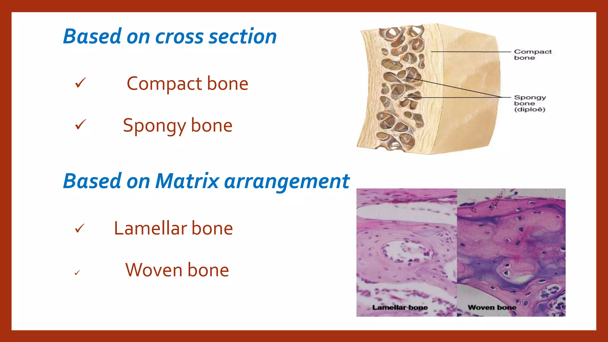
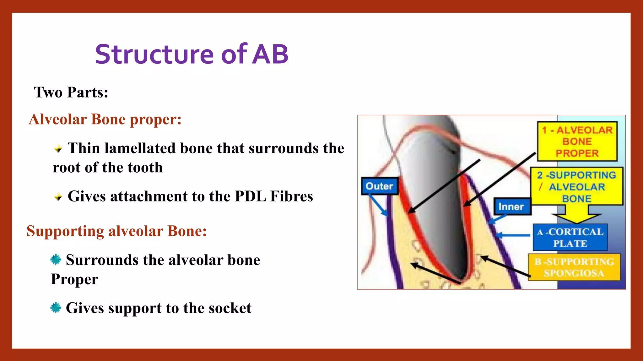
GOOD MORNING…
GOOD MORNING…
ALVEOLAR
BONE
Presented by
DR NAVEEN PARVATHAREDDY
I MDS, NARAYANA DENTAL COLLEGE
contents
• Bone – introduction, classification, composition, functions , histology & bone cells
• Introduction of AB
• Development of AB
• Structure of AB
• Blood supply & nerve supply of AB
• Functions of AB
• Age changes of AB
• Clinical considerations of AB
• Therapeutic considerations of AB
• Summary
• References
Introduction
BONE – It is a living specialized
connective tissue with calcified inter
cellular substance, which makes up
the body skeleton and is one of the
hardest structures of human body.
Based on
location
CLASSIFICATION OF BONES
Axial skeleton Appendicular skeleton
80 126+
206
Based on shape
Based on developmental origin
Intra-membranous Intracartilagenous / Endochondral
Based on cross section
 Compact bone
 Spongy bone
Based on Matrix arrangement
 Lamellar bone
 Woven bone
Woven bone
•Immature(Woven) Bone •Mature (Lamellar )Bone
•Collagen fibers oriented in
many directions
•Fibers are orderly arranged
•Interfibrillar space is more •Interfibrillar space is less
•H&E- Matrix shows blue •H&E- Matrix shows Acidophilic
•Deposition & Mineralization
rates are fast.
•Deposition & Mineralization
rates are slow.
• Bone sialoprotein present •Osteocalcin present
Based on Maturity
Immature(Woven) Bone Mature (Lamellar )Bone
Mineral density is lower &water
content higher
Low water content
Entirely removed by osteo
clasts.
Portion of lamellar matrix
resorbed at one time
Matrix vesicles participate in
mineralization
Collagen mediated mechanism
is involved in calcification
Composition
Non collagenous proteins
osteocalcin

sialoprotein

phosphoprotein

osteonectin

bone specific protein

bone morphogenic protein
functions
•Supportive
•Anchorage
•Protective
•Hematopoietic
•Storage
•Locomotive
Bone histology
: thin , delicate
connective tissue membrane
:
• circumferential lamellae:
these lamellae are arranged in
parallel layers surrounding bone.
• concentric lamellae: these
lamellae are arranged in
concentric layers.
• Interstitial lamellae: these are
present in between osteons.
• TRABECULAE- spongy bone
consists of large slender
spicules
haversian canal
and concentric lamellae are together
called as osteon.
the vascular canal
present at the centre of concentric
lamellae.
adjacent haversian
systems are connected by volkmann’s
canals.Contain vascular network
osteocytes are present in
the lacunae at the junctions of
lamellae.
 this line has more regular
appearence which denotes period of rest
during formation of bone. Deliniates
haversion system
 irregular lines which indicate
previous area of bone resorption.
Hemopoietic tissue
• Red marrow – young bone
Spongy bone of long and flat bones
Contain stem cells of fibroblasts and blood cells
• Yellow marrow – old bone
Seen in epiphysis of long bones
Loss of hemopoietic potential
Increased accumulation of fat cells
Bone cells
Osteogenic cells:
1. Osteoprogenitor cells.
2. Osteoblasts.
3. bone lining cells
4. Osteocytes.
Osteoclasts
 these are
angular shaped cells with
centrally placed nuclei.
 these cells
differentiate in to
osteoblasts which form
bone.
osteoblasts
• Derivation
• Reservoir
• Morphology
• functions
Bone lining cells
• The osteoblasts that have
completed their function and lie
on bony surface as bone lining
cells
• These cells contain few cell
organelles
osteocytes
• As the osteoblasts form bone matrix ,
they get entrapped within the matrix
they secrete are called osteocytes
• Number of osteoblasts that become
osteocytes , depends on rapidity of bone
formation
• Woven bone, repair bone shows more
osteocytes
• Osteocytes are 1o times more than
osteoblasts
• Half life
• Lacunae , canaliculi
osteoclasts
• It is a type of bone cell that
removes bone tissue by removing
mineralized matrix of bone
• Osteoclasts lie in resorption bays
• Multinucleated
• Exhibits 2 zones
• Acid phosphatase
Alveolar bone
Alveolar Bone (Process) is the part
of maxilla & mandible that forms &
supports the sockets (alveoli) of the
teeth
• Also known as Processes
Alveolaris
• Pars Alveolaris
Development of AB
• At the end of 2nd month of iu life, maxilla and
mandible forms a groove
• As tooth germs start to develop , bony septa form
gradually
• As root develop, alveolar process increases in height
• During rapid growth period, chondroid bone is
formed
alv. Process forms with development and eruption of teeth
Morphology of Alv. Bone depends on
Size
Shape
position of teeth
alv bone attains max thickness
Alv. bone undergoes atrophy
If teeth congenitally missing – Alv. Bone not developed
Boundaries
No distinct boundary
Certain areas Alveolar bone fused with basal bone
Anterior maxillary region
Posterior Mandibular region.
Structure of AB
ALVEOLAR
BONE
ALVEOLAR
BONE
PROPER
LAMELLATED
BONE
BUNDLE BONE
SUPPORTING
ALVEOLAR
BONE
CORTICAL
PLATE
SPONGY
BONE
Structure of AB
Two Parts:
Alveolar Bone proper:
Thin lamellated bone that surrounds the
root of the tooth
Gives attachment to the PDL Fibres
Supporting alveolar Bone:
Surrounds the alveolar bone
Proper
Gives support to the socket
Alveolar bone proper
 That lines the socket or alveolus
 Forms the inner wall
 Consists of two parts
 0.1 – 0.4 mm thick
 Otherwise called as cribriform plate
Cribriform plate
Anatomical name
Resembles a fine holed sieve
Perforated by – foramina
Transmit nerves &Vessels
Interdental septum
Inter –radicular septum
Lamellated bone
 Lamellar bone consists of
osteons
 Concentric lamellae along with
central blood vessel forms an
osteon
 Continuous with supporting
alveolar bone
 Mature bone
Bundle bone
Term bundle - Sharpey’s fibre
These fibers are inserted at 90 deg angle
into ABP
Consists of fibrils in the intercellular
substance and are arranged at right angles
to sharpey’s fibers
These fibrils are less in number compared
to lamellated bone and therefore it appears
dark in H&E
Silver stain
Lamina dura
Radiologic name
appears as radiopaque line
Is lost
Alveolar Crest
alveolus
distance b/n CEJ and free border
of ABP
ABP meets cortical plates
Most cervical rim = alveolar crest
It is slightly apical to CEJ in
healthy patients
If neighburing teeth is inclined
Alveolus
Rim
Floor
Alv. crest
Fundus
Supporting alveolar bone
Two Parts
Cortical Bone
Spongiosa
Supporting alveolar bone
Cortical Bone
Compact bone
Forms inner &outer plates of the
alveolar process
Variations:
Maxilla –thinner
Mandible -Thicker
Premolar, Molar region –Thicker
Anterior region -Thin
Buccal cortical plate – Thick
Lingual Cortical plate -Thinner
Relation to Local anaesthesia
 maxilla
perforated by many
openings
Infiltration is sufficient
 Mandible
 Dense cortical plate
Nerve Blocks required
SpSpongiosaongiosa
Fills space b/n ABP and Cortical
bone
Variations;
Maxilla –More
Mandible -Less
In ant region –both jaws :
Spongiosa is absent
Cortical plate is fused with ABP
Spongy bone
Spongy bone
Two types –R/A
Type I
Trabeculae –
Regular
Horizontal
Thick
Ladderlike
Mandible
Trajectorial pattern
Along lines of stress
Type II
Irregular
Fine/Delicate
Maxila
No trajectory pattern , More
marrow space
Nutrient Canals
Zuckerkandl & Hirschfeld
Interdental &Inter-radicular septum
Contain –BV, lymph vessels and nerves
Appear as radioluscent lines or shadows
Parallel to long axis of tooth
Surface structures
Ju'ga alveolaria mandibulae
Ju'ga alveolaria maxillae
Blood supply
Nerve supply
Lymphatic drainage
Bony defects
• Fenestration
• Dehiscence
• Central buttressing
• Peripheral buttressing
• Osseous craters
Functions of AB
• Houses the roots of teeth
• Anchorage
• Helps to move teeth
• Helps to absorb and distribute occlusal forces
• Supplies vessels to pdl
• Protects developing permanent teeth
• Organizes eruption of
Age changes
• Alveolar sockets appear jagged and
uneven
• Marrow spaces have fatty infiltration
• In Edentulous jaws
• Loss of maxillary bone
• Internal trabecular arrangement is
more open,
• Distance b/n alveolar crest and CEJ
• Brittleness
• Osteoporosis
• Alveolar crest slope distally due to mesial tilting of teeth
• With loss of teeth ,resorption of alveolar ridge
Pattern of bone resorption
•Maxilla
•Mandible
•Alv crest
Clinical considerations
Maxillary teeth
Fractured tooth
 Abscess
loss of lamina dura
AB loss
Orthodontic Treatment
Effect of hormones
•Proximity of AB to sinus cavities or major nerves
•Dental implants
•Surgical procedures
•Periodontal disease
Therapeutic considerations
• Bone grafting – used to bone deposition in periodontal surgery
• Autografts, allografts , xenografts
• Guided tissue regeneration – collagen is used to support natural bone
formation
• Enamel matrix proteins – used as an adjunct to periodontal surgery
summary
References
 ORBAN'S ORAL HISTOLOGY : S.N. BHASKAR.
TENCATE’S ORAL HISTOLOGY
HISTOLOGY (ATEXT AND ATLAS), MICHEAL H. ROSS,WOJCIEH PAWLINA
HAM’S GENERAL HISTOLOGY
GLICKMANN'S CLINICAL PERIODONTOLOGY : CARRANZA.
BASIC HISTOLOGYTEXT AND ATLAS- LUIZ CARLOS JUNQUEIRA, JOSE CARNEIRO
ORAL STRUCTURAL BIOLOGY : H.E.SCHROEDER. .
 TEXT BOOK OF HISTOLOGY : ROSS & REITH.
INTERNET ACCESS
THANK YOU…

AlVEOLAR BONE dr naveen reddy

  • 1.
  • 2.
    ALVEOLAR BONE Presented by DR NAVEENPARVATHAREDDY I MDS, NARAYANA DENTAL COLLEGE
  • 3.
    contents • Bone –introduction, classification, composition, functions , histology & bone cells • Introduction of AB • Development of AB • Structure of AB • Blood supply & nerve supply of AB • Functions of AB • Age changes of AB • Clinical considerations of AB • Therapeutic considerations of AB • Summary • References
  • 4.
    Introduction BONE – Itis a living specialized connective tissue with calcified inter cellular substance, which makes up the body skeleton and is one of the hardest structures of human body.
  • 5.
    Based on location CLASSIFICATION OFBONES Axial skeleton Appendicular skeleton 80 126+ 206
  • 6.
  • 7.
    Based on developmentalorigin Intra-membranous Intracartilagenous / Endochondral
  • 8.
    Based on crosssection  Compact bone  Spongy bone Based on Matrix arrangement  Lamellar bone  Woven bone
  • 9.
  • 10.
    •Immature(Woven) Bone •Mature(Lamellar )Bone •Collagen fibers oriented in many directions •Fibers are orderly arranged •Interfibrillar space is more •Interfibrillar space is less •H&E- Matrix shows blue •H&E- Matrix shows Acidophilic •Deposition & Mineralization rates are fast. •Deposition & Mineralization rates are slow. • Bone sialoprotein present •Osteocalcin present Based on Maturity
  • 11.
    Immature(Woven) Bone Mature(Lamellar )Bone Mineral density is lower &water content higher Low water content Entirely removed by osteo clasts. Portion of lamellar matrix resorbed at one time Matrix vesicles participate in mineralization Collagen mediated mechanism is involved in calcification
  • 12.
  • 13.
  • 14.
  • 15.
    Bone histology : thin, delicate connective tissue membrane
  • 16.
    : • circumferential lamellae: theselamellae are arranged in parallel layers surrounding bone. • concentric lamellae: these lamellae are arranged in concentric layers. • Interstitial lamellae: these are present in between osteons. • TRABECULAE- spongy bone consists of large slender spicules
  • 17.
    haversian canal and concentriclamellae are together called as osteon. the vascular canal present at the centre of concentric lamellae. adjacent haversian systems are connected by volkmann’s canals.Contain vascular network
  • 18.
    osteocytes are presentin the lacunae at the junctions of lamellae.
  • 19.
     this linehas more regular appearence which denotes period of rest during formation of bone. Deliniates haversion system  irregular lines which indicate previous area of bone resorption.
  • 20.
    Hemopoietic tissue • Redmarrow – young bone Spongy bone of long and flat bones Contain stem cells of fibroblasts and blood cells • Yellow marrow – old bone Seen in epiphysis of long bones Loss of hemopoietic potential Increased accumulation of fat cells
  • 21.
    Bone cells Osteogenic cells: 1.Osteoprogenitor cells. 2. Osteoblasts. 3. bone lining cells 4. Osteocytes. Osteoclasts
  • 23.
     these are angularshaped cells with centrally placed nuclei.  these cells differentiate in to osteoblasts which form bone.
  • 24.
  • 25.
    Bone lining cells •The osteoblasts that have completed their function and lie on bony surface as bone lining cells • These cells contain few cell organelles
  • 26.
    osteocytes • As theosteoblasts form bone matrix , they get entrapped within the matrix they secrete are called osteocytes • Number of osteoblasts that become osteocytes , depends on rapidity of bone formation • Woven bone, repair bone shows more osteocytes • Osteocytes are 1o times more than osteoblasts • Half life • Lacunae , canaliculi
  • 27.
    osteoclasts • It isa type of bone cell that removes bone tissue by removing mineralized matrix of bone • Osteoclasts lie in resorption bays • Multinucleated • Exhibits 2 zones • Acid phosphatase
  • 28.
    Alveolar bone Alveolar Bone(Process) is the part of maxilla & mandible that forms & supports the sockets (alveoli) of the teeth • Also known as Processes Alveolaris • Pars Alveolaris
  • 29.
    Development of AB •At the end of 2nd month of iu life, maxilla and mandible forms a groove • As tooth germs start to develop , bony septa form gradually • As root develop, alveolar process increases in height • During rapid growth period, chondroid bone is formed
  • 30.
    alv. Process formswith development and eruption of teeth Morphology of Alv. Bone depends on Size Shape position of teeth alv bone attains max thickness Alv. bone undergoes atrophy If teeth congenitally missing – Alv. Bone not developed
  • 31.
    Boundaries No distinct boundary Certainareas Alveolar bone fused with basal bone Anterior maxillary region Posterior Mandibular region.
  • 32.
    Structure of AB ALVEOLAR BONE ALVEOLAR BONE PROPER LAMELLATED BONE BUNDLEBONE SUPPORTING ALVEOLAR BONE CORTICAL PLATE SPONGY BONE
  • 33.
    Structure of AB TwoParts: Alveolar Bone proper: Thin lamellated bone that surrounds the root of the tooth Gives attachment to the PDL Fibres Supporting alveolar Bone: Surrounds the alveolar bone Proper Gives support to the socket
  • 34.
    Alveolar bone proper That lines the socket or alveolus  Forms the inner wall  Consists of two parts  0.1 – 0.4 mm thick  Otherwise called as cribriform plate
  • 35.
    Cribriform plate Anatomical name Resemblesa fine holed sieve Perforated by – foramina Transmit nerves &Vessels
  • 36.
  • 37.
    Lamellated bone  Lamellarbone consists of osteons  Concentric lamellae along with central blood vessel forms an osteon  Continuous with supporting alveolar bone  Mature bone
  • 38.
    Bundle bone Term bundle- Sharpey’s fibre These fibers are inserted at 90 deg angle into ABP Consists of fibrils in the intercellular substance and are arranged at right angles to sharpey’s fibers These fibrils are less in number compared to lamellated bone and therefore it appears dark in H&E Silver stain
  • 39.
    Lamina dura Radiologic name appearsas radiopaque line Is lost
  • 40.
    Alveolar Crest alveolus distance b/nCEJ and free border of ABP ABP meets cortical plates Most cervical rim = alveolar crest It is slightly apical to CEJ in healthy patients If neighburing teeth is inclined Alveolus Rim Floor Alv. crest Fundus
  • 41.
    Supporting alveolar bone TwoParts Cortical Bone Spongiosa Supporting alveolar bone
  • 42.
    Cortical Bone Compact bone Formsinner &outer plates of the alveolar process Variations: Maxilla –thinner Mandible -Thicker Premolar, Molar region –Thicker Anterior region -Thin
  • 43.
    Buccal cortical plate– Thick Lingual Cortical plate -Thinner Relation to Local anaesthesia  maxilla perforated by many openings Infiltration is sufficient  Mandible  Dense cortical plate Nerve Blocks required
  • 44.
    SpSpongiosaongiosa Fills space b/nABP and Cortical bone Variations; Maxilla –More Mandible -Less In ant region –both jaws : Spongiosa is absent Cortical plate is fused with ABP
  • 45.
  • 46.
    Two types –R/A TypeI Trabeculae – Regular Horizontal Thick Ladderlike Mandible Trajectorial pattern Along lines of stress
  • 47.
  • 48.
    Nutrient Canals Zuckerkandl &Hirschfeld Interdental &Inter-radicular septum Contain –BV, lymph vessels and nerves Appear as radioluscent lines or shadows Parallel to long axis of tooth
  • 49.
    Surface structures Ju'ga alveolariamandibulae Ju'ga alveolaria maxillae
  • 50.
  • 51.
    Bony defects • Fenestration •Dehiscence • Central buttressing • Peripheral buttressing • Osseous craters
  • 52.
    Functions of AB •Houses the roots of teeth • Anchorage • Helps to move teeth • Helps to absorb and distribute occlusal forces • Supplies vessels to pdl • Protects developing permanent teeth • Organizes eruption of
  • 53.
    Age changes • Alveolarsockets appear jagged and uneven • Marrow spaces have fatty infiltration • In Edentulous jaws • Loss of maxillary bone • Internal trabecular arrangement is more open, • Distance b/n alveolar crest and CEJ
  • 54.
    • Brittleness • Osteoporosis •Alveolar crest slope distally due to mesial tilting of teeth • With loss of teeth ,resorption of alveolar ridge
  • 55.
    Pattern of boneresorption •Maxilla •Mandible •Alv crest
  • 56.
    Clinical considerations Maxillary teeth Fracturedtooth  Abscess loss of lamina dura AB loss Orthodontic Treatment Effect of hormones
  • 57.
    •Proximity of ABto sinus cavities or major nerves •Dental implants •Surgical procedures •Periodontal disease
  • 58.
    Therapeutic considerations • Bonegrafting – used to bone deposition in periodontal surgery • Autografts, allografts , xenografts • Guided tissue regeneration – collagen is used to support natural bone formation • Enamel matrix proteins – used as an adjunct to periodontal surgery
  • 59.
  • 60.
    References  ORBAN'S ORALHISTOLOGY : S.N. BHASKAR. TENCATE’S ORAL HISTOLOGY HISTOLOGY (ATEXT AND ATLAS), MICHEAL H. ROSS,WOJCIEH PAWLINA HAM’S GENERAL HISTOLOGY GLICKMANN'S CLINICAL PERIODONTOLOGY : CARRANZA. BASIC HISTOLOGYTEXT AND ATLAS- LUIZ CARLOS JUNQUEIRA, JOSE CARNEIRO ORAL STRUCTURAL BIOLOGY : H.E.SCHROEDER. .  TEXT BOOK OF HISTOLOGY : ROSS & REITH. INTERNET ACCESS
  • 61.