Downloaded 143 times

![CBCT displays multiple views
of the maxillofacial complex with a
single scan, giving dentists access to
anterior, coronal, and axial images,
in addition to a 3D reconstruction
of the bony skeleton. These images
can be rotated, allowing dentists to
visualize multiple planes and angles,
including some that are not available
with 2D radiography.108,109
CBCT
images are self-corrected for magnifi-
cation, producing orthogonal images
with a practical 1:1 measuring ratio;
as a result, CBCT is considered a
more accurate option than pan-
oramic and traditional 2D images.110
Applications in TMJ disorders
Diagnostic imaging of the TMJ
is crucial for proper diagnosis of
diseases and dysfunctions associated
with joint conditions. According to
Tsiklakis et al, although CT is read-
ily available, it is not very popular
in dentistry, due in large part to
its high cost and the high dose of
radiation involved.111
CBCT makes
it possible to examine the joint
space and the true position of the
condyle within the fossa, which is
instrumental in revealing possible
dislocation of the joint disk.2,111
CBCT’s accuracy and lack of
superimposition makes it possible
to measure the roof of the glenoid
fossa and visualize the location
of the soft tissue around the
TMJ, which can offer a workable
diagnosis and reduce the need for
MRI.112-114
According to Tsiklakis
et al, MRI “is considered one of the
most useful investigations since it
provides images of both soft tissue
and bony components.”111
While
MRI is recommended for TMJ
soft tissue evaluation, CBCT offers
lower doses of radiation. However,
it should be emphasized that unlike
CT and MRI, CBCT does not
provide soft tissue detail.
These advantages outlined above
have made CBCT the best imaging
device for cases involving trauma,
fibro-osseous ankylosis, pain,
dysfunction, and condylar cortical
erosion and cysts.71,86,115-117
Applications in periodontics
According to Vandenberghe et al,
2D intraoral radiography is the
most common imaging modality
used for diagnosing bone morphol-
ogy, such as periodontal bone
defects. However, the limitations of
2D radiography could cause den-
tists to underestimate the amount
of bone loss or available bone due
to projection errors and has led to
errors in identifying reliable ana-
tomical reference points.118
These
findings confirm the observation by
Misch et al that 2D radiographs are
inadequate for detecting changes in
bone level or determining the archi-
tecture of osseous defects.119
CBCT
provides accurate measurement
of intrabony defects and allows
clincians to assess dehiscence,
fenestration defects, and periodon-
tal cysts.2,120-122
While CBCT and
2D radiographs are comparable in
terms of revealing interproximal
defects, only 3D imaging such as
CBCT can visualize buccal and
lingual defects.2,6,118,119,123
CBCT has been used to obtain
detailed morphologic descriptions
of bone as accurately as direct
measurement with a periodontal
probe.2,118,119
CBCT can also be
used to assess furcation involve-
ment of periodontal defects
and allow clinicians to evaluate
postsurgical results of regenerative
periodontal therapy.2,6,123
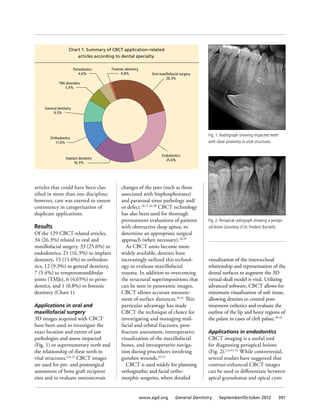
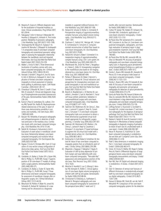
Fig. 12. Multiple endodontically treated teeth with a history of periapical surgery.
Table 1. Typical doses (in MsV)
produced by dental radiological
procedures.11
Procedure Dosage
Intraoral (F speed,
rectangular collimator)
0.001
Intraoral (E speed,
round collimator)
0.004
Full-mouth set (E speed,
round collimator)
0.080
Lateral ceph (F speed,
rare-earth screen)
0.002
Dental panoramic technique
(F speed, rare-earth screen)
0.015
Cone beam CT, both jaws 0.068
Hospital CT, both jaws 0.600
(Reprinted from Dental Update, by permission
of George Warman Productions [UK] Ltd.)
Information Technology/Computers Applications of CBCT in dental practice
394 September/October 2012 General Dentistry www.agd.org](https://image.slidesharecdn.com/e31edbfe-60aa-406f-aee6-87baa08e6955-150707020709-lva1-app6891/85/CBCT-5-320.jpg)
![While CBCT offers numerous
advantages over 2D radiography,
there are inherent limitations
requiring precise attention to selec-
tion criteria and indications. For
example, CBCT is susceptible to
motion artifacts (including artifacts
unique to CT technology) and beam
hardening around dense objects; in
addition, CBCT has low contrast
resolution and a limited ability to
visualize internal soft tissues. Many
new CBCT units contain flat-panel
detectors that are less prone to beam
hardening artifacts, so they are able
to provide more detailed informa-
tion. However, due to lack of
consistency between manufacturers,
CBCT cannot generate accurate HU
measurements and is therefore unre-
liable for quantifying bone density.4
In the authors’ opinion, it is
crucial to respect the as low as
reasonably achievable (ALARA)
radiation dose concept. This tenet
should not be misconstrued as a
reason to avoid using CBCT units
with higher doses that will provide
the necessary information. There are
no strict protocols regarding when
the technology is to be used; rather,
individual dentists, oral radiologists,
and neuro-radiologists must actively
monitor their practice’s protocols.
Interpreting these images requires
extensive anatomical knowledge of
areas that have traditionally been in
the realm of dentistry and neuro-
radiology.4
Possessing the knowledge
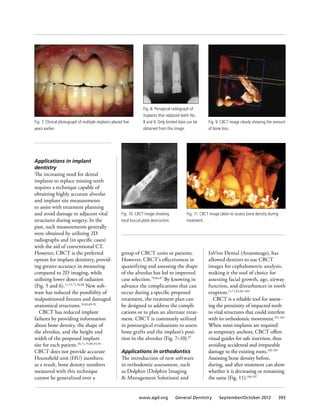
Table 2. Basic principles concerning the use of CBCT in dental applications.126
• CBCT examinations must not be carried out unless a history and
clinical examination have been performed.
• CBCT examinations must be justified for each patient to demonstrate
that the benefits outweigh the risks.
• CBCT examinations should potentially add new information to aid the
patient’s management.
• CBCT should not be repeated routinely on a patient without a new
risk/benefit assessment having been performed.
• When accepting referrals from other dentists for CBCT examinations,
the referring dentist must supply sufficient clinical information (results
of a history and examination) to allow the CBCT practitioner to
perform the justification process.
• CBCT should be used only when the question for which imaging is
required cannot be answered adequately by lower dose conventional
(traditional) radiography.
• CBCT images must undergo a thorough clinical evaluation (radiological
report) of the entire image dataset.
• Where it is likely that evaluation of soft tissues will be required as
part of the patient’s radiological assessment, the appropriate imaging
should be conventional medical CT or MRI, rather than CBCT.
• CBCT equipment should offer a choice of volume sizes, and examina-
tions must use the smallest volume that is compatible with the clinical
situation if this provides a lower radiation dose to the patient.
• Where CBCT equipment offers a choice of resolution, the resolution
compatible with adequate diagnosis and the lowest achievable
radiation dose should be used.
• A quality assurance program must be established and implemented
for each CBCT facility, including equipment, techniques, and quality
control procedures.
• Aids to ensure accurate positioning (light beam markers) must always
be used.
• All new installations of CBCT equipment should undergo a critical
examination and detailed acceptance tests before use to ensure optimal
radiation protection for staff, patients, and members of the public.
• CBCT equipment should undergo regular routine tests to ensure
that radiation protection, for both practice/facility users and
patients, has not deteriorated significantly.
• For staff protection from CBCT equipment, the guidelines detailed
in Section 6 of the European Commission document Radiation
protection 136. European guidelines on radiation protection in
dental radiology should be followed.
• All those involved with CBCT must have received adequate
theoretical and practical training for the purpose of radiological
practices and relevant competence in radiation protection.
• Continuing education and training after qualification are
required, particularly when new CBCT equipment or techniques
are adopted.
• Dentists responsible for CBCT facilities who have not previously
received “adequate theoretical and practical training” should
undergo a period of additional theoretical and practical training
that has been validated by an academic institution (university or
equivalent); where national specialist qualifications in dentomaxil-
lofacial radiology exist, the design and delivery of CBCT training
programs should involve a dentomaxillofacial radiologist.
• For dentoalveolar CBCT images of the teeth, their supporting
structures, the mandible, and the maxilla up to the floor of the
nose (for example, 8 cm X 8 cm or smaller fields of view), the
clinical evaluation (radiological report) should be made by a
specially trained dentomaxillofacial radiologist or, when this is not
possible, an adequately trained general dental practitioner.
• For non-dentoalveolar small fields of view (for example, temporal
bone) and all craniofacial CBCT images (fields of view extending
beyond the teeth, their supporting structures, the mandible
[including the TMJ], and the maxilla up to the floor of the nose),
the clinical evaluation (radiological report) should be made by
a specially trained dentomaxillofacial radiologist or a clinical
(medical) radiologist.
(Reprinted with permission of the British Institute of Radiology.)
Information Technology/Computers Applications of CBCT in dental practice
396 September/October 2012 General Dentistry www.agd.org](https://image.slidesharecdn.com/e31edbfe-60aa-406f-aee6-87baa08e6955-150707020709-lva1-app6891/85/CBCT-7-320.jpg)


This article reviews the clinical applications of cone beam computed tomography (CBCT) in dentistry as described in 129 peer-reviewed publications from 1998 to 2010. The literature review found that CBCT has been used for oral and maxillofacial surgery, endodontics, implant dentistry, orthodontics, temporomandibular joint disorders, periodontics, and forensic dentistry. Specifically, CBCT provides advantages over 2D imaging such as a lack of superimposition, accurate measurements, and 3D visualization which has made it useful for applications like dental trauma assessment, implant planning, endodontic diagnosis, and orthognathic surgery planning.