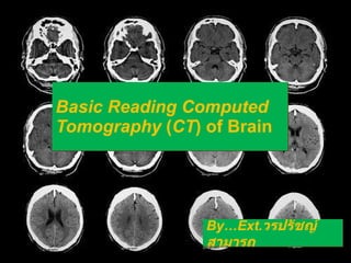
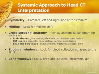
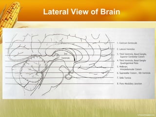
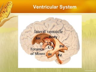
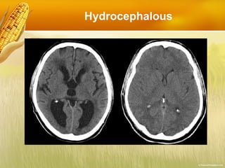
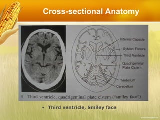
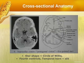
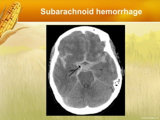
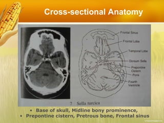
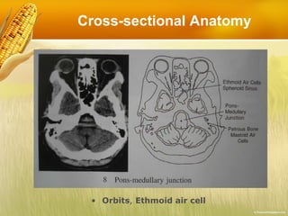
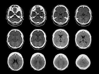
This document provides guidance on interpreting a basic computed tomography (CT) scan of the brain. It outlines a systematic approach by comparing left and right sides, looking for midline shifts, and reviewing cross-sectional anatomy at each slide. Key anatomical landmarks are identified, including gray matter, white matter, ventricles, cisterns, sulci and fissures. Specific pathologies like subdural hematomas and physiologic calcifications are also addressed.