Downloaded 174 times

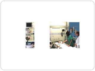

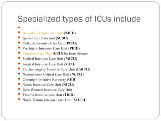


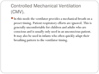


Critical care units such as intensive care units (ICUs) and critical care units (CCUs) provide specialized intensive care for critically ill patients. There are many different types of ICUs depending on medical specialty, such as neonatal ICUs, cardiac ICUs, and surgical ICUs. ICUs are equipped with mechanical ventilators and extensive monitoring equipment to support vital organ functions. Mechanical ventilation is often required to assist patients who cannot breathe adequately on their own. There are various modes of mechanical ventilation to meet patients' different respiratory needs.


![Thyroid ppt [autosaved]](https://cdn.slidesharecdn.com/ss_thumbnails/thyroidpptautosaved-170310134424-thumbnail.jpg?width=640&height=640&fit=bounds)


![Simulation_lecture_11_mechanical_ventillation[1].pptx](https://cdn.slidesharecdn.com/ss_thumbnails/simulationlecture11mechanicalventillation1-240330192829-1f83f7bc-thumbnail.jpg?width=640&height=640&fit=bounds)