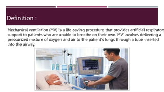
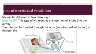
2. OUTLINE
:
– definition of mechanical ventilation
– indication of mechanical ventilation
- Types of mechanical ventilation
4 - Modes of mechanical ventilation
- Parameters of mechanical ventilation
- Alarms of mechanical ventilation
3. Complications of mechanical ventilation
7- definition of mv weaning
9- Weaning criteria
10 - Weaning methods
11- Signs of mv weaning
failure
12- extubation
13- Nursing care plan
OUTLINE:
4. Mechanical ventilation (MV) is a life-saving procedure that provides artificial respiratory
support to patients who are unable to breathe on their own. MV involves delivering a
pressurized mixture of oxygen and air to the patient's lungs through a tube inserted
into the airway.
Definition :
5. Indication for mechanical
ventilation
MV is indicated for patients with a variety of respiratory conditions, including:
Acute respiratory failure
Chronic obstructive pulmonary disease (COPD)
Asthma
Neuromuscular disorders
Pneumonia
Severe chest trauma
Postoperative respiratory failure
6. Types of mechanical ventilation
MV can be delivered in two main ways:
Invasive MV: This type of MV requires the insertion of a tube into the
airway.
The tube can be inserted through the nose (endotracheal intubation) or
through the mouth (orotracheal intubation).
7. Modes of Mechanical ventilation
Noninvasive MV (NIV):
This type of MV does not require the insertion of a tube into the
airway
NIV is delivered through a mask or nasal cannula that fits snugly
over the patient's nose and mouth.
8. Modes of mechanical ventilation
There are many different modes of MV, each with its own advantages and
disadvantages. The most common modes of MV include:
Control mandatory ventilatin ( CMV)
Ventilation is completely provided by the ventilation with a Preset VT , RR and fio
2
Ventilator totally controls the patient’s ventilation i.e. the ventilator initiates and
controls both the volume delivered and the
frequency of breath.
No interaction by the patient
advantage
rests muscles of respiration
Disadvantage
requires sedation
9. Assist /control ventilation (A/C)
A/C delivers the preset volume in response to the patient’s own inspiratory
effort (initiate the breath).
This means that any inspiratory attempt by the patient triggers a ventilator
breath. Patient may be sedated to limit the number of breath due to
hyperventilation
Advantage
reduce work of breathing , allows patient to modify
minute ventilation by increasing respiratory rate
Disadvantage
potential adverse hemodynamic affects
or inappropriate hyperventilation as dy- synchronomy
Or respiratory alkalosis
10. ynchronized Intermittent mandatory ventilation (SIMV)
SIMV was developed as a result of the problem of high respiratory rates
associated with A/C.
SIMV delivers the preset volume and rate while allowing the patient to breathe
spontaneously in between ventilator breaths i.m Additional spontaneous breatht at
tidal volume and rate determined by patient
Each ventilator breath is delivered in synchrony with the patient’s breaths, yet the
patient is allowed to completely control the spontaneous breaths.
Advantage
Each ventilator breath is delivred in synchrony with the patient’s breath .
SIMV is used as a primary mode of ventilation as well as a weaning mode
Disadvantage :
May increase the work of breathing and respiratory muscle
11. Pressure support ventilation (PSV)
PSV is preset pressure that augments the patient’s spontaneous inspiratory effort
and
decreases the work of breathing
With PSV, the patients breathe spontaneously and is assisted with every breath to a
preset
inspiratory pressure.
the patient also regulates his own respiratory rate and tidal volume
PSV is used for patients with a stable respiratory status and is often used with SIMV
toovercome the resistance of breathing through ventilator circuits and tubing
advantage
the flow rate , inspiraory time and frequency are variable and
determined by the patient
disadvantage
requires spontaneous respiratory effort
12. CPAP
CPAP is similar to PEEP except that it works only forpatients who are breathing
spontaneously.
No mandatory breaths are delivered in this mode.
Allows spontaneous breathing at elevated baseline pressure
Patient controls rate and tidal volume
CPAP can also be administered using a mask and CPAP machine for patients who do
not require mechanical ventilation, but who need respiratory support, for example,
patients with sleep apnea
CPAP allows the nurse to observe the ability of the patient to breathe
spontaneously while still on the ventilator.
Advantages
Mode used primarily for weaning from mechanics ventilation
CPAP can be used for intubated and nonintubated patients
Disadvantage
Requires spontaneous respiratory effort
Delivered volumes affected by changes in lung compliance
13. Pressure-Controlled Ventilation Mode(PCV)
In pressure-controlled ventilation the breathing gasflows under constant pressure
into the lungs duringthe selected inspiratory time.
Similar to CMV, in PCV the rate is fixed and cannotbe increased by the patient
effort, but a difference isthat it is the peak inflation pressure rather than VTthat is a
set.
Advantage
decrease the risk of barotrauma.
It is used when the patient has persistent oxygenation
problems despite a high FiO2 and high levels of PEEP
14. Bi PAP
BiPAP is a noninvasive form of mechanical ventilation provided by
means of a nasal mask or nasal prongs, or a full-face mask.
The system allows the clinician to select two levels of positive-
pressure support:
An inspiratory pressure support level
(referred to as IPAP)
An expiratory pressure called EPAP (PEEP).
Recommendation: in acute hypercapnia, respiratory muscle rest,
immunosuppressed individuals with infection.
15. Parameters of Mechanical Ventilation
The parameters of MV are adjusted to meet the individual needs of each patient.
Common parameters include:
Tidal volume (TV orVT)
the volume of air inhaled at each breath expressedin milliliters. The tidal volume is
about 6-8 mL/kg ofideal body weight for patients with healthy lungs.
For unhealthy lungs, such as in ARDS or ALI, 4-6mL/kg is used as a protection
strategy for the lungs
16. Respiratory rate (RR
the number of breaths delivered By the Ventilator per minute. RR is
commonly set between 12and 20 breath / minute.
17. Fraction of inspired oxygen (FiO2) is the percentage of O2 delivered to the
patient.
The FiO2 ranges from 0.21 (room air) to 1(100% O2).
18. Positive end-expiratory pressure PEEP
is the amount of pressure applied at the end of
exhalation phase of respiratory cycle to help keep the
alveoli open .
The initial PEEP for patients admitted to ICU is usually
between 5 cm and 10 cm H2O.
19. Alarm of mechanical ventilation
Mechanical ventilators in a healthcare setting are equipped with various alarms to alert
healthcare providers to potential issues or changes in a patient's condition. These
alarms are critical for patient safety. Common alarms associated with mechanical
ventilation include:
High Pressure Alarm: This alarm activates when the peak airway pressure exceeds a
set limit. It can indicate issues like airway blockage, patient coughing or biting on the
endotracheal tube, or changes in lung compliance.
20. Low Pressure Alarm: This alarm sounds when the pressure in the breathing circuit drops
below a predefined threshold. It might suggest disconnection of the patient from the
ventilator or a leak in the system.
Low Exhaled Tidal Volume (Vt) Alarm: This alarm triggers when the measured exhaled
tidal volume falls below the set minimum level. It could indicate issues with patient-
ventilator synchrony or problems with lung compliance.
High Exhaled Tidal Volume (Vt) Alarm: This alarm activates when the measured exhaled
tidal volume exceeds the set maximum level. It can be a sign of increased airway
resistance or patient coughing.
Low Minute Ventilation (MV) Alarm: This alarm is activated when the minute
ventilation (the product of tidal volume and respiratory rate) falls below a
predefined threshold. It might suggest hypoventilation or disconnection.
High Respiratory Rate Alarm: This alarm sounds when the patient's respiratory rate
exceeds the set maximum limit. It could indicate distress or discomfort.
21. Low Oxygen Concentration Alarm: This alarm activates if the inspired oxygen concentration (FiO2) falls
below the set minimum level, indicating a potential issue with the oxygen supply.
High Oxygen Concentration Alarm: This alarm triggers when the FiO2 exceeds the set maximum limit,
which might be due to a malfunction in the oxygen delivery system.
Apnea Alarm: Some ventilators are equipped with apnea detection, which can sound an alarm if the
patient stops breathing for a defined duration.
Battery Alarm: Ventilators often have backup power sources, and an alarm can indicate a low battery,
prompting the need for immediate attention or connection to a power source.
Disconnect Alarm: This alarm activates when the patient becomes disconnected from the ventilator
circuit, indicating a potential emergency situation.
22. Complications of mechanical ventilation
MV can be associated with a number of complications
including:
Ventilator-associated pneumonia (VAP)
Barotrauma
Lung collapse
Airway injury
Infection
Thromboembolism
23. Definition of mv weaning
MV weaning is the process of gradually reducing the ventilator settings
and allowing the patient to breathe on their own. Weaning is a complex
process that must be carefully managed to avoid complications.
24. Weaning criteria
1- Adequate Oxygenation: The patient should have a satisfactory oxygenation
status, typically defined by a PaO2 (partial pressure of arterial oxygen) greater
than 60 mm Hg on FiO2 (fraction of inspired oxygen) less than 40%.
2 - Adequate Ventilation: The patient's CO2 levels, measured as PaCO2 (partial
pressure of arterial carbon dioxide), should be within an acceptable range, often less
than 45 mm Hg.
Hemodynamic Stability: The patient should have stable blood pressure, heart rate,
and overall cardiovascular status.
3 - Sufficient Mental Alertness: The patient should be awake, alert, and able to follow
commands. Some healthcare providers use tools like the Richmond Agitation-
Sedation Scale (RASS) to assess consciousness.
25. 4- Low Ventilatory Support: The patient should be on minimal levels of ventilatory
support (e.g., low FiO2 and positive end-expiratory pressure (PEEP)).
5- Adequate Respiratory Muscle Strength: The patient should have sufficient respiratory
muscle strength to support spontaneous breathing. This can be assessed by measuring
parameters like the vital capacity and negative inspiratory force (NIF).
6- Chest X-ray: A chest X-ray should show no worsening of lung pathology or
complications that might require continued mechanical ventilation.
7- Spontaneous Breathing Trial (SBT): A common practice is to perform a spontaneous
breathing trial, during which the patient is temporarily disconnected from the ventilator t
see if they can sustain breathing on their own. The most common method is a T-piece
trial or a low-level pressure support trial.
26. 8- Assessment of Rapid Shallow Breathing Index (RSBI): RSBI is calculated by
dividing the respiratory rate by the tidal volume. An RSBI less than 105 is
often considered a good predictor of successful weaning.
It's important to note that the weaning process should be individualized to
the patient's condition, and these criteria are used as guidelines rather than strict
rules. The decision to wean a patient from mechanical ventilation should always
be made by a healthcare professional based on a comprehensive assessment of
the patient's clinical status.
27. There are a number of different weaning methods, each with its own advantages and
disadvantages. The most common weaning methods include:
Pressure support ventilation (PSV) weaning: PSV weaning involves gradually reducing th
level of pressure support provided by the ventilator.
Volume-controlled ventilation (VCV) weaning: VCV weaning involves gradually reducing
tidal volume delivered by the ventilator.
T-piece weaning: T-piece weaning involves connecting the patient to a T-piece connecto
which allows them to breathe on their own while still receiving some support from the
ventilator.
Weaning methods
30. Intubation for Mechanical Ventilation
Intubation is the process of inserting a tube (endotracheal tube) into the airway
through the nose or mouth. This is done to protect the airway and to deliver oxygen
and other medications to the lungs. Intubation is often necessary for patients who
are unable to breathe on their own, such as those who are unconscious or have
severe respiratory failure.
31. Nursing care for ETT
1- Monitor the patient's vital signs and respiratory status closely.
2- Keep the endotracheal tube clean and suction it as needed.
3- Prevent the endotracheal tube from becoming kinked or
obstructed.
4- Provide oral hygiene and humidification.
5- Assess the patient's ability to communicate.
6-Educate the patient and their family about endotracheal tube care.
32. Extubation
Extubation is the process of removing the
endotracheal tube from a patient's airway. It is a
critical step in the weaning process and should
only be performed when the patient is ready.
33. Nursing care for mechanical ventilation
Assessment: Regular and thorough assessment of the patient is essential. This
includes monitoring vital signs, oxygen saturation, lung sounds, and assessing the
ventilator settings and alarms.
Ventilator Settings: Nurses should be familiar with the ventilator settings and
alarms. They need to ensure that the ventilator is functioning correctly and that the
settings are appropriate for the patient's condition.
Oral Care: Proper oral hygiene is vital to prevent ventilator-associated
pneumonia (VAP). This involves regularly cleaning the patient's mouth and
providing mouth care.
Suctioning: Suctioning of the airway is necessary to remove secretions and
maintain airway patency. It should be done when clinically indicated and with
proper technique to prevent complications.
34. Positioning: Turning and repositioning the patient helps prevent complications like
pressure ulcers and ventilator-associated lung injury. Patients should be positioned to
promote optimal lung expansion.
Medication Management: Administering medications as prescribed, including sedatives,
analgesics, and neuromuscular blocking agents, if required. Nurses need to monitor the
effects of these medications carefully.
Communication: Patients on mechanical ventilation are often unable to speak. Nurses
should ensure there is a means of communication in place, such as a communication
board or assistive devices.
Weaning and Extubation: Nursing plays a key role in assessing a patient's readiness to
be weaned from the ventilator and extubated. This involves regular assessments and
collaboration with the medical team.
35. Psychosocial Support: Being on a ventilator can be distressing for patients and their
families. Nurses should provide emotional support and keep families informed about
the patient's condition.
Infection Control: Strict adherence to infection control practices, including hand
hygiene, is vital to prevent healthcare-associated infections.
Monitoring for Complications: Keep an eye out for complications such as barotrauma,
pneumothorax, or ventilator-associated events and alert the medical team if any issues
arise.
Documentation: Thorough and accurate documentation of all care provided, changes
in the patient's condition, and ventilator settings is essential for continuity of care.
Collaboration: Effective communication and collaboration with other healthcare
professionals, including respiratory therapists and physicians, are critical in managing
mechanically ventilated patients
36. What’s the vab bundle?
The term "VAB bundle" typically refers to a set of protocols or best practices used in
the context of mechanical ventilation in a healthcare setting, particularly in the
intensive care unit (ICU). VAB stands for "Ventilator-Associated Pneumonia Bundle."
These bundles are designed to reduce the risk of patients developing ventilator-
associated pneumonia (VAP), which is a serious and potentially life-threatening
infection that can occur when a patient is on a ventilator.
me common components of a VAB bundle may include:
1-Elevation of the head of the bed to 30-45 degrees to reduce the risk of
aspiration.
2-Daily "sedation vacations" or assessment of sedation levels to reduce the use
of sedative medications when not necessary.
37. 3-Daily assessment of a patient’s readiness to extubate and early weaning
from mechanical ventilation when appropriate.
4-Good oral hygiene and regular mouth care to reduce the risk of
bacterial colonization in the mouth and subsequent aspiration.
5-Regular changes of ventilator circuit tubing to prevent biofilm
formation.
6- Strict hand hygiene and infection control measures to reduce the risk
of cross-contamination.
7-Minimizing the duration of mechanical ventilation when possible.