Perioperative Arrythmias and management
•Download as PPTX, PDF•
8 likes•4,086 views

Anaesthesia and arrythmias

Recommended
Recommended
More Related Content
What's hot
What's hot (20)
Similar to Perioperative Arrythmias and management
Similar to Perioperative Arrythmias and management (20)
More from Dr Nandini Deshpande
More from Dr Nandini Deshpande (7)
Recently uploaded
Recently uploaded (20)
Perioperative Arrythmias and management
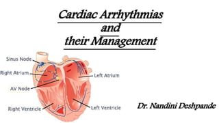
- 1. Cardiac Arrhythmias and their Management Dr. Nandini Deshpande
- 2. Normal ECG
- 3. Arrhythmias AHA defines an arrhythmia as: The term "arrhythmia" refers to any change from the normal sequence of electrical impulses. The electrical impulses may happen too fast, too slowly, or erratically – causing the heart to beat too fast, too slowly, or erratically. When the heart doesn't beat properly, it can't pump blood effectively. When the heart doesn't pump blood effectively, the lungs, brain and all other organs can't work properly and may shut down or be damaged.
- 4. Clinical Manifestations Many arrhythmias go unnoticed by the patient and are picked up incidentally on a routine physical examination or EKG. Most Common manifestation is palpitation, an awareness of one's own heartbeat. More serious symptoms occur when the arrhythmia compromises the heart's ability to pump blood effectively. Among these are light- headedness and syncope. Rapid arrhythmias can increase the oxygen demands of the myocardium and cause angina (chest pain). The sudden onset of an arrhythmia in a patient with underlying cardiac disease can also precipitate congestive heart failure. Sometimes, the first clinical manifestation of an arrhythmia is sudden death.
- 5. Basic Types of Arrhythmias The electrical activity follows the usual conduction pathways, but it is too fast, too slow, or irregular. These are arrhythmias of sinus origin. The electrical activity originates from a focus other than the sinus node. These are called ectopic rhythms. The electrical activity is trapped within an electrical racetrack whose shape and boundaries are determined by various anatomic or electrical myocardial configurations. These are called reentrant arrhythmias. The electrical activity follows anomalous accessory conduction pathways that bypass the normal ones, called pre-excitation syndromes. The electrical activity originates in the sinus node and follows the usual pathways but encounters unexpected blocks and delays. These conduction blocks.
- 6. Arrhythmias of Sinus Origin
- 7. Sinus Tachycardia and Sinus Bradycardia Normal sinus rhythm is the normal rhythm of the heart. Depolarization originates spontaneously within the sinus node. The rate is regular and between 60 and 100 beats per minute. If the rhythm speeds up beyond 100, it is called sinus tachycardia; if it slows down below 60, it is called sinus bradycardia. Normal Variants: Strenuous exercise, for example, can accelerate the heart rate well over 100 beats per minute, whereas resting heart rates below 60 beats per minute are typical in well-conditioned athletes. Pathological: Sinus tachycardia can occur in patients with fever, congestive heart failure, severe lung disease, or hyperthyroidism. Sinus bradycardia can be caused by medications, eg beta-blockers, calcium channel blockers, and opioids.
- 8. (A) Sinus tachycardia. Each beat is separated by two and one-half large squares for a rate of 120 beats per minute. (B) Sinus bradycardia. More than seven large squares separate each beat, and the rate is 40 to 45 beats per minute.
- 9. Peri-operative Sinus bradycardia Causes Autonomic disturbance including vasovagal stimulation. Hypoxia Hypothermia Endotracheal suctioning Increased intracranial pressure Hypothyroidism Drugs like B blockers, CCB’s
- 11. Peri-operative Sinus tachycardia[Narrow regular] Causes Pain Fever Hypercarbia Hypovolemia Inadequate anesthetic depth Drugs like sympathomimetics, antimuscarinics Correction of these conditions is the first step in the treatment of sinus tachyarrhythmia. If required beta blockers may be used after these factors have been worked at. Most commonly used agents are esmolol and metoprolol.
- 12. Sinus Arrest, Asystole Sinus arrest occurs when the sinus node stops firing. If nothing else were to happen, the EKG would show a flat line without any electrical activity, and the patient would die. Prolonged electrical inactivity is called asystole. Rx- ACLS protocol.
- 13. Ectopic Rhythms
- 14. Premature Atrial Complex (PAC) These arise from ectopic pacemaking tissue within the atria. An abnormal (non-sinus) P wave is followed by a normal QRS complex. The abnormal P wave may be hidden in the preceding T wave. •Multiple PACs, showing Bigeminy •This hidden PAC gives a peaked appearance to the T wave (circled). •It is followed by a compensatory pause. •No QRS complex, indicating blocked PAC.
- 15. Premature Atrial Complex (PAC) Causes 1. Anxiety. 2. Sympathomimetics. 3. Beta-agonists. 4. Excess caffeine. 5. Hypokalemia. 6. Hypomagnesaemia. 7. Digoxin toxicity. 8. Myocardial ischemia Clinical Significance : Frequent PACs may cause palpitations and a sense of the heart “skipping a beat”. In patients with underlying predispositions (e.g LA enlargement, IHD, WPW), it may be the trigger for the onset of a re-entrant tachyarrhythmia — e.g. AF, flutter, AVNRT or AVRT. Usually no treatment is required. May use Beta blockers or Calcium Channel blockers.
- 16. Premature Ventricular Complex (PVC) Causes Anxiety Sympathomimetics Beta-agonists Excess caffeine Hypokalemia Hypomagnesaemia Digoxin toxicity Myocardial ischemia Multifocal PVCs Ventricular Bigeminy
- 17. When to worry about PVCs? PVCs posing an increased risk for triggering ventricular tachycardia: Frequent PVCs. Runs of consecutive PVCs, especially three or more in a row. Multiform PVCs, in which the PVCs vary in their site of origin and hence in their appearance. PVCs falling on the T wave of the previous beat, called the “R-on-T” phenomenon. Although PVCs meeting one or several of these criteria are associated with an increased risk for developing a life-threatening arrhythmia, there is no evidence that suppressing these PVCs with antiarrhythmic medication reduces mortality in any setting.
- 18. PAC PVC Abnormal p waves Normal p waves Normal QRS complex Abnormal QRS complex Problems in the atria Problems in the ventricles No compensatory pause Compensatory pause is present Rarely life threatening Life threatening if progresses to VT
- 19. AV Nodal Reentrant Tachycardia [Narrow regular] AVNRT is a very common arrhythmia. Its onset is sudden, usually initiated by a premature supraventricular beat , and its termination is just as abrupt. AVNRT is an absolutely regular rhythm, with a rate between 150 and 250 bpm. It is most often driven by a reentrant circuit looping within the AV node. Retrograde P waves may sometimes be seen in leads II or III, or we may see a pseudo-R′ disturbance in V1 lead. QRS complex is usually narrow. It can occur in perfectly normal hearts. Persons with AVNRT typically present with palpitations, shortness of breath, dizziness, or syncope. Not uncommonly, alcohol, coffee, or just sheer excitement can elicit this rhythm.
- 21. SINUS TACHYCARDIA AVNR TACHYCARDIA HR- 100-150/bpm HR-150-250/bpm P waves and QRS both present, regular RR interval P waves not present, regular RR interval Gradual onset Abrupt onset Physiological as well as pathological Always pathological
- 22. Atrial Flutter [Narrow regular] It may occur in normal hearts . The atrial activation in atrial flutter is absolutely regular but is at a rate of 250 to 350 bpm, generated by a reentrant circuit that usually runs around the tricuspid valve. Discrete P waves may not be seen. Instead saw-toothed pattern may be seen. The AV node cannot handle the extraordinary number of atrial impulses bombarding it, some impulses just bump into a refractory node. This phenomenon is called AV block.
- 23. Atrial Flutter - associations 1. Hypertension 2. Obesity 3. Diabetes mellitus 4. Electrolyte imbalances 5. Alcohol intoxication 6. Drug abuse, particularly cocaine and amphetamines 7. Pulmonary disease (e.g., chronic obstructive pulmonary disease and pulmonary embolism) 8. Thyrotoxicosis 9. Various underlying cardiac conditions, both congenital (e.g., atrial septal defect) and acquired (e.g., rheumatic valvular disease, coronary artery disease, and congestive heart failure)
- 24. Atrial Fibrillation [Narrow irregular] In atrial fibrillation, atrial activity is completely chaotic, and the AV node may be bombarded with more than 500 impulses per minute! Multiple tiny reentrant circuits whirl around in totally unpredictable fashion. No true P waves can be seen. The AV node allows only occasional impulses to pass through at variable intervals, generating an irregularly irregular ventricular rate. This irregularly irregular appearance of QRS complexes in the absence of discrete P waves is the key to identifying atrial fibrillation. Associations similar to flutter.
- 27. Ventricular Tachycardia[Wide regular] A run of three or more consecutive PVCs is called ventricular tachycardia. The rate is usually between 120 and 200 beats. Sustained VT — lasting more than 30 seconds—or VT associated with hemodynamic instability are emergencies, presaging cardiac arrest and requiring immediate treatment. Polymorphic ventricular tachycardia is more commonly associated with acute coronary ischemia, infarction, profound electrolyte disturbances, and conditions causing prolongation of the QT interval. Uniform ventricular tachycardia is more often seen with healed infarctions.
- 28. Ventricular Tachycardia MECHANISMS Enhanced automaticity (ectopic activity) Enhanced trigger activity Re-entry Drugs that cause QT Prolongation Clarithromycin Erythromycin Metaclopramide Haloperidol TCA Methadone Droperidol Electrolytes • Hypokalaemia Hyperkalaemia • Hypomagnesaemia Hypocalcaemia Hypothermia Structural heart disease • LV dysfunction • Coronary artery disease • MI • HOCM
- 30. Ventricular Tachycardia Supraventricular Tachycardia Clinical Clues Clinical history Diseased heart Usually healthy heart Carotid massage No response May terminate Cannon A waves May be present Not seen EKG Clues AV dissociation May be seen Not seen Fusion beats May be seen Not seen Initial QRS deflection May differ from normal QRS complex Same as normal QRS complex
- 31. Ventricular Fibrillation[Wide irregular] Ventricular fibrillation is a preterminal event seen solely in dying hearts. The EKG tracing jerks about spasmodically, there are no true QRS complexes. In VF, the heart generates no cardiac output, and cardiopulmonary resuscitation and electrical defibrillation must be performed. Common precipitants of ventricular fibrillation include Myocardial ischemia/infarction Heart failure Hypoxemia or hypercapnea Hypotension or shock Electrolyte imbalances Overdoses of stimulants, especially when used in combination. In many cases, ventricular fibrillation is preceded by ventricular tachycardia
- 35. First degree AV Blocks The depolarisation at AV node is held up for longer than the usual. PR interval is longer than 0.2 seconds. Every QRS complex is preceded by a single P wave. It can also be an early sign of degenerative disease of the conduction system or a manifestation of myocarditis or drug toxicity. By itself, it does not require treatment. .
- 36. Second degree AV Blocks Wenckebach Block Almost always due to a block within the AV node. The delay is variable increasing with each ensuing impulse. Each successive atrial impulse encounters a longer delay in the AV node until one impulse (usually every third or fourth) fails to make it through. Progressive lengthening of the PR interval is seen and then a dropped beat is seen. The sequence repeats itself, over and over, and often with impressive regularity.
- 37. Second degree AV Blocks Mobitz Type II Block- Usually due to a block below the AV node. The EKG shows two or more normal beats with normal PR intervals and a dropped beat. The cycle is then repeated. The ratio of conducted beats to nonconducted beats is may or not be constant. Mobitz type II block is far more serious that type1, often signifying serious heart disease and capable of progressing suddenly to third-degree heart block. Mobitz type II heart block requires insertion of a pacemaker.
- 38. Third degree AV Blocks No atrial impulses at all make it through to activate the ventricles. For this reason, it is often called complete heart block / AV dissociation. The site of the block can be either at the AV node or lower. The ventricles generate an escape rhythm. The atria continue to contract atria at 60 to 100 bpm and ventricles at 30 to 45 bpm. May be seen in MI / degenerative diseases / Lyme’s – reversible.
- 39. Management of Blocks • 1st degree – nothing unless symptomatic and other causes of symptoms excluded • 2nd degree (Mobitz type I) – nothing unless symptomatic and other causes of symptoms excluded • 2nd degree (Mobitz type II) – pacemaker • 3rd degree – pacemaker
Editor's Notes
- Sinus tachycardia is the most common arrhythmia occurring in the perioperative period
- Virtually all myocardial cells have the ability to behave as pacemakers. Ordinarily, the fastest pacemaker drives the heart, and under normal circumstances, the fastest pacemaker is the sinus node. The sinus node overdrives the other pacemaker cells by delivering its wave of depolarization throughout the myocardium before its potential competitors can complete their own, more leisurely, spontaneous depolarization
- Atrial ectopics, atrial extrasystoles, atrial premature beats, atrial premature depolarisations. The P wave typically has a different morphology and axis to the sinus P waves.
- Atrial ectopics, atrial extrasystoles, atrial premature beats, atrial premature depolarisations. The P wave typically has a different morphology and axis to the sinus P waves.
- Two different morphologies of PVCs is sen “Discordance” is seen.
- In most situations, we don't have to worry at all.
- Regular rhythm 2nd panel shows the typical psudo R’ aftre QRScomplex