
More Related Content
What's hot
What's hot (20)
Similar to Cranial remodelling orthosis
Similar to Cranial remodelling orthosis (20)
More from POLY GHOSH
More from POLY GHOSH (12)
Recently uploaded
Recently uploaded (20)
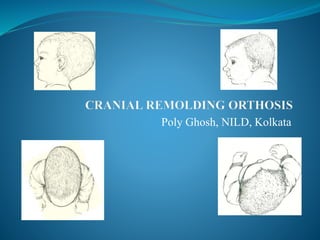
Cranial remodelling orthosis
- 1. Poly Ghosh, NILD, Kolkata
- 2. INTRODUCTION The term plagiocephaly derives from the Greek plagios meaning oblique and cephalo referencing the head. The term deformational plagiocephaly, deformational Brachiocephaly and deformational Scaphocephaly refer to cranial deformities. Cranial deformity features:- • deviations in proportion and/or symmetry of the neurocranium(i.e skull) • Accompanied by misalignment of the bones of the viscerocranium(i.e face).
- 3. Cont… Cranial remoulding orhoses are usually in the shape of an adjustable helmet or band that progressively molds the shape of the infants cranium by applying corrective forces to prominences while leaving room for growth in adjacent flattened areas. A Cranial remoulding orthoses may be requested for the treatment of positional or postsurgical synostosis in paediatric patient. An asymmetrically shaped head may be synostotic(craniosynostosis) or nonsynostotic(deformational). Positional cranial deformity also known as deformational or nonsynostosis cranial deformity. The nonsynostosis or deformational cranial deformity refer to cranial deformities that are recognized in infancy and develop from both prenatal and postnatal factors.
- 4. Synostosis defined as premature closures of the sutures of the cranium, may result in functional deficits secondary to increasing intracranial pressure in an abnormally or asymmetrically shaped cranium. The type and degree of craniofascial deformity depends on the type of synostosis. Cranial deformity may be plagiocephaly ,brachiocephaly and scapho cephaly.
- 5. Clinical anatomy of skull • The shape and structure of the skull bones are affected by genetic, metabolic, and mechanical factors because deformation or misalignment of one unit affects the alignment and shape of adjoining structures • The skull of the infant has nine bones of the neurocranium • The viscerocranium consists of 14 facial bones. • Alignment and orientation of viserocranium structures are interdependent with the alignment and orientation of the neurocranium.
- 6. Pathophysiology Causes:- 1. Abnormalities in brain shape or development- includes microcephaly, macrocephaly and in uterocerebrovascular accident. 2. Abnormalities in bone or suture development- craniosyntosis, apert syndrome, crouzon syndrome 3. Prenatal and postnatal deforming forces.
- 7. Common head deformities:- Plagiocephaly Most common cranial anomaly Asymmetry of rt and lt side of skull Asymmtery of the neurocranium and viscerocranium Unilateral occipital flattening Ipsilateral anterior ear progression Ipsilateral forehead bossing Contralateral forehead depression contralateral occipital bossing Fascial asymmetry Congenital muscular torticollis Altered alignment of eyes, cheeks, nose, ,mouth and chin
- 8. Brachiocephaly Less common Present with primary disruption in cranial proportion Disproportion of the neurocranium and viscerocranium B/L occipital flattening B/L forehead bossing Increased height of cranial vault Associated weakness of neck musculature Increased cranial width Decreased cranial length Increased cranial vault height
- 9. Scaphocephaly Disproportion of the neurocranium and viscerocranium B/L parietal flattening Anterior forehead bossing Posterior occipital bossing Associated weakness of neck musculature Increased cranial length Decreased cranial width
- 10. Cont… Asymmetrical brachiocephalic head shape deformity- common to both plagiocephaly and brachiocephaly
- 11. Factors for creating cranial deformities Prenatal factors- Deforming forces in utero- Restrictive in utero environments Fetal constraints Sustained abnormal positioning Deforming forces during birth process- Vaginal deliveries Cesarean deliveries Postnatal factors- Premature infants due to increased plasticity of the underdeveloped cranial structure 1. Sustained supine positioning in compliance with the AAP Back to sleep program 2. Sustained supine positioning in neonatal intensive care units 3. Sustained supine positioning during normal daily infant/caregiver routines 4. Congenital muscular torticollis 5. Neck muscle asymmetry 6. Unilateral neck involvement 7. Cranial hemivertebrae
- 12. Evaluation of cranial deformities Degree of asymmetry Degree of disproportion Plasticity of bony structure Amount of translation of bony plates Cellular disruption and alterations in suture development Soft tissue involvement and/or contributions from neck musculature Genetic predisposition Effect of neurocranial structure and alterations on adjacent viscerocranial structure
- 13. Historical perspective Intentional cranial deformation Different cultures applied a variety of measures to alter the shape of infants skull Boards, vines, cloth bandages and even weight of stones were applied to the head of the infants to create a culturally desired shape. Early and sustained application of intentional and specific force would produce long standing changes in cranial shape. In 1979, clarren et al, reported that many infants were treated for many different congenital or hereditory disorders of the brain, bone or sutures. Cranial remolding orthoses were being used postoperatively and infants diagnosed with nonsynostosis deformity treated with cranial orthosis and treatment concept was based on intentional cranial deformation.
- 14. Cont… In early to mid 1990 – significant increase in the no. of infants with asymmetrical and disproportional skull deformities coincided with AAP back to sleep program. Haung et al, outlined specific anatomy of deformational plagiocephaly versus unilateral lambdoid synostosis. Asymmetry and unilteral occipital flattening in both condition Parallelogram head shape in plagiocephaly and trapezoid shape in lambdoid syndrome. Anterior ear progression in plagicephaly and posterior ear progression in lamdoid synostosis With the increasing no. of infants with these deformities orthotics management with cranial remolding orthosis was developed and refined.
- 15. Management Cranial distortion in newborn is common and generally dissolved within the first 12 weeks of life. Early identification of cranial deformities creates the opportunity for altering external forces. Treatment recommendations- 1. Repositioning 2. Therapeutic efforts 3. Orthosis- a cranial deformity is considered for orthotic intervention when the infants abnormal skull proportion remains or fails to improve despite early intervention of repositioning and therapeutic efforts during the first 3 months after birth
- 16. Repositioning technique Goal- better distribution of external forces acting on the developing skull. Time- first 3months of life Techniques = Tummy time Strategic positioning during diapering, feeding, carrying and handling Limitation on time spent in car seats, carriers and swings Positioning of nursery furniture relative to bright areas
- 17. Anthropometic evaluation Anthrometric skull measurements establish a baseline for clinical documentation of improvement or progression of the skull deformity. Common clinical measurements- 1. Cranial circumference 2. Cranial base 3. Cranial width 4. Cranial length 5. Cranial vault 6. Orbitotragial depth 7. Cranial base measurements. 8. Cranial vault asymmetry 9. Orbitotragial depth asymmetry(OTDA) 10. CVAI 11. Cephalic index(CI) 12. Cranial base asymmetry(CBA)
- 18. Cranial circumference and cranial width measurements Taken at the equator Euryon to euryon(EU- EU)
- 19. Cranial length measurement Glabella to opisthocranion (g –op)
- 20. Cranial vault Frontozygomaticus to contralateral euryon
- 21. Orbitotragial depth(upper face) and cranial base(lower face) Orbitotragial depth= exocanthion to tragion(ex-t) Cranial base= Subnation to tragion(sn- t)
- 22. Cranial width=78% cranial length
- 23. Normative values of cranial development 2cm of circumferential growth/ month in the first 3 months 1 cm of circumferential growth/ month between 4 and 6 months Approx 0.5 cm of circumferential growth/ month between 6 and 12 months After 12 months cranial growth slows significantly
- 24. Therapy technique Continued cranial deformation Developmental delay Weakness and tightness of neck musculature Hypotonicity Asymmetry of nuchal fold Persistent positional preference Limitations in active and passive neck ROM
- 25. Orthotic management Indication- moderate to severe deformities Disruption in cranial symmetry and/or proportion result from the application external forces acting on developing cranial structure. Deformation of the infant skull occurs in response to the amount , direction, mode and frequency of the forces applied. Temporomandibular jt. Or orbital alignment may be affected. Function- 1. Presumes growth 2. Balance the static and dyanamic forces acting on the developing structure to ensure functional competence of both the neurocranium and viscerocranium.
- 26. Goal Achieve maximum correction of the deformity, Establish symmetry and/or proportion of the skull, Provide stimulus to cranium growth, Encourage passive expansion of the cranium.
- 27. General consideration for orthosis in pediatric population 1. Time of onset of condition 2. Duration of deforming forces 3. Degree of severity 4. Degree of correctability 5. Diagnosis/etiology 6. Remaining growth of physiological structures 7. Overall health of physiological structures 8. Developmental level
- 28. Examination Patient history:- Prenatal and postnatal factors Birth history:- Gestational age, Weight , Length, Fetal positioning, Delivery Associated medical conditions:- Skin sensivity, head shape at birth and age when head deformity was first noted. Developmental observation:- preferred sleeping postion, repositioning efforts by caregivers, acquisition of developmental milestone
- 29. Cont…. Physical examination of the infants skull and face 1. Visual observation from all angles 2. Manual palpation of cranial structure Documentation of Asymmetrical and displaced skull and fascial features Nonvertical orientation of the head relative to the trunk Neck musculature ROM Digital photographs and anthrometric measurements
- 30. Discussion with caregivers An overview of orthotic treatment program Determination of treatment goals and expectatations Importance of supervised prone position Casting or scanning process Fitting and follow up schedule Orthotic cleaning regimen Wearing schedule Signs indicating the need for adjustments Situations requiring removal of the cranial remolding orthosis
- 31. Fabrication of cranial remolding orthosis Step -1 Measurement procedure:- Goal- to obtain an accurate model with identifiable anatomical landmarks necessary for rectification and fabrication of custom CRO. 1. Casting- produce an accurate negetive model of infants head 2. Scanning- produce digital representation of infants head.
- 33. Step -2 Rectification- Goal- to create a new model with greater symmetry and/ or proportion than currently exists. Modification- areas of flattening are expanded to allow for planned growth and Areas of bossing are maintained to resist growth. Various orthosis use slightly different rectification technique but basic theory is same.
- 34. Cranial mapping instrument Adapted from a bremer pediatric halo crown Was developed to measure deformational head shapes. The instrument measures 1. The position and amount of depression or flatness, 2. The amount of displacement, 3. The orientation of head shape within the instrument. 4. Allows the orthotist to create a blueprint and establish the predicted shape of the skull after orthotic treatment.
- 35. Creating the blueprint After a plaster impression of the infant's head is made, it is essential to critique the positive model for quality and biomechanical accuracy. The model must be assessed 1. to ensure that it represents the exact model of the cranium 2. to ensure that all anatomic positions and marks are captured. The anthropometric data collected during the evaluation must be compared with the cast
- 36. Procedure Clean and remove excess plaster from the model. Remove all ridges and unnatural bumps using smooth finishing. The positive model must be free of plaster irregularities. The indelible pencil marks are transferred and reinforced on the mold
- 37. Cont… Mark the following bony prominences and landmarks: 1. Glabella (the center at which the eyebrows meet); 2. Tuberosity of the frontal bone (left and right); 3. Frontozygomatic bone (at the level of the tragus); 4. Pinna beginning from the anterior inferior lobule to the posterior inferior lobule (called auricle or trumpet); 5. External occipital protuberance; 6. The inferior edge of occipital bone; and 7. Sagittal and coronal lines dividing the mold into quadrants.
- 38. Establish the trimlines Mark the glabella and make a horizontal line around the cast at the level of the supra orbital margin (eyebrow) extending to the superior edge of the pinna. This serves as a primary reference line for anterior trim lines.
- 39. Lateral Trimline for Temporal Extension
- 40. Cont…. In the sagittal plane, draw a vertical line 1 cm lateral to the supraorbital margin and another 1 cm distal to the frontozygomatic bone. These lines connect to create the anterior trim line. The width of the temporal extension is maintained with 0.5-cm clearance anterior to tragus. A width approximately 3 to 3.5 cm seems to be appropriate to distribute any pressure exerted at the temporal area. Draw another line starting from the frontozygomatic line following the mark of pinna to the posterior inferior lobule. The line terminates 12 mm distal to the inferior border of occipital bone
- 42. Cont…. The posterior trimline is established by joining two points drawn with the anatomic curvature to facilitate neck extension without impingement.
- 43. Length and Width Dimensions Once the trimlines are established, measure the length and width dimensions of the cast. Compare these measurements with the anatomic measurements recorded in the craniometry form.
- 44. Establishing symmetry Symmetry is an attribute of a shape, an exact correspondence of form on opposite sides of a dividing line or plane. Mild asymmetry is usually found in human craniofacial bones and is present in both affected and unaffected groups. 2 The left and right side differences occur in variable degrees and could affect appearance. Absolute symmetry could be considered when each half of the skull is exactly the same, like in a mirror image.
- 45. Establishing proportion (displacement and orientation) Proportion is the relationship of various parts of the skull to the overall whole. Maintaining proportion of the skull is an essential element of cranial molding therapy. a sense of proportion by using a simple cranial mapping instrument is developed.
- 46. Cont…. The anterior and posterior center pins are placed at marks of glabella and occipital protuberance and joined. the lateral lines are joined at the midline from the center of the superior border of the left pinna to the superior border of the right pinna. The geometric center of the instrument is marked by length and width lines. Quadrant II is displaced laterally compared with the geometric center . The ear displacement is measured from the centerline and documented.
- 47. Craniometry form It was developed to document the linear measurements of the cranium. The distance is measured from the inner border of the craniometry mapping instrument to the outer surface of the positive mold. amount of asymmetry present in the mold. Repeat the measurements at the glabella, lateral one third of the eyebrow, ears, lateral one third of occipital bone, and occipital protuberance. Make further notations regarding the skin condition, muscle tightness, range of motion, facial asymmetry, and any other relevant information.
- 49. Factors affecting modification specification AGE OF THE INFANT The skull expands as the brain grows so that normal head growth is a sign of healthy brain growth. The philosophy behind the cranial molding orthosis is that the brain and the skull grow very quickly during the first year of life. This growth curve is quite steep in the early months and then starts to level off after 6 months of age. The cranial molding orthosis is designed to take advantage of that rapid growth. Therefore, the sooner the treatment is initiated, the better the result.
- 50. DEGREE OF SEVERITY Degree of severity affects potential outcomes and protocols of the orthotic treatment program. In general, moderate to severe deformities can be treated satisfactorily with a cranial molding orthosis.
- 51. HEAD SHAPE The cranial base forms the platform on which the rest of the skull grows and attaches, and it provides and protects the crucial foramina through which the brain connects to the face and the rest of the body. variations in the shape of the human neurocranium are influenced by variations in the shape of the neurocranium growth and endocranial expansion driven by brain growth. During normal growth in humans, the upper half of the neurocranium enlarges and the cranial growth assumes that overall shape. This integrated growth occurs through many processes, like sutural expansion, deposition, and drift. Length and width changes of the skull occur through coronally oriented and sagittally oriented sutures.
- 52. Types of cranial remoulding orthoses The design and application of a cranial remolding orthosis does not alter the magnitude of the intrinsic brain growth but merely its direction. 1. Types- Active or dynamics and passive 2. Rigid or flexible 3. Hinged or circumferential Most cranial orthoses are passive. Active part - ongoing growth of the infants brain and skull and the extent to which orthotist is involved in directing head growth. Different design variations include- 1. Variety of plastics and lining materials 2. Trimlines 3. Strapping 4. Construction of the inner liner
- 53. Postoperative use of cranial orthosis- Goal- to maintain and enhance surgical procedure. To allow for reduction of swelling. To allow initial healing of suture site. Protective, preventing inadverent trauma to the skull. To discourage growth along specific suture lines. To maintain or improve the corrected head shape.
- 55. Principle of cranial remoulding orthosis to resist growth in undesired areas and directions and promotes growth in desired areas and directions.
- 56. Factors for improvements in CRO Effective fit of the orthosis Treatment provided during periods of cranial growth Compliance with wearing schedules. Age Design appropriate follow up
- 57. Effective fit maintaining good suspension with room for growth, appropriate trims, no redness after 15 minutes' wear time, control of rotation.
- 58. Age at the initiation of treatment Cranial orthoses are approved for use an infants , aged 3 to 6 months. Moderate to severe deformities who have not shown improvement after atleast 6 to 8 weeks of repositioning( less than 6 months of age) Best results have been observed in infants with 4 to 12 months age as a result of greater malleability of the skull and rapid brain growth during that period.
- 59. Wearing duration Last from 3 to 6 months Factors of length and the results of treatment program- 1. Biomechanics of skull growth 2. Neuromuscular maturation 3. Chronological age at initiation of treatment 4. Severity of deformity 5. Type of cranial deformity 6. Presence or absence of congenital muscular torticollis 7. Other neck weakness or asymmetry 8. Cranial growth pattern
- 60. Effect of torticollis on average of length of treatment If torticollis is resolved it is unlikely to affect the duration of cranial orthosis treatment. If unresolved, then duration of cranial orthosis treatment should be increased by 2 to 4 weeeks as a prophylactic measures.
- 61. Wearing schedule May be immediate or implemented over a short period of time. Depends on- 1. developmental level 2. Fit of the orthosis 3. Regional medical practices Termination of treatment- an acceptable of improvements in symmetry and/or proportion has been obtained. Average wearing schedule:- 22 to 23 hours per day for 6 weeks to 6 months. Having a full time schedule captures the redirection of growth to the fullest.
- 62. Follow up To evaluate acceptable cranial growth patterns and dimensions and documents the maintenance of orthotic outcomes. To ensure proper fit and function of the orthosis Document changes in anthropometric measurements Performs necessary adjustments and modification to the orthosis Verify compliance and understanding of the treatment program Provide guidance and support to caregivers
- 63. Contraindications Hydrocephalus craniosynostosis Children less than 3 months of age with positional plagiocephaly Cranial remolding orthoses are contraindicated after 2 years of age.
- 64. Craniosynostosis The second most common group of infants with plagiocephaly is infants diagnosed with craniosynostosis presenting as early closure of one or more cranial sutures. In any case, infants with asymmetrical head shapes will undergo definitive diagnostic testing to identify the cause of the deformation. DIAGNOSIS:- Physical examination X-ray Computed tomography (CT) scan, magnetic resonance imaging (MRI).
- 65. Treatment SURGERY :- Performed between 3 and 9 months of age. ORTHOSES:- Infants with craniosynostosis are contraindicated for cranial remolding orthoses until the affected suture(s) have been surgically addressed. Orthoses can be used postoperatively for protection of the surgical site and/or continued remodeling.
- 66. Treating Torticollis Torticollis is the third most common musculoskeletal deviation in the newborn after dislocated hips and clubfeet. 80-85% of all infants with positional plagiocephaly present with some degree of torticollis caused by an asymmetrical tightness of the sternocleidomastoid muscle. The overall management of positional plagiocephaly requires the coordinated treatment of torticollis 1. to prevent the child from continuing to rest on the same area of posterior cranial flatness 2. to develop bilateral head, neck, and trunk symmetry.
- 67. TREATMENT Provided before, during, and after the orthotic treatment program. Infants under 3 months of age :- 1. Passive stretch to the sternocleidomastoid, upper trapezius, and ipsilateral trunk muscles. 2. Specific handling and positioning instructions are provided 3. Supervised "tummy time" is used to provide active and passive stretch to the neck musculature.
- 68. CONT… The child reaches 4 to 6 months of age:- EXECRCISES:- 1. Stretch and massage to the affected muscle is provided . 2. Encourage age-appropriate developmental exercises. 3. emphasize head and neck mobility, 4. equal weight bearing, and midline activities ORTHOSES:- the cranial orthosis may be removed during these exercises. Occasionally, a cervical orthosis may be needed to augment the orthotic treatment program. Cervical orthoses block lateral flexion to the affected side and prevent the head from rotating to the opposite shoulder. Weaning time- Usually the orthosis is worn before orthotic treatment is initiated or whenever the cranial orthosis is not in place.
- 69. Current research A systematic review of literature support the need for orthotic treatment of moderate and severe skull deformity. 1988, Rekate found that prenatal and postnatal factors contributed in skull deformities and he also found three primary treatment options – observation and repositioning, mechanical intervention and surgery. A review by Lima in 2004 reiterated the lack of consistent terminology and definitions. Difficulty in establishing the incidence of skull deformities in young infants remained, although many citations suggested common risk factors and early intervention.