
Recommended
More Related Content
What's hot
What's hot (20)
Similar to Cranoficial anomalies and craniosynostosis
Similar to Cranoficial anomalies and craniosynostosis (20)
Recently uploaded
Recently uploaded (20)
Cranoficial anomalies and craniosynostosis
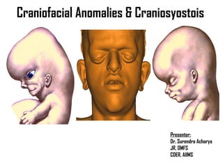
- 1. Craniofacial Anomalies & Craniosyostois Presenter: Dr. Surendra Acharya JR, OMFS CDER, AIIMS
- 2. Brief development of face and skull Introduction Classification Etiology Cranisynostosis Screening and pre op evaluation Management Complications Conclusion References
- 3. Embryogenesis of face and skull
- 4. Embryogenesis of face and skull
- 5. Embryology of face and cranium
- 6. Embryology of face and cranium
- 7. Embryology of face and cranium
- 8. Embryology of face and cranium
- 9. Embryology of face and cranium
- 10. Embryology of face and cranium
- 11. Growth And Development of Cranifacial Complex: An Epigenetic Viewpoint The functional matrix theory: Cranial vs. cephalic. Functional cranial component(FCC): functional matrix(FM) + skeletal unit(SU). Functional matrices: Periosteal and capsular(Tenon’s capsule). Active and passive growth. The classical triad of cranial growth process: Role of suture Role of deposition and resorption Role of cephalic cartilage Neural regulation of functional matrices
- 12. Craniofacial anomalies Malformation: A morphological defect of an organ, part of an organ, or larger region resulting from an intrinsically abnormal developmental process. The risk of recurrence depends on the etiology of the malformation. Congenital anomalies: All forms of developmental defects present at birth, whether caused by genetic, chromosomal, or environmental factors. Dysmorphology: The study of congenital anomalies. The term dysmorphic describes an individual with obvious multiple and severe malformations.
- 13. Craniofacial anomalies Sequence: Pattern of multiple anomalies derived from a single structural defect or mechanical factor (Mobius sequence). Developmental field: A group of cells or a region that responds as a coordinated unit to embryonic interaction; defects in developmental fields result in multiple malformations (formation of the lens depends on interaction with the optic cup). Many malformations are field defects. Syndrome: Pattern of anomalies thought to be pathologically related. The term implies a single cause, not a field defect.
- 14. Anomalies: Something that deviates from what is standard, normal, or expected. Craniofacial abnormalities: Are birth defects of the face or head, affecting 2-3% of all babies. Some, like cleft lip and palate, are among the most common of all birth defects. Others are very rare. Most of them affect how a person's face or head looks. Craniofacial anomalies
- 15. Craniofacial anomalies ‘‘Craniofacial anomalies (CFA) are a diverse group of deformities in the growth of the head and facial bones. Anomaly is a medical term meaning "irregularity" or "different from normal." These abnormalities are congenital (present at birth) and there are numerous variations- some are mild and some are severe and require surgery’’.
- 16. Craniofacial anomalies Congenital anomalies (CA) are a major cause of infant mortality and childhood morbitity, affecting 2-3% of all babies. Approximately 1% of these newborns have syndromes or multiple anomalies; CFA are often a component part. Syndromes that have cleft lip and/or cleft palate as one of the features are of interest in the quest for etiologic and pathogenetic factors, and it is estimated that 30% of cleft cases are syndromic. Conversely, therefore, approximately 70% are non-syndromic
- 18. Craniofacial anomalies Proposed new classification system: Based on extensive experiences in treatment of craniofacial anomalies, classification suggested as below: 1. Cleft: centric and acentric 2. Synostosis: symmetric and asymmetric 3. Atrophy: hypoplasia 4. Hypertrophy: neoplasia 5. Unclassified
- 19. Classification of craniofacial anomalies Craniofacial anomalies can be broadly divided into three main subgroups: Craniofacial clefts Craniosynostoses Hemifacial microsomia Deformational plagiocephaly Treacher collins syndrome Miscellaneous craniofacial anomalies
- 21. Cephalic index
- 22. Cleft lip and palate Cleft lip and/or cleft palate Cleft lip and cleft palate are the most common congenital craniofacial anomalies seen at birth. Cleft lip. An abnormality in which the lip does not completely form. The degree of the cleft lip can vary greatly, from mild (notching of the lip) to severe (large opening from the lip up through the nose).
- 23. Cleft palate
- 25. Kernahan and Stark classification: 1.Primary palate includes those structures anterior to the incisive foramen (lip, pre-maxilla, anterior septum). 2.Secondary palate includes those structures posterior to the incisive foramen (lateral palatine shelves, soft palate, and uvula)
- 26. Timing of cleft lip and palate repair 1. Age 3 months - Repair of CL (and placement of ventilation tubes) 2. Age 6 months – Pre surgical orthodontics, if necessary; first speech evaluation 3. Age 9 months - Speech therapy begins 4. Age 9-12 months - Repair of CP (placement of ventilation tubes if not done at the time of CL repair) 5. Age 1-7 years - Orthodontic treatment 6. Age 7-8 years - Alveolar bone graft 7. Older than 8 years - Orthodontic treatment continues
- 27. Craniosynostosis The premature fusion of the sutures between the growth plates in an infant's skull that prevents normal skull expansion which can cause an abnormally shaped skull. Premature closure of all the sutures can cause microcephaly (an abnormally small head), which prevents the normal growth of the brain and results in mental retardation.
- 28. Craniosynostosis
- 29. Craniosynostosis 1. Primary: a. Isolated (mostly sporadic) b. Associated anomalies: I. limb defects(mostly single gene defect) II. Limb + others defects (single gene + chromosomal syndromes) 2. Secondary: a. CNS malformation b. Metabolic disorder c. Others disorder
- 30. Types of craniosynostosis Primary craniosynostosis : found as an idiopathic developmental error occurring in otherwise normal individuals. It also occurs as part of complex syndromes involving other developmental aberrations; such syndromes often show Mendelian inheritance. It should be noted, however, that there is no familial incidence in the large majority of cases of primary craniosynostosis.
- 31. Types of craniosynostosis 2. Secondary craniosynostosis :A failure of brain growth as in microcephaly or an encephaloclastic process occurring during the first years of life will result in premature fusion of the cranial sutures. A similar process may also be seen when severe hydrocephalus has been treated with a low-pressure shunt. Metabolic craniosynostosis
- 32. Types of craniosynostosis 3. Metabolic craniosynostosis : results from premature sutural fusion determined by obvious biochemical disorders such as the mucopolysaccharidoses, rickets, hypophosphatasia or hypercalcaemia.
- 33. Symptomatology of craniosynostosis Raised intracranial pressure Exorbitism and orbitostenosis Orbital hypertelorism Orbital hypotelorism Orbital dystopia Midface hypoplasia Airway restriction Effects on speech Impair normal mastication.
- 34. Ocular complications Proptosis or exophthalmos Increased inner and outer canthal distances Increased interpupillary distance palpebral fissure size position of the lacrimal puncta obliquity of the palpebral fissure and asymmetry of orbits and orbital structures.
- 35. Some common syndrome with Craniosynostosis I. Single gene disorders: Autosomal dominant: 1. Crouzon syndrome (coronal, Sagital) 2. Apert syndrome (coronal, sagital and lamdoid 3. Pfeiffer syndrome: type 1(coronal and sagital), type 2- all sutures type 3- all sutures Autosomal recesive: 1. Carpenenter syndrome- all sutures
- 36. Some common syndrome with Craniosynostosis II. Chromosomal disorders: 1. Duplication 3q syndrome- coronal 2. Deletion 13q syndrome- metopic III. Teratogenic syndrome: 1. Valporic acid embryopathic- all 2. Fetal aminopterin syndrome- coronal and lambdoidal
- 37. Craniosynostosis PATHOGENESIS: The causes of craniosynostosis are known to be heterogeneous: Chromosomal Monogenetic and teratogenic syndromes; Nutritional deficiency Other syndromes of unknown genesis.
- 38. Craniosynostosis PATHOGENESIS: Most cranial and facial sutures do not close until adulthood, but a few synostose spontaneously early in development (premaxial, maxiofacial and metopic). The reason for this predictably age-related synostosis is unknown, but it has been proposed to be related to the functional environment of a particular suture and the need for adaptive skeletal change in the craniofacial area.
- 39. Causes for craniosynostosis Complex Multisuture Synostosis Secondary Synostosis Hematologic disorders • Congenital hemolytic icterus • Polycythemia vera • Sickle cell anemia • Thalassemia Iatrogenic disorders • Hydrocephalus with shunt Malformations • Encephalocele • Holoprosencephaly • Microcephaly Metabolic disorders • Hyperthyroidism • Rickets (various forms) • Mucolipidosis III
- 40. Causes for craniosynostosis Complex Multisuture Synostosis Secondary Synostosis Mucopolysaccharidoses and related disorders • D-mannosidase deficiency • ß-glucuronidase deficiency • Hurler syndrome • Morquio syndrome • Mucolipidosis III Teratogens • Aminopterin, Cyclophosphamide, Fluconazole,Retinoidse • Valproate
- 41. Ocular finding in craniosynostosis Vision Proptosis (sometimes called exorbitism) Strabismus Corneal exposure Restricted eye ball movement Increased ICP papilledema, optic atrophy, corneal exposure, or amblyopia secondary to strabismus or anisometropia Poor visual acquity Dacrocystitis
- 42. Proptosis
- 43. Evaluation 1. Assessment of craniofacial shape. 2. Movement or lack of movement of the calvarial bones during infancy. 3. The presence or absence of sutural ridging. 4. Can be confirmed by an X-ray of the skull or head CT examination. 5. In some instances, particularly with sagittal or coronal involvement, plain X- rays may suffice. If radiographic interpretation is equivocal, CT imaging is mandatory. 6. 25-26 weeks ultrasound detects syndrome
- 45. Syndromic craniosynostosis key points Syndromic craniosynostosis is rare, occurring in 1:30,000 to 1:100,000 live births. Fibroblast growth factor receptor and tumor growth factor-b receptor mutations have been reported to be associated with many forms of syndromic craniosynostosis.
- 46. Syndromic craniosynostosis key points Intracranial hypertension, developmental delays, and strabismus are more frequent in syndromic forms of craniosynostosis than isolated synostosis. Distraction osteogenesis is a useful adjunct in syndromic synostosis to increase intracranial volume and is helpful with fronto-orbital and midface advancements.
- 47. Syndromic craniosynostosis Addressing decompression by increasing intracranial volume and decreasing intracranial pressure before 1 year of age is a common goal through an interdisciplinary team approach.
- 48. Syndromic craniosynostosis More liable to have ventricular expansion, hydrocephalus, expanded subarachnoid space, and cerebellar tonsillar herniation compared with patients with sporadic single suture synostoses. Increased intracranial pressure (ICP) is more likely to occur in patients with syndromic craniosynostosis and multisuture synostosis.
- 49. Secondary Craniosynostosis Causes: 1.congenital 2.metabolic 3.iatrogenic 4.infectious Whatever the cause, the resultant underdeveloped brain fails to drive normal calvarial bone growth, cranial sutures fuse prematurely, and the head often remains microcephalic.
- 50. Secondary Craniosynostosis Blood disorders like thalassemia, sickle cell anemia, polycythemia vera can also cause cranial sutures to fuse prematurely. Iatrogenic craniofacial anomalies can occur in patients receiving ventricular shunts in infancy and early childhood. In children with patent cranial sutures over shunting of CSF leads to shunt-induced craniosynostosis and frequently microcephaly. Trauma and neoplasms rare causes of acquired craniofacial deformities.
- 51. Crouzon syndrome It is also called craniofacial dysostosis, has autosomal dominant transmission, but up to 50% of cases occur sporadically, representing fresh mutations. Clinical features 1. Cranium : exihibit calvarial deformity, the brachycephalic deformities predominate, but many cases of the syndrome show no obvious calvarial deformity, even when there are marked radiological abnormalities.
- 52. 2. Face: Midface hypolasia with relative mandibular prognathism, drooping lower lip and short upper lip are typical features. The nasal bridge is often flattened, and the tip of the nose may appear beak-like. There is deviation of the nasal septum in 35% and obstruction of the nasopharynx in 30% of cases. 3. Oral findings : A narrow high-arched palate, crowding of the dental arches and an anterior open bite. Ectopic eruption of the maxillary first molar teeth occurs in about half of the patients, and 35% are obligate mouth breathers. exhibit a cleft palate(3%) and 10% have a bifid uvula. Crouzon syndrome
- 53. 4. Eyes: Proptosis secondary to the shallow orbits. Divergent strabismus, nystagmus and hypertelorism are frequently found. Exposure conjunctivitis (50%), keratitis (10%), poor vision (45%) and optic atrophy (25%); rarely there is luxation of the globes. 5. Ears: More than 50% of patients have a conductive hearing loss associated with malformed auditory ossicles, and some 15% patients have atresia of the auditory canals. 6. Other anomalies : Stiffness of the joints, especially the elbows. Cervical spine anomalies occur in 30% of patients, and 85% exhibit calcification of the stylohyoid ligament Crouzon syndrome
- 54. Crouzon syndrome
- 55. Genetics: It has a clear autosomal dominant mode of inheritance, with about 67% of cases being familial. 44 Variability of expression characterizes Crouzon syndrome. Most of the mutations associated with Crouzon syndrome are located in FGFR2. Crouzon syndrome
- 56. Crouzon syndrome
- 57. It is characterized by craniosynostosis of coronal suture, midface hypoplasia and syndactyly of extremities. Its transmissions is believed to be autosomal dominant, but most cases are sporadic and caused by high neonatal mortality and reduced fitness of affected individuals. Blank estimated a frequency of 1 in 160,000 population, but Cohen believed the prevalence may be higher. Prenatal diagnosis is possible. Apert syndrome
- 58. Apert syndrome 1. Face : The facial dysplasia is severe, especially in older patients. maxilla is grossly hypoplastic, while the nose and mandible are relatively prominent. Facial asymmetry is sometimes present, and can be very pronounced. 2. Oral findings : high arched palate, constricted and may have a median furrow. The soft palate is cleft with bifid uvula. The maxillary dental arch V-shaped, with severe dental crowding and bulging alveolar ridges. A skeletal class III malocclusion and an anterior open bite. Retarded dental eruption is common. All these deformities, together with mental impairment, frequently combine to impair speech.
- 59. Apert syndrome 3. Skeletal system: syndactyly of digits two, three and four is always found; in addition, digits one and five may be joined to digits two and four respectively. The interphalangeal joints of the fingers are stiff, while fingernails of the mid-digital hand mass may be continuous or partly continuous. In the feet, toes two, three and four are joined by soft-tissue syndactyly; toes one and five may either be joined by soft-tissue syndactyly to the second and fourth toes respectively.
- 60. Apert syndrome The upper extremities are shortened, and there may be aplasia or ankylosis of several joints, especially the elbow, shoulder and hip. Progressive synostosis of the bones of the hands, feet and cervical spine have been reported 4. Eyes: Hypertelorism is common, and there is usually some degree of proptosis. All degrees of orbitostenosis are seen, but it is not generally as severe as in Crouzon syndrome. The palpebral fissures may show an antimongoloid slant, congenital glaucoma, abnormal postiotion, origin ans insertion of extra occular muscles.
- 61. Apert syndrome
- 62. Apert syndrome Fronto orbital advancement
- 64. Apert syndrome Middle third advancement A:Le Fort III osteotomy line; B: external distractor device applied in a patients after a Le Fort III procedure. C: internal distractor device applied in a patient after Le Fort III osteotomies.
- 65. Apert syndrome
- 66. Apert syndrome
- 67. Pfeiffer’s syndrome In 1964, Rudolf Arthur Pfeiffer It is similar to Apert syndrome but also is characterized by very shallow orbits (already part of Apert) and short broad thumbs and toes. Corneal exposure is an important problem in these children. Types: Pfeiffer’s syndrome types 2 and 3 are more common than 1
- 69. Pfeiffer’s syndrome It is similar to Apert syndrome but also is characterized by very shallow orbits (already part of Apert) and short broad thumbs and toes. Corneal exposure is an important problem in these children. Types: Pfeiffer’s syndrome types 2 and 3 are more common than 1
- 70. Pfeiffer’s syndrome Type 1: exhibit a spectrum of craniofacial involvement ranging from moderate to severe midfacial hypoplasia. Skeletal features encompass broad and medially deviated thumbs and great toes; variable degrees of brachydactyly can occur as well. Others include hearing loss or hydrocephalus. Intellect is usually normal
- 71. Type 2: Craniofacial involvement in Pfeiffer’s syndrome type 2 is more severe that in type 1, with cloverleaf skull and extreme proptosis (often to the point of inability to close eyelids laryngotracheal abnormalities, hydrocephalus, seizures, and increased risk for early death.Ankylosis of elbow and knee. Type 3: is almost identical to type 2; however, the skull shape is turribrachycephalic. Pfeiffer’s syndrome
- 72. • Craniosynostosis with turribrachycephaly (short and tall head) • Ocular hypertelorism (wide spread eyes) • Midface defficiency . • Hypoplstic maxilla • Prominent lower jaw • Dental abnormalities
- 73. Broad Thumbs
- 74. Broad great toes
- 75. Pfeiffer syndrome 1. Cranium: Turricephaly is the commonest deformity, being associated with premature fusion of the coronal sutures. Other sutures may be involved, and cases with trigonocephaly and cloverleaf skull. Intelligence is usually normal, but mental retardation does occur, being most severe in those cases associated with clover-leaf skull.
- 76. Pfeiffer syndrome 2. Face: Maxillary hypoplasia with relative mandibular prognathism is common, and the ears are frequently low-set. Facial asymmetry, orbital hypertelorism, antimongoloid palpebral fissures, proptosis and strabismus have all been reported. Oral findings include a high-arched palate, dental malocclusion and, rarely, a bifid uvula.
- 77. Pfeiffer syndrome 3. Hands and feet : The thumbs and great toes are broad, with varus deformity. In some patients the great toes may be shortened. Cutaneous syndactyly is usually present, involving digits two and three, and at times three and four, of both hands and feet. Clinodactyly and symphalangism of both hands and feet have been reported. Other skeletal anomalies described include fused cervical vertebrae,
- 78. Pfeiffer syndrome 4. Other anomalies : Other features occasionally seen are pyloric stenosis, bicuspid aortic valve, hypoplasia of the gallbladder, single umbilical artery, umbilical hernia, preauricular tags, choanal atresia and hearing loss.
- 79. Genetics: Inheritance is autosomal dominant with complete penetrance and variable expressivity. Mutations causing Pfeiffer’s syndrome have been found on FGFR1 and FGFR2. A single common missense mutation, Pro250Arg, has been associated in five unrelated families with a relatively mild form of Pfeiffer’s syndrome Pfeiffer’s syndrome
- 80. Carpenter’s syndrome It is also known as acrocephalopolysyndactyly type II, is a variation of Apert syndrome characterized by preaxial polydactyly on the side of the thumb or big toe, syndactyly, brachycephaly, synostosis, obesity, hypogonadism, mental retardation, shallow orbits, proptosis, and laterally placed intercanthi. GENETICS Inheritance is autosomal recessive and the molecular basis is unknown to date. In all cases, examination of the parents has been normal.
- 85. A condition in which the tissues on one side of the face are underdeveloped, affecting primarily the ear (aural), mouth (oral), and jaw (mandibular) areas. Sometimes, both sides of the face can be affected and may involve the skull, as well as the face. Hemifacial microsomia
- 86. Hemifacial microsomia is also known as Goldenhar syndrome, brachial arch syndrome, facio-auriculo-vertebral syndrome, oculo-auriculo-vertebral spectrum, or lateral facial dysplasia. Defect of first and second brachial arch Hemifacial microsomia
- 87. Deformational (or positional) plagiocephaly. A misshapen (asymmetrical) shape of the head (cranium) from repeated pressure to the same area of the head. Plagiocephaly literally means "oblique head" (from the Greek "plagio" for oblique and "cephale" for head).
- 89. History and clinical examination Routine investigations Cross matching and blood arrangement Anesthetic consideration X- ray skull, AP, Lateral, Oblique, Vertex NCCT face and CECT or MRI face and neck 3 D virtual planning Pre operative evaluation and preperation
- 90. Anesthetic challenges Nasal fiberoptic bronchoscopy was contraindicated and regional nerve block was not a feasible option because of obstructed breathing pattern. Oral fiberoptic bronchoscopy-guided intubation with good airway anesthesia and sedation was successfully managed in the next attempt.
- 91. Anesthetic challenges These patients often have nasal airway obstruction due to mid-face hypoplasia and a high arched palate. incidence of C2,3 spinal fusion. Orbit manipulation during these procedures can cause marked bradycardia. Anesthetic concerns in caring for these complicated patients are airway abnormalities, adequate vascular access for hemorrhage and monitoring and also heat loss. Preoperative sedation to a patient with a compromised airway should be individualized and used cautiously.
- 92. Anesthetic challenges Airway management difficulty should be predicted and planned for in advance to avoid urgent problems. Options should include availability of oral airways, oversized masks, fiberoptic bronchoscope, a variety of laryngoscope blades and lighted stylets. Control of the airway may involve awake, fiberoptic intubation. Inhalation induction of anesthesia is preferred over intravenous. Paralysis with neuromuscular agents should be avoided until controlled mask ventilation is assured.
- 93. Anesthetic challenges Blood loss during these procedures can be excessive with 1 to 2 blood volumes often required. Adequate vascular access with heated intravenous solutions are mandatory. The use of erythropoietin injections and additional iron supplements started 3 weeks prior to elective craniofacial surgery has been shown to modify transfusion requirements. An arterial line is required to monitor the hemodynamic and laboratory values in these patients.
- 94. Anesthetic challenges Use of central venous catheters to monitor volume status and precordial doppler to detect venous air emboli should be individualized to the procedure and patient. Postoperative care must continue in a closely monitored environment (intensive care unit) to continually access hemodynamic and respiratory parameters. Pediatric patients with craniofacial abnormalities continue to provide the anesthesiologist with some of the most challenging cases that we see today. These difficult cases need careful planning and cooperation from the many professionals involved in the care of these patients.
- 95. Treatment of the craniosynostoses The aims of early craniofacial surgery: 1. To allow the brain to expand normally 2. To provide a normal shape to the forehead and skull 3. To provide eye protection by reducing the exorbitism . 4. To prevent or minimize the problem of impaired facial growth in facial stenosis.
- 96. Timing of surgery for patients with syndromic craniosynostosis depends on the presenting symptoms, such as the degree of exorbitism and signs and symptoms of elevated ICP. There is no clear consensus on the ideal operative window for syndromic craniosynostosis; however, delaying surgery beyond 1 year results in a higher likelihood of elevated ICP, cognitive deficits, and behavioral problems.
- 97. Timing of surgery Less then 6 month or less tha 12 months for primary surgery isolated nonsyndromic craniosynostosis. Craniofacial clinic visit: at presentation; during any preoperative planning; and postoperatively at 2 weeks, 3 months, 12 months, and annually until the patient reached the age of 6 years.
- 98. Timing of surgery for Pfeiffer syndrome Mostly early craniosynostosis released to be done for increased ICP. Hydrocephalus need to be treated by putting a stent. Lefort III distraction when patient is 5-7 years of age. Orthognathic surgery is indicated after skeletal maturity. There is no review for optimum timing for surgery. The potential for brain compression, loss of vision through exposure keratoconjunctivitis, significant airway compromise, and psychosocial maladjustment may dictate the timing of surgery
- 99. Apert syndrome Step 1 — from birth to age 2 Step 2 — growth period Step 3 — adult age. Monobloc advancement or Le Fort III osteotomy and facial bipartition are performed at 6-7 years, midface Le Fort III advancement at 9-12 years.
- 100. 1. Prone position and 2. The neck in neutral position. 3. A zig-zag bicoronal 4. Incision was created, and extensive scalp flaps were developed in the subgaleal plane. 5. Barrel-stave osteotomies were then created in the frontal, parietal, temporal, and occipital squamous bones, which were then outfractured 6. Tension, and a Jackson-Pratt drain was left in the subgaleal space at closure. Operative technique
- 101. It can be used to advance the mandible, midface, orbits, and skull including lower level and higher level Le Fort osteotomies, PCD, or fronto-facial (Monobloc) advancements using either external or internal distractors. Cranial distraction has been used to increase microcephalic skulls or provide added volume in the case of shunt-induced craniosynostosis. Typically an open bicoronal approach is used because eventual anterior cranial vault reconstruction (ACVR) with FOA and frontal cranioplasties are usually required months after distraction Distraction osteogenesis
- 102. It can be used to advance the face alone when coupled with Le Fort I, II, and III osteotomies or to advance the face and frontal skull as in Monobloc advancement, which can be used as early as 4 years of age if deemed necessary because of neurosurgical, ophthalmologic, or airway issues or later in the teenage years to improve exorbitism, midface hypoplasia, and skeletal occlusal relationships that are poor. Distraction osteogenesis
- 103. Mid face advancements can be done between 4 years of age and adulthood to address skeletal hypoplasia and functional problems related to sleep-disordered breathing, OSA, malocclusion, and feeding.
- 104. A 6 year old with syndromic facies; no exorbitism; but mild midface hypoplasia, anterior open bite, and anterior crossbite with relative normocephaly (cephalic index 85%). 3D CT images reveal pansynostosis, lateral plain film shows thumb-printing (nonspecific for increased ICP), and sagittal CT shows Chiari malformation (asymptomatic). Genetic testing was negative for common fibroblast growth factor receptor and TWIST mutations.
- 105. Distraction osteogenesis External arm of distractor removal after 3-5 weeks and distractor removal after 2-3 months at the time of ACVR
- 106. Frontal, lateral, vertex, and occipital views with three-dimensional computed tomography demonstrating nonsynostotic positional brachycephaly, although mild metopic ridging is evident without significant trigonocephaly.
- 107. A 2 year old syndromic patient with hydrocephalus s/p shunt who developed secondary multisuture craniosynostosis with resultant severe brachyturricephaly and underwent PCD. (A–H) Postshunt and predistraction photographs and 3D CT. (I) Intraoperative photograph demonstrating posterior craniotomies and distractor placement. (J) Immediately postoperative 3D CT scan. (K, M) Right, left, vertex 3D CT scan after consolidation phase. (L, N, O) Four weeks postdistractor removal, lateral and vertex photographs. s/p, status post.
- 108. Axial CT scan. (A) Predistraction and (B) postdistraction demonstrating improved intracranial dimensions and improvement in appearance of ventricles and gyri/sulci.
- 109. A 5 year old with benign intracranial hypertension, increased ICP, and pansynostosis but relative normocephaly who underwent VP shunt but had persistent ICP elevation. (A) Right lateral photograph after VP shunt. (B) Right lateral 3D CT scan depicting pansynostosis before shunt. (C) Right lateral 3D CT scan after completion of PCD consolidation. (D) At time of distractor removal noted improvement in intracranial volume but severe occipital shelf deformity. (E) One week postoperative after removal of distractors and posterior cranial vault reconstruction with autologous bone graft repositioning to improve occipital shape. VP, ventriculoperitoneal.
- 110. A 13 year old with autosomal-dominant FGFR craniosynostosis. (A) Left lateral preoperative photograph demonstrating severe brachycephaly, midface hypoplasia, and exorbitism. (B) Preoperative left lateral 3D CT showing pansynostosis. (C) Two weeks postoperative with rigid external distractor in place for Monobloc distraction. (D, E) Left lateral photograph and 3D CT 2.5 years postoperative demonstrating improved midface and decreased proptosis despite some deficient cranial bone in region of frontal cranial distraction.
- 111. A 2-month-old girl with brachyturricephaly and bicoronal synostosis, with midface hypoplasia.
- 112. A 9 month old with partial bicoronal synostosis, midface hypoplasia, and exorbitsim with underlying spontaneous FGFR2 mutation. (A, C, E) Preoperative frontal, left lateral, and vertex photographs at 9 months. (B, D, F). Frontal, left lateral, and vertex 3D CT after ICP pressure monitor performed. Results revealed a mild elevation of ICP. (G) Left lateral intraoperative photograph at time of PCD showing left internal posterior cranial distractor. (H) Left lateral skull radiograph after latency phase before beginning active distraction. (I) Left lateral 3D CT midway through active distraction when patient developed CSF leak, external ventricular drain placed, and endoscopic third ventriculostomy performed shortly thereafter. (J) Left lateral skull radiograph at conclusion of 30-mm active distraction. (K, L) One year after distractors removal and ACVR.
- 113. Risks of distraction osteogenesis: Second surgery Infection Soft tissues laceration around the distracters Dural tear CSF leak Steps off deformity Non union Distraction osteogenesis
- 114. Sagital synostosis(scaphocephaly) Golf tee deformity- frontal bossing with biparietal narrowing. Early surgery gives best results. Surgical techniques are: Pi squeeze technique Zenker’s solution Strip craniotomy Conceptual modification of the pi technique is the T technique Reverse pi and Y technique Radical cranial reconstructive surgery for older child
- 125. Lamdoidal synostosis ▣Timing of surgery 6-12 months ▣Surgical techniques: Radial fan cuts Transposition technique Tiara technique
- 126. Lamdoidal synostosis
- 127. Lamdoidal synostosis
- 128. Lamdoidal synostosis
- 129. Oxycephaly
- 130. Oxycephaly Characterized by pointed skull Classifications: Type I congenital form with limb anomalies Type II delayed oxycephaly Type III false oxycephaly Features: absence of facial anomalies, normal ethmoidal complex, major claverial anomalies, absence of temporal overgrowth. Increased ICP, mentall retardation, exorbitism, visual loss, hydrocephalus, epilepsy,
- 131. Oxycephaly
- 132. Oxycephaly Surgical technique: linear craniotomies and fragmentation of vault Total craniotomies Craniofacial technique Shaving, skin incision, periosteum and temporal muscle, frontal osteotomies, supraorbital ridge bandeu, z plasty,
- 133. Plagiocephaly
- 134. Plagiocephaly
- 135. Plagiocephaly
- 144. Management total facial deficiency ▣Lefort III osteotomy ▣Distraction osteogenesis ▣Monobloc ▣Facial bipartiton ▣Fronto-orbital advancement
- 145. Lefort III osteotomy Gillies and Harrison performed the first successful Le Fort III. Twenty years later, Tessier revisited, refined, and eventually popularized the operation.
- 147. Surgical Technique
- 148. • Infiltration of LA with Adr (1:200000) • Oro-endotracheal intubation is preferred) • Hypotensive anesthesia • Intraoperative steroid and antifibrinolytic injection. Step 1: Anesthesia and Preparation:
- 149. Step 2: Tarsorapphy
- 150. Step 3: Surgical approach 1) Bicoronal 2) Subcilliary /subtarsal 3) Maxillary vestibular approach.
- 151. Surgical Technique Subcranial Lefort III osteotomy 1. Zygomatic osteotomy 2. Lateral orbital wall osteotomy 3. Frontozygomatic sutures 4. Orbital floor 5. Nasofrontal suture 6. Medial orbital wall 7. Vomer(nasal septal) osteotomy 8. Pterygoid dysjunction 9. Disimpaction by forceps 10. Resuspention and forced duction test 11. Distractor placement
- 152. Step 4: Zygomatic osteotomy
- 154. Step 5 : Lateral orbital wall osteotomy
- 155. Step 6: Nasofrontal osteotomy
- 156. Step 7: Medial orbital wall osteotomy
- 157. Step 8: Nasal Septal Osteotomy
- 158. Step 9: Pterygoid Dysjucntion
- 159. Step 10: Maxillary Dysimpaction
- 162. General guidelines For sub-cranial Le Fort III external distractor placement are as follows: (1) Adjust the transverse width of the device, allowing the lateral arm to extend wider than the scalp or hair by 2 cm bilaterally. (2) The halo should be aligned parallel to the Frankfort horizontal plane with the vertical bar component approximately 3 cm anterior to the upper lip. (3) Hand-turn the cranial screws sequentially bilaterally until bone is Contacted on a minimum of four screws per side. (4) Place the two horizontal bars at the desired vector of anterior distraction corresponding to the distraction plates and/or intraoral appliance. Closure and Final Distractor Placement
- 164. • DO was initiated after 1 week. • The rate of distraction was 1 mm per day in 2 daily activations. • The duration of DO depended on the desired advancement. • During the distraction period, vector modifications took place when necessary in patients treated with an external distractor. • In all patients a consolidation period of three months after distraction was respected Lefort 3 Distraction protocol
- 165. Hemorrhage result primarily from the pterygomaxillary dysjunction and down-fracture Atypical fracture that extends to the skull base, orbit and pterygoid plate Unexpected skull fracture. Lethal subarachnoid hemorrhage after Le Fort III osteotomy followed by fracture of base of skull Intra-operative Complication
- 166. CSF leak Orbital hemoatoma Chemosis Minor complications include Cutting the infra-orbital nerve Fracturing of zygoma during mobilization Partial exposure of the nasal bone Intra-operative Complication
- 167. Major complications include respiratory distress requiring tracheotomy, Subgaleal hematoma Cerebro-spinal fluid leakage and fistula Visual loss after retro-orbital hemorrhage Strabismus Partial anosmia, Post operative complication related to Lefort III osteotomy
- 168. Ptosis Esthetic Unavoidable most important shortcoming is the tendency of medial canthi to drift away from nose. Widening of intercanthal distance Post operative Complications late complication related to Lefort III osteotomy
- 169. Pin loosening of halo frame (20%) Pin infection (7%) Intracranial Migration of pin Complication related to rigid external distraction device
- 170. Risks of early craniotomy ▣Blood loss ▣Intravascular air ▣Cardiovascular effects ▣Infection ▣Pressure necrosis
- 171. Genetic counseling
- 172. Genetic counseling
- 173. Genetic counseling
- 174. Take home message It can be said that diagnostic and therapeutic planning in patients with craniosynostosis emphasizes the need to integrate various specialties. It is necessary to frame the patient’s clinical picture upon his arrival. Very important are also a multi-disciplinary plan and clinical programmed check-ups in accordance to a rational therapeutic clinical widely agreed upon and verified in its efficiency. As noted in this paper establishing an integrated and tailored surgery timing, scheduling, combining and coordinating actions to be taken at different stages of the patient’s age reduces the number of general anesthesia thus simplifying therapy for both patients and their families.
- 175. References Craniofacial Anomalies: Growth and Development From a Surgical Perspectives By James Tait Goodrich, Craig D. Hall. Maria T. et al. Treatment timing and multidisciplinary approach in Apert syndrome. Anand R. et al. Advances in the Treatment of Syndromic Midface Hypoplasia Using Monobloc and Facial Bipartition Distraction. Semin Plast Surg. 2014;28:179–183. Distraction osteogensis of facial skeleton:-William H Bell Distraction of Craniofacial Skelton :- Mc-carthy Orthognathic surgery: Principle & Practice:- J.C Posnick Atlas of oral and Maxillofaical surgery :- D. Kademani Atlas of oral and maxillofacial surgery clinics of north America, Volume 16 september 2008 Various article
Editor's Notes
- (genetic, teratogenic).
- b/o etiology anatomy and tratment principles
- causes: Combination of genes. Environmental. Folic acid deficiency.
- Medicine.net
- These measurements serve two purposes. First, they prevent errors of, secondary to abnormal size, shape,or position of bony and soft tissue changes in orbital structures recording false impressions such as pseudohypertelorism caused A more accurate and reproducible method of measuring orbital separation is the radiologic measurement of the bony intraorbital distance. by soft tissue changes in the canthal area such as telecanthus. In addition, they provide useful data for the study of the syndrome characteristics and serve as a baseline if reconstructive surgery is performed.
- monobloc
- Type I
- All sutures
- All sutures
- All sutures
- All sutures
- Plagiocephaly, also known as flat head syndrome, is a condition characterized by an asymmetrical distortion (flattening of one side) of the skull. It is characterized by a flat spot on the back or one side of the head caused by remaining in a supine position for too long.
- Plagiocephaly, also known as flat head syndrome, is a condition characterized by an asymmetrical distortion (flattening of one side) of the skull. It is characterized by a flat spot on the back or one side of the head caused by remaining in a supine position for too long.
- Plagiocephaly, also known as flat head syndrome, is a condition characterized by an asymmetrical distortion (flattening of one side) of the skull. It is characterized by a flat spot on the back or one side of the head caused by remaining in a supine position for too long.
- Four wall osteotomy
- rotation
- rotation
- rotation
- , and the tube is secured with wire adjacent to the incisal edge of the mandibular central incisors or is sutured to the labiomental fold region.(south pole tube fixation
- A tunneling technique for exposure of
- 1cm
- The first osteotomy is the zygomatic osteotomy via a vertical cut at the body of the zygoma.
- The nasal septal osteotome is used to separate the nasal septum from the cranial base. The direction of the osteotome is parallel to the base of the skull and directed toward the posterior nasal spine as palpated transorally
- The inferior half of the pterygomaxillary junction is separated bilaterally using a curved pterygoid osteotome approach via the temporal fossa The inferior half of the pterygomaxillary junction is separated bilaterally using a curved pterygoid osteotome approach via the temporal fossa
- , can be managed by re-impacting the maxilla