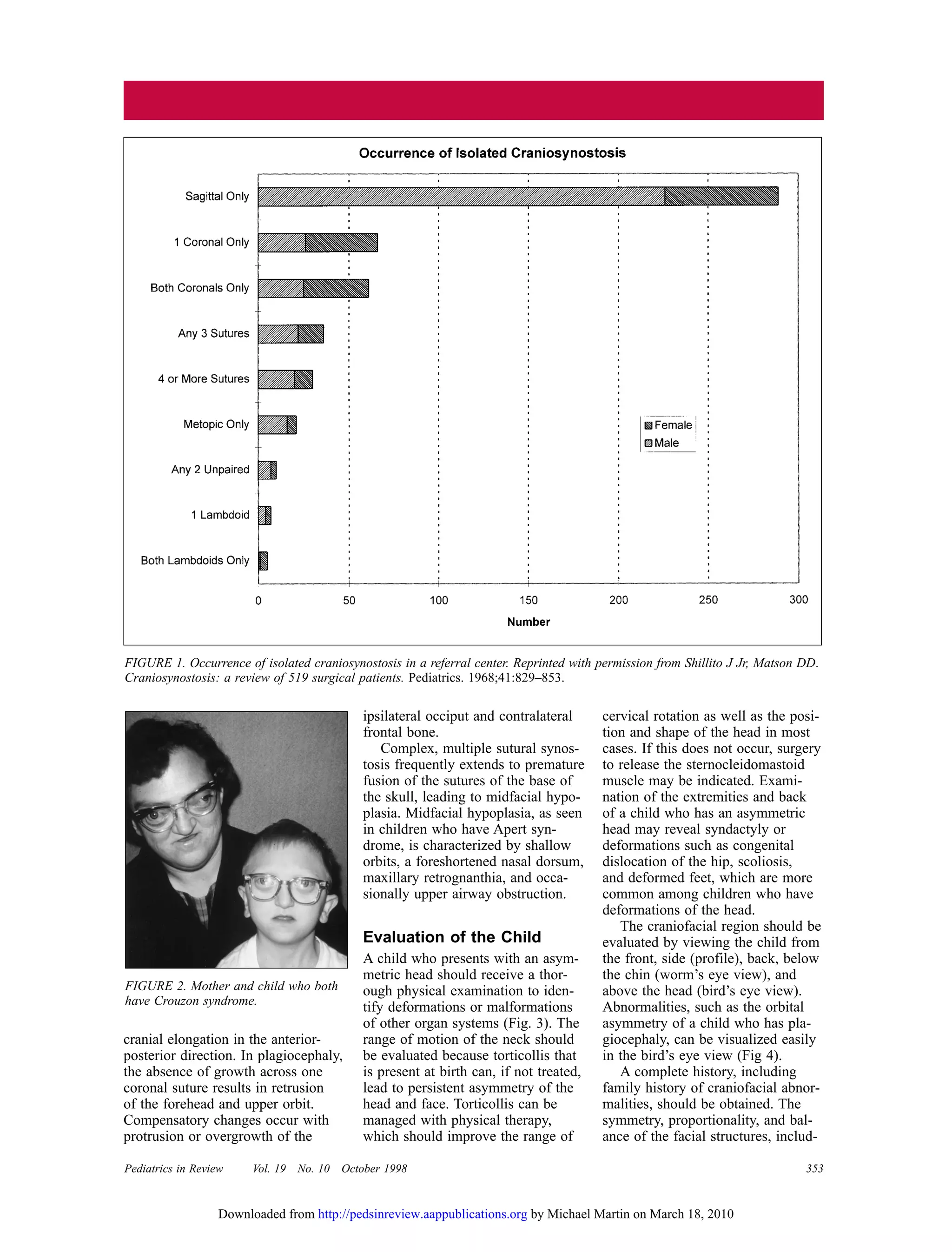
2. Consultation with
352 Pediatrics in Review Vol. 19 No. 10 October 1998
Occurrence
Asymmetry of the head is a com-
mon occurrence in infants and may
have a number of etiologies, includ-
ing positional deformity, simple
craniosynostosis, complex cranio-
facial synostosis, metabolic bone
disease, depressed skull fracture,
microcephaly due to brain damage,
and hydrocephalus. Deformations
of the head attributable to prenatal
compression usually resolve by the
time a child is 2 months of age.
However, a number of centers have
reported a recent increase in the
occurrence of cranial deformations
in infants older than 6 months,
which is attributed to the current
recommendation that children sleep
on their backs to avoid sudden
infant death syndrome.
Synostosis of a single suture may
lead to only cosmetic changes, but
synostosis of several sutures may be
life-threatening and lead to increased
intracranial pressure, loss of vision,
and severe deformities that affect
social interactions. Primary cranio-
synostosis, which is defined as
synostosis without an underlying
abnormality of the brain or meta-
bolic defect, occurs in 1 in 2,000
births. Secondary craniosynostosis,
such as would be due to cerebral
atrophy, is somewhat less common.
Figure 1 shows the occurrence of
isolated craniosynostosis in a refer-
ral center. (“Isolated” in this context
means involvement of the sutures
only without an identified genetic or
chromosomal abnormality such as
Apert syndrome.) Involvement of
the sagittal suture is the most com-
mon form and occurs more fre-
quently in males than in females.
Synostosis of a single suture is more
common than synostosis of multiple
sutures.
Etiology
Secondary craniosynostosis may be
due to a structural cerebral abnor-
mality such as atrophy or hypoplasia
and may be related to other con-
ditions such as hyperthyroidism,
severe anemia (eg, thalassemia), or
a metabolic condition (eg, muco-
polysaccharidosis). Primary cranio-
synostosis has been related to
intrauterine constraint, such as
occurs with twinning or with uterine
abnormalities such as a bicornuate
uterus. Exposure to maternal smok-
ing or high altitude during gesta-
tion has been associated with an
increased occurrence of cranio-
synostosis. Genetic abnormalities
such as Apert syndrome and chro-
mosomal anomalies such as abnor-
malities of chromosome 13q can
cause craniosynostosis. Some occur-
rences of isolated craniosynostosis
are familial (eg, involvement of
both coronal sutures), but most
cases do not have a clear etiology.
A number of discrete genetic con-
ditions produce craniosynostosis,
such as Crouzon syndrome (Fig. 2).
One of the most exciting develop-
ments in the understanding of cran-
iosynostosis has come through the
discovery that at least four genetic
syndromes associated with cranio-
synostosis (Table 1) are related
to abnormalities of fibroblast
growth factor receptor 2. In fact,
all four of the syndromes shown
in Table 1, which are autosomal
dominant, have abnormalities of
the same region of chromosome
10, despite having significant
differences in phenotype. Although
these entities account for only 5%
of children who have craniosynos-
tosis, this knowledge may increase
the understanding of other types
of craniosynostosis as well as the
role of fibroblast growth factors
and may lead to interventions to
prevent or ameliorate the effects
of these genetic anomalies.
Growth of the Skull
Cranial growth occurs as a direct
result of the growing brain. If all
cranial sutures are open, the brain
grows normally without areas of
restriction, and skull growth and
shape occur normally. Without
adequate stimulation from the brain
and other intracranial soft tissues,
the skull will not grow normally.
For example, an infant who has
severe cerebral atrophy will develop
microcephaly and premature closure
of all sutures because of the lack of
stimulation of the growing brain
upon the surrounding skull.
Once a suture becomes fused,
growth perpendicular to that suture
becomes restricted, and the fused
bones act as a single bony structure.
With failure of growth across a
single fused suture, compensatory
growth occurs at the remaining
open sutures to accommodate the
growing brain. These compensatory
changes allow continued unrestricted
brain growth without the develop-
ment of increased intracranial pres-
sure in most instances. This combi-
nation of restricted perpendicular
growth and compensatory changes
produces the typical deformities
seen with each type of single suture
synostosis. In scaphocephaly, for
instance, there is no growth per-
pendicular to the sagittal suture,
which results in bitemporal narrow-
ing. Compensatory changes cause
Pediatric Approach to Craniosynostosis
Gregory S. Liptak, MD, MPH* and Joseph M. Serletti, MD†
*Associate Professor of Pediatrics.
†Associate Professor of Surgery, University
of Rochester Medical Center, Rochester, NY.
by Michael Martin on March 18, 2010http://pedsinreview.aappublications.orgDownloaded from
3. Pediatrics in Review Vol. 19 No. 10 October 1998 353
cranial elongation in the anterior-
posterior direction. In plagiocephaly,
the absence of growth across one
coronal suture results in retrusion
of the forehead and upper orbit.
Compensatory changes occur with
protrusion or overgrowth of the
ipsilateral occiput and contralateral
frontal bone.
Complex, multiple sutural synos-
tosis frequently extends to premature
fusion of the sutures of the base of
the skull, leading to midfacial hypo-
plasia. Midfacial hypoplasia, as seen
in children who have Apert syn-
drome, is characterized by shallow
orbits, a foreshortened nasal dorsum,
maxillary retrognanthia, and occa-
sionally upper airway obstruction.
Evaluation of the Child
A child who presents with an asym-
metric head should receive a thor-
ough physical examination to iden-
tify deformations or malformations
of other organ systems (Fig. 3). The
range of motion of the neck should
be evaluated because torticollis that
is present at birth can, if not treated,
lead to persistent asymmetry of the
head and face. Torticollis can be
managed with physical therapy,
which should improve the range of
cervical rotation as well as the posi-
tion and shape of the head in most
cases. If this does not occur, surgery
to release the sternocleidomastoid
muscle may be indicated. Exami-
nation of the extremities and back
of a child who has an asymmetric
head may reveal syndactyly or
deformations such as congenital
dislocation of the hip, scoliosis,
and deformed feet, which are more
common among children who have
deformations of the head.
The craniofacial region should be
evaluated by viewing the child from
the front, side (profile), back, below
the chin (worm’s eye view), and
above the head (bird’s eye view).
Abnormalities, such as the orbital
asymmetry of a child who has pla-
giocephaly, can be visualized easily
in the bird’s eye view (Fig 4).
A complete history, including
family history of craniofacial abnor-
malities, should be obtained. The
symmetry, proportionality, and bal-
ance of the facial structures, includ-
FIGURE 1. Occurrence of isolated craniosynostosis in a referral center. Reprinted with permission from Shillito J Jr, Matson DD.
Craniosynostosis: a review of 519 surgical patients. Pediatrics. 1968;41:829–853.
FIGURE 2. Mother and child who both
have Crouzon syndrome.
by Michael Martin on March 18, 2010http://pedsinreview.aappublications.orgDownloaded from
4. 354 Pediatrics in Review Vol. 19 No. 10 October 1998
ing the nose, ears, and eyebrows,
which constitute the “aesthetics” of
the face and skull, should be evalu-
ated. A systematic approach that
enables thorough evaluation involves
separating the face and skull into
regions and evaluating each region
separately. These regions are: 1) the
forehead and upper orbit, 2) the
orbito-naso-zygomatic area, 3) the
maxillomandibular area, and 4) the
posterior cranial vault.
The physical examination can
suggest whether craniosynostosis is
present. This is sometimes obvious
from the shape of the skull. Certain
skull shapes are characteristic for
their affected suture. For example, a
keel-shape deformity of the central
forehead indicates trigonocephaly
or metopic synostosis (Fig. 5a);
a flattened occipital area may indi-
cate premature closure of the lamb-
doidal sutures (Fig. 5b); a long,
narrow cranium suggests scapho-
cephaly or sagittal synostosis (Fig.
5c); and a unilaterally recessed fore-
head and upper orbit suggests pla-
giocephaly or unilateral coronal syn-
ostosis (Fig. 5d).
The presence of a palpable bony
ridge in the location of a cranial
suture strongly suggests synostosis.
If computed tomography (CT) is not
TABLE 1. Autosomal Dominant Genetic Syndromes
Associated With Abnormalities of Fibroblast Growth Factor Receptor 2 (FGFR2)
LOCATION OF ABNORMALITY
CONDITION (CHROMOSOME/GENE) PRIMARY CHARACTERISTICS
Apert syndrome 10q25.3-q26/FGFR2 Exon 7(U) Acrocephaly, brachycephaly, flat face,
(Acrocephalosyndactyly, large fontanelle, shallow orbits,
type I) hypertelorism, down-slanting pal-
pebral fissures, maxillary hypoplasia,
narrow palate, syndactyly, short
fingers, broad thumbs and great
toes, fusion of cervical vertebrae,
mental retardation (in 50%), acne
Crouzon syndrome 10q25.3-q26/FGFR2 Exon 9(B) Shallow orbits, hypertelorism, frontal
(Craniofacial dysostosis) bossing, maxillary hypoplasia,
curved beak-shaped nose, A-shaped
palate, conductive hearing loss
Jackson-Weiss syndrome 10q25.3-q26/FGFR2 Exon 9(B) Shallow orbits, hypertelorism, mid-
(Cephalo-syndactyly) facial hypoplasia, syndactyly,
broad great toe
Pfeiffer syndrome 10q25.3-q26/FGFR2 Exon 9(B) Brachycephaly, full high forehead,
(Acrocephalosyndactyly, and 8p11.2-p12/FGFR1 Exon 5 hypertelorism, up-slanting palpebral
type V) fissures, small nose, broad thumbs
and great toes, syndactyly
Asymmetric Head
Physical Examination
No TorticollisTorticollis
No CraniosynostosisCraniosynostosis
May be Positional
Evaluate for Central Nervous
System Abnormality such as
Hydrocephalus or Atrophy
Single Gene
Syndrome such
as Apert
Responds to
Treatment
No Response
Physical Examination
Isolated Limb
Abnormalities
Chromosome
Disorders such
as Deletion 13q
Limb and/or
Other Abnormalities
Isolated
Craniosynostosis
Rule Out Metabolic
Condition such as
Hyperthyroidism
No Other
Abnormalities
Radiography of Head
FIGURE 3. Flow diagram for evaluation of a child who has an asymmetric head.
by Michael Martin on March 18, 2010http://pedsinreview.aappublications.orgDownloaded from
5. Pediatrics in Review Vol. 19 No. 10 October 1998 355
readily available and the diagnosis
cannot be ascertained by physical
examination, plain radiographs of
the skull may be helpful. Abnormali-
ties seen on radiography include
sclerosis of the suture line, bony
bridging and beaking or heaping up
of bone along the suture line, or
indistinctness of the suture. These
abnormalities may involve only a
small part of the suture line and
require a thorough search. The entire
suture line may not be visible on a
single film, resulting in a falsely
negative interpretation. Infants
whose suture lines are primarily
cartilaginous may have no apparent
abnormalities on radiography, but
distinctive abnormalities in the
shape of the skull can be identified
on the films. CT scanning with
three-dimensional reconstruction has
become the most useful radiologic
examination in identifying the skull
shape and the presence or absence
of involved sutures (Fig. 6). If there
is concern about the anatomy or
structure of the cerebrum, magnetic
resonance imaging may be helpful.
If indicated, chromosome analysis
or referral to a geneticist may help
to determine the underlying cause
of the craniosynostosis.
Differentiating Lambdoidal
Synostosis from
Positional Deformation
in Plagiocephaly
Differentiating positional from struc-
tural changes is important for deter-
mining the treatment and prognosis
for a child who presents with poste-
rior plagiocephaly. There is contro-
versy over whether prolonged posi-
tional changes can lead to physical
changes in the lambdoidal sutures,
called “functional synostosis.” First,
true synostosis of the lambdoidal
suture is rare, whereas positional
deformity of the skull is becoming
increasingly common. Second, chil-
dren who have lambdoidal synosto-
sis usually have distinctive features,
including: 1) a palpable ridge over
the suture line, 2) an ipsilateral
occipitomastoid bulge, 3) posterior
displacement of the ipsilateral ear
(in positional deformities, the ipsi-
lateral ear is displaced anteriorly),
4) tilt in the base of the skull when
viewed from the back of the child’s
FIGURE 4. “Bird’s eye” view of a child who has plagiocephaly, illustrating orbital
asymmetry.
FIGURE 5. Four categories of craniosynostosis and the sutures involved. Plagio-
cephaly may result from synostosis of the lamboidal as well as the coronal suture.
Figure 5C demonstrates the forces that cause the skull to grow abnormally.
by Michael Martin on March 18, 2010http://pedsinreview.aappublications.orgDownloaded from
6. 356 Pediatrics in Review Vol. 19 No. 10 October 1998
head, and 5) trapezoidal shape of the
head when viewed from the vertex.
(In positional deformity, the head is
shaped like a parallelogram when
viewed from the vertex.) If these
features are absent, true synostosis
is less likely.
Response to treatment can assist
with the differentiation. If the head
is only mildly deformed and torti-
collis is absent, the child should
receive range of motion exercises
for the neck and repositioning of
the head while sleeping. If the child
responds to physical therapy and
changes in positioning, synostosis is
extremely unlikely. If the child does
not respond within 4 to 6 weeks, a
plain radiograph of the head should
be obtained. If this shows closed or
poorly visualized sutures, a CT scan
of the head with three-dimensional
reconstruction should be obtained. If
the radiograph shows patent sutures,
but the head is not improving, the
child should be referred to a plastic
surgeon or neurosurgeon for further
treatment.
Complications of
Craniosynostosis
One of the most serious effects of
craniosynostosis is increased intra-
cranial pressure, which generally
is not a problem if one suture is
involved, but becomes more likely
as the number of involved sutures
increases. This increased pressure
may be acute, leading to signs and
symptoms such as headache and
vomiting, or they may be more sub-
tle and eventually lead to papill-
edema and optic atrophy with loss
of vision.
Craniosynostosis can have devas-
tating effects on the visual system.
If the bony orbits are shallow, prop-
tosis may occur, which in addition
to its cosmetic effect, may lead to
exposure keratopathy. Asymmetry
of the orbits may lead to ptosis or
strabismus, both of which can result
in amblyopia. Papilledema and optic
atrophy, which initially may be
asymptomatic, may occur due to
increased intracranial pressure.
Hypertelorism, the result of coronal
synostosis, may affect conjugate
vision and cosmesis.
Children who have midfacial
hypoplasia that results from cranio-
synostosis of the sutures of the base
of the skull may have problems with
the upper airway, leading to stridor
and apnea. For example, sleep apnea
has been reported in a significant
percentage of children who have
Apert syndrome. Midfacial hypo-
plasia may lead to dental problems
such as malocclusion. Occasionally,
involvement of the lower airway, as
with Pierre-Robin sequence, can
lead to lower airway obstruction.
Some conditions, such as Crouzon
syndrome, are associated with
abnormalities of hearing. Children
who have associated cleft palate
may have an increased incidence
of ear infections as well. Rarely,
children will have difficulty with
swallowing if the mandible is
involved.
Craniosynostosis severe enough
to cause noticeable deformity of the
skull and/or face can raise psycho-
social concerns. For example, bond-
ing and attachment in infancy may
be impaired by the appearance of a
child who has severe multiple cran-
iosynostoses. School-age children
who have unusual facial appearances
often encounter ridicule from peers.
Treatment
NONSURGICAL TREATMENT
Children who have positional defor-
mities without evidence of cranio-
synostosis often benefit from a trial
of physical therapy to maximize
range of motion of the neck and
changes in sleeping position, such as
using a sand bag against the child’s
trunk and changing the position of
the bed so the child has to turn his
or her head the opposite way from
the favored position to interact with
individuals. If these are unsuccess-
ful, molded plastic helmets can be
used to reshape the head. Rarely,
surgery may be needed.
SURGERY
The goals of surgical treatment in
children who have true craniosynos-
tosis are to: 1) normalize intracranial
pressure; 2) allow normal growth of
the brain and skull; 3) improve ocu-
lar, phonetic, nasal, and dental func-
tioning; and 4) maximize psycholog-
ical functioning. Current surgical
therapy includes resection of the
synostosed suture and correction
of the skull and upper facial abnor-
malities that result from the re-
stricted growth and compensatory
changes. Older techniques, such
as surgical separation at the suture
site with interposition of a nonreac-
tive plastic material, has resulted
in late residual deformities and
are discouraged.
Fronto-orbital osteotomies and
craniotomies with cranial vault
remodeling are the most common
procedures for surgical correction in
these patients. This surgery usually
is performed by a team consisting
of a plastic surgeon, neurosurgeon,
and pediatric anesthesiologist. These
procedures have become common-
place in many medical institutions
and are associated with very low
morbidity rates and with hospital
stays of 4 to 5 days. Multiple fused
sutures or syndromic synostosis
require more complex osteotomies
(Fig. 7).
During the first year of life, the
brain achieves a significant portion
of its adult volume and weight (Fig.
8). Therefore, surgery to free sutures
and allow normal brain growth to
expand the skull in normal direc-
tions needs to be performed at this
early age. Multiple staged operative
procedures are usually necessary
with the more serious syndromic
craniosynostoses such as Apert syn-
drome. For example, a child may
have initial surgery at 6 months
of age, with a second procedure
at 4 years of age and a third one at
age 13. A number of studies have
shown that cranial volume increases
FIGURE 6. Three-dimensional recon-
struction of a CT scan showing fusion of
the metopic suture and trigonocephaly.
by Michael Martin on March 18, 2010http://pedsinreview.aappublications.orgDownloaded from
7. Pediatrics in Review Vol. 19 No. 10 October 1998 357
at a normal rate following this type
of surgery, parallel to normal growth
curves. Children also develop a sig-
nificantly improved appearance of
the face and skull.
Children who have complex cran-
iosynostosis should be treated by
a craniofacial team that includes
plastic surgery, neurosurgery, oph-
thalmology, oral surgery, otorhino-
laryngology, and pediatric anesthesia
and critical care. The team also
should include individuals familiar
with the developmental, nutritional,
and airway-related aspects of these
conditions, such as a pediatrician,
as well as someone who can help
the family with the psychological
issues and provide support and
access to resources, including
financial counseling.
POTENTIAL COMPLICATIONS
Surgical complications are illustrated
in Table 2. Although complications
from most of these procedures have
been exceedingly rare, the potential
for eye or brain injury always is
present. A leak of cerebrospinal
fluid may occur, as might meningo-
cele in which the meninges protrude
through one of the openings created
in the skull. Syndrome of inappro-
priate antidiuretic hormone secretion
and panhypopituitarism also may
occur following this surgery.
ANCILLARY EVALUATIONS
Table 3 lists activities crucial to
the evaluation and treatment of a
child who has severe craniofacial
abnormalities, such as Apert or
Crouzon syndrome. Because many
of these diagnoses now are being
made in the prenatal period through
the use of routine ultrasonography,
facilities for prenatal genetic coun-
seling and anticipatory guidance
should be provided. Genetic analysis
through amniocentesis or chorionic
villi sampling may be indicated.
During the neonatal period, the child
should be evaluated thoroughly to
identify other conditions, with spe-
cial attention to the areas indicated
in the table.
Between 2 and 9 months of age,
the child’s skull and face should be
evaluated by the craniofacial team.
The cervical spine may need to be
evaluated, as in children who have
Apert syndrome, and may exhibit
partial fusion. Referral to an ortho-
dontist or oral surgeon should be
made at this time. The primary
surgery usually is performed during
FIGURE 7. Illustration of lateral views of the skull before and after surgical remod-
eling in a child who has Crouzon syndrome. The dotted line indicates the areas in
which bone will be cut and reshaped. The bone is reassembled as shown in the sec-
ond illustration to remodel the skull. The osteotomized segments of the cranium and
upper orbit are held in their corrected position with stainless steel wires or small
plates and screws.
FIGURE 8. Increase in weight of brain by age. Reprinted with permission from
Copoletta JM, Wolbach JB. Body length and brain weight of infants and children.
Am J Pathol. 1933;9:55–70.
TABLE 2. Complications
of Surgery to Correct
Craniosynostosis
• Blood loss
• Subdural hematoma
• Subgaleal hemorrhage
• Cerebrospinal fluid leak
• Meningocele
• Infection
• Intravascular air
• Pressure necrosis of skin
• Retinal damage
• Corneal drying
• Strabismus from damage to
trochlea or canthal tendons
• Syndrome of inappropriate
antidiuretic secretion
• Panhypopituitarism
• Risks from anesthesia
by Michael Martin on March 18, 2010http://pedsinreview.aappublications.orgDownloaded from
8. 358 Pediatrics in Review Vol. 19 No. 10 October 1998
this period. Children who have
genetic or chromosomal abnormali-
ties, as well as those who have
primary brain anomalies, should
undergo developmental evaluations.
Throughout the entire treatment
period, psychosocial issues such as
stress, support, and grieving should
be addressed. A national organiza-
tion, FACES, the National Associa-
tion for the Craniofacially Handi-
capped (PO Box 11082, Chatta-
nooga, TN 374001; (800) 332-2373)
provides support to parents of chil-
dren who have severe involvement.
For children who have simple iso-
lated synostosis, further interven-
tions usually are not necessary, but
additional surgery may be necessary
for those who have midface hypo-
plasia or more complex abnormali-
ties, even through adolescence.
Conclusion
Asymmetry of the infantile head is a
common occurrence. Because the
optimal period for surgical correc-
tion of craniosynostosis is in the
first year of life, pediatricians need
to be able to differentiate positional
deformations from premature closure
of sutures as early in the child’s life
as possible. If craniosynostosis is
diagnosed, its etiology should be
ascertained and referral to an appro-
priate team made expeditiously.
Ongoing surveillance of the child
who has craniosynostosis for issues
related to genetic counseling, cogni-
tive development, airway manage-
ment, and nutrition, as well as psy-
chological issues such as attachment
and interaction with peers should be
provided by the pediatrician. The
primary care pediatrician should
assist the surgical team in the pre-
and postoperative periods. The pedi-
atrician can improve the child’s care
and outcomes by knowledgeable
coordination of care.
SUGGESTED READING
Becker LE, Hinton DR. Pathogenesis of cran-
iosynostosis. Pediatr Neurosurg. 1995;22:
104–107
Huang MH, Gruss JS, Clarren SK, et al.
The differential diagnosis of posterior
plagiocephaly: true lambdoidal synostosis
versus positional molding. Plast Recon-
struct Surg. 1996;98:765–776
Opitz JM. Terminology of craniofacial anom-
alies. Birth Defects. 1990;25:27–37
Pollack IF, Losken HW, Fasick P. Diagnosis
and management of posterior plagio-
cephaly. Pediatrics. 1997;99:180–185
TABLE 3. Evaluation and Treatment of Complex Craniosynostosis
CHILD’S AGE FACTORS TO CONSIDER POSSIBLE CONSULTATIONS
Prenatal Anticipatory guidance regarding the future Genetics
Genetic factors
Neonatal Airway—upper airway abnormalities; consider Otorhinolaryngologist
tracheostomy
Nutrition—consider gastrostomy Dietitian
Neurologic—rule out increased intracranial pressure; Neurosurgeon, Plastic surgeon
consider head ultrasonography or computed
tomography, skull surgery, or ventriculoperi-
toneal shunt
Ophthalmologic—evaluate vision and extraocular Ophthalmologist
motion; consider eye lubricants
Auditory—evaluate hearing Audiologist
Extremities—evaluate for malformations Orthopedist
Psychosocial issues—evaluate bonding and attachment Social worker, Psychologist
2 to 6 Months Primary Surgery Plastic surgeon, Radiologist
Skull and face—evaluate using three-dimensional
computed tomography
Neck—evaluate cervical spine as indicated Orthopedist
Airway—evaluate for stridor and apnea Otorhinolaryngologist
Ears—watch for acute otitis media; evaluate hearing Audiologist
Dentition—observe for abnormalities of palate and Orthodontist or Oral surgeon
midface, occlusion
Development—evaluate milestones Early intervention specialists
(eg, physical therapist,
occupational therapist)
Psychosocial issues—stress and support, financial Psychologist
issues, grieving
4 years Secondary Surgery, eg, Le Fort III midface advancement
Adolescence Additional Surgeries, eg, jaw surgery, Le Fort advance-
ment, or maxillomandibular osteotomies or genioplasty
Identify transitions to adulthood Psychologist, Transitional
program
by Michael Martin on March 18, 2010http://pedsinreview.aappublications.orgDownloaded from
9. Pediatrics in Review Vol. 19 No. 10 October 1998 359
PIR QUIZ
11. Recently an increase in the occur-
rence of cranial deformations in
infants older than 6 months has
been reported. This increase is
attributed to:
A. Depressed skull fracture.
B. Metabolic bone disease.
C. Microcephaly due to brain
damage.
D. Positional deformity.
E. Simple craniosynostosis.
12. An isolated keel-shape deformity
of the central forehead in an infant
indicates:
A. Plagiocephaly or unilateral
coronal synostosis.
B. Premature closure of all of the
sutures.
C. Premature closure of the lamb-
doidal sutures.
D. Scaphocephaly or sagittal
synostosis.
E. Trigonocephaly or metopic
synostosis.
13. Which of the following diagnostic
procedures is most useful in identi-
fying the abnormal skull shape and
which of the sutures is involved?
A. Cerebral arteriography.
B. Computed tomography of the
head.
C. Cranial ultrsonography.
D. Magnetic resonance imaging
of the head.
E. Plain radiographs of the skull.
The American Academy of Pediatrics
(AAP) is accredited by the Accredita-
tion Council for Continuing Medical
Education (ACCME) to sponsor con-
tinuing medical education for physi-
cians. Pediatrics in Review (PIR) was
planned and produced in accordance
with the ACCME Essentials.
The AAP designates this activity
for up to 38 hours in Category 1 of
the Physician’s Recognition Award of
the American Medical Association
(3 hours per completed print issue of
PIR and 2 hours per completed com-
pact disc issue of PIR). Each physician
should claim only those hours of
credit that he/she actually spent in
the educational activity.
PIR Quiz: A short quiz can be
found at the end of each article in
PIR. Use the PIR Quiz Card (bound
into the January issue) to record your
answers. Each question has a single
best answer. The answers to the ques-
tions appear on the inside front cover
of each issue.
1998 Credit Deadline: February
28, 1999. If you want to receive
CME credit in 1998, the PIR Quiz
Card must be received in the PREP
Office by February 28, 1999. Credit
reply material received after February
28, 1999, will be applied to the fol-
lowing year.
Expiration of Credit: December
31, 2000. Credit for completing the
1998 PIR will be awarded for up to
2 years. Credits will be posted to the
year in which they are submitted.
Verification of Credit will be
mailed by: April 30, 1999. You will
receive a complimentary transcript
by April 30, 1999, containing a sum-
mary of CME credits earned in 1998
through AAP programs. If you require
a transcript at any other time of the
year, there will be a $25 processing fee.
Mail form to: American Academy
of Pediatrics - PREP Office,
141 Northwest Point Boulevard,
PO Box 927, Elk Grove Village, IL
60009-0927
PREP Education Award: The
AAP PREP Education Award recog-
nizes Academy Fellows and Candidate
Fellows who earn a minimum of
150 AAP-approved CME credits over
3 consecutive years. The Award will
be mailed July 1999 to all individuals
who qualify. To qualify for the PREP
Education Award, an Academy Fellow
or Candidate Fellow must:
• Earn a minimum of 75 credit hours
through participation in PREP or
PREP: The Course, and
• Earn the remaining credit hours
(75 hours) through other Academy-
sponsored or -approved CME
activities, including AAP Spring
Session of Annual Meeting, AAP
CME courses, ACQIP, Pediatric
UPDATE Audiocassette Tape
Program, or other AAP-approved
courses.
Other Organizations Granting
Credit: PIR has been approved for
credit as follows:
• American Academy of Pediatrics
(AAP); up to 38 hours of credit
toward the AAP PREP Education
Award
• American Osteopathic Association
(AOA); up to 12 hours, Category
2-B
• National Association of Pediatric
Nurse Associates and Practitioners
(NAPNAP); up to 38 contact
hours
• Canadian Paediatric Society has
approved PREP as one method
for pediatricians to demonstrate
maintenance of competence
(MOCOMP)
• PREP has been reviewed and
accepted by the American Academy
of Family Physicians (AAFP) for
up to 38 Prescribed hours. Term
of approval begins January 1998.
Enduring materials are approved
for 1 year with the option to
request renewal.
Earning CME Credit–Completing the PIR Quiz
by Michael Martin on March 18, 2010http://pedsinreview.aappublications.orgDownloaded from
10. DOI: 10.1542/pir.19-10-352
1998;19;352Pediatr. Rev.
Gregory S. Liptak and Joseph M. Serletti
Consultation with the Specialist: Pediatric Approach to Craniosynostosis
& Services
Updated Information
http://pedsinreview.aappublications.org/cgi/content/full/19/10/352
including high-resolution figures, can be found at:
Subspecialty Collections
http://pedsinreview.aappublications.org/cgi/collection/genetics_dysmorphology
Genetics/Dysmorphology
collection(s):
This article, along with others on similar topics, appears in the following
Errata
http://pedsinreview.aappublications.org/cgi/content/full/20/1/20
or:next pageAn erratum has been published regarding this article. Please see
Permissions & Licensing
http://pedsinreview.aappublications.org/misc/Permissions.shtml
entirety can be found online at:
Information about reproducing this article in parts (figures, tables) or in its
Reprints
http://pedsinreview.aappublications.org/misc/reprints.shtml
Information about ordering reprints can be found online:
by Michael Martin on March 18, 2010http://pedsinreview.aappublications.orgDownloaded from
11. DEPARTMENT OF CORRECTIONS
Erratum
As many readers have informed us
via letter or telephone, there is an
error in the answer key of the Octo-
ber 1998 issue (referring to the arti-
cle on craniosynostosis). The an-
swers to questions 11 and 12 (page
359) are: #11: D, not E; #12: E,
not A.
PIR QUIZ
Quiz also available online at
www.pedsinreview.org.
5. Spinal cord injury is characterized
by each of the following except:
A. Absence of the bulbocavernosus
reflex.
B. Hypertension.
C. Loss of rectal tone.
D. Transient areflexia.
6. A 5-year-old boy is running, falls,
and hits his head on the edge of a
coffee table. He arrives in the
emergency department alert and
crying. He turns his head to either
side to watch his mother as he is
being strapped to the papoose board.
The most appropriate management is
to:
A. Observe him.
B. Obtain radiographs of his
cervical spine.
C. Obtain radiographs of his skull.
D. Place him in a cervical collar.
7. A 10-year-old boy arrives in the
emergency department after a motor
vehicle accident. He is having
difficulty breathing, has lacerations
of the head and neck, cannot move
his right arm, and has a blood
pressure of 70/40 mm Hg. The
initial step in management is to:
A. Evaluate his cervical spine.
B. Infuse a bolus of normal saline.
C. Obtain radiographs of his right
arm and shoulder.
D. Secure his airway.
8. The most common clinical finding in
patients who have atlantoaxial rotary
displacement is:
A. Impaired gag reflex.
B. Mandibular pain.
C. Paresthesias.
D. Torticollis.
SPORTS MEDICINE
Neck Injuries
20 Pediatrics in Review Vol. 20 No. 1 January 1999
by Michael Martin on March 18, 2010http://pedsinreview.aappublications.orgDownloaded from