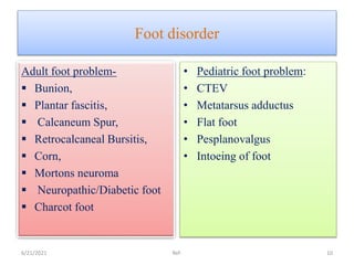
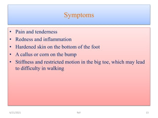
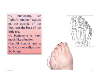
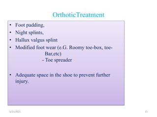
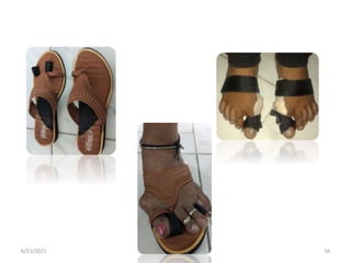
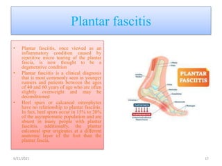
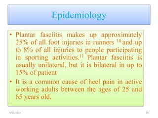
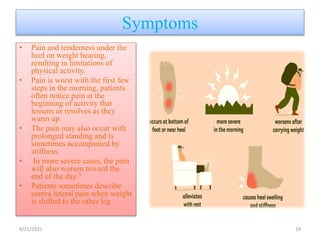
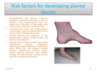
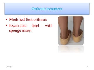
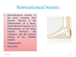
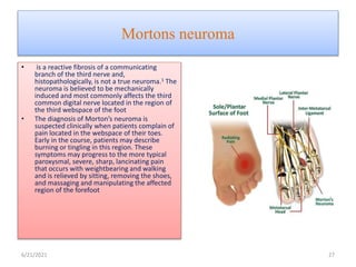
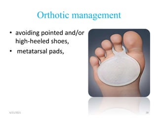
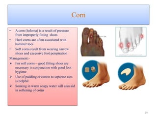
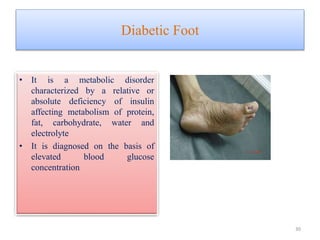
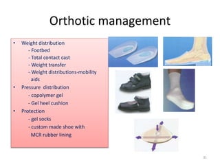
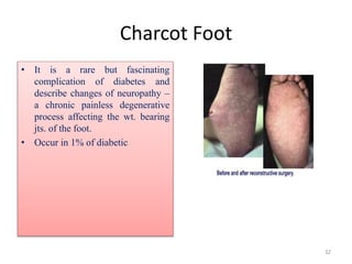
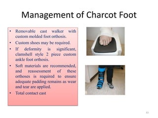
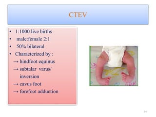
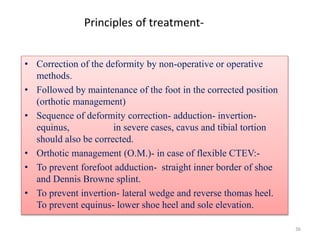
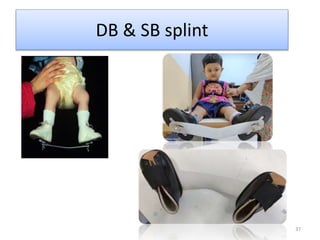
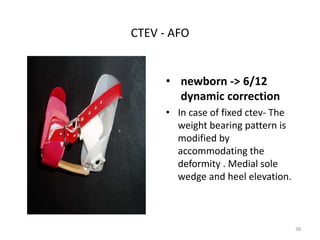
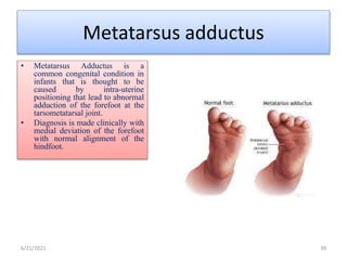
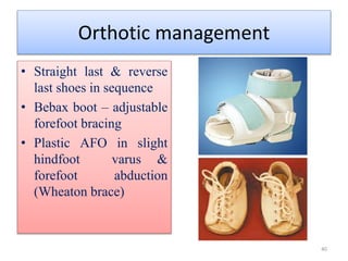
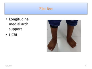
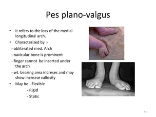
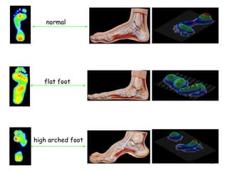
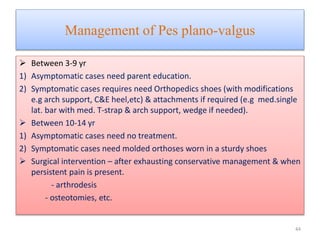
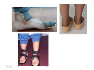
4. Foot anatomy
• The feet are flexible structures of
bones, joints, muscles, and soft
tissues that let us stand upright and
perform activities like walking,
running, and jumping.
• The foot contain :
1. 26 bones
2. 2 Sesamoid Bones
3. 33 joints
4. 19 muscles
5. 107 ligaments
6. 3 Arches
6/21/2021 Ref: Huson A. Functional anatomy of the foot. Disorders of the foot and ankle. 1991;1:409-31. 4
5. • Foot are divided into three parts:
1. Forefoot
2. Midfoot
3. Hindfoot
6/21/2021 Ref: Huson A. Functional anatomy of the foot. Disorders of the foot and ankle. 1991;1:409-31. 5
7. Plantar Fascia
• An important stabilizer in the foot
where a great deal of foot pathology
begins.
• It originates from the plantar surface of
the calcaneus and attaches to the plantar
surfaces of the five metatarsal heads and
proximal phalanges of the toes.
• It acts as a major stabilizer of the foot
• It helps maintain the arch of the foot and
is an antipronator.
• In its function of maintaining the
congruity of the relationship between
the calcaneus and the metatarsal heads,
it resists the torsion movement of the
forefoot in relation to the hindfoot
during pronation. Most of the eversion
of pronation occurs in the mid and
forefoot while the calcaneus remains
stable in the hindfoot.
7
8. Evaluation of foot disorders
Quality,
Location,
Radiation,
Severity,
Duration,
exacerbating and/or relieving factors related to the pain
A family history of congenital or acquired disorders
history of previous injuries,
occupational and recreational activities,
any recent changes in the type or amount of activity.
6/21/2021
Ref: Ayub, A., Yale, S. H., & Bibbo, C. (2005). Common foot disorders. Clinical medicine &
research, 3(2), 116–119. https://doi.org/10.3121/cmr.3.2.116 8
9. Contd..
• Current or previous
treatment(s).
• Observation :
• Patient’s gait
• foot position
• Inspection of his/her footwear
6/21/2021 9
11. Bunions
• Bursa sac that develops over the
first metatarsophalangeal (MTP)
joint as a result of a hallux valgus
deformity of this joint
• Women are prone to develop
• Etiology:
• Primary in the absence of any
known underlying diseases or
secondary to a variety of
metabolic, structural or
inflammatory disorders.
• Ill fitted shoe
6/21/2021 Ref: 11
12. Pathophysiology
• Genetic predisposition from
ligamentous laxity and
hyperpronation of the foot.
• The MTP joint gets larger and
protrudes from the inside of the
forefoot.
• complex relationship of the first ray
biomechanics and hindfoot
mechanics influence bunion
development.
• any disorder resulting in first MTP
joint inflammation may weaken
MTP soft tissue restraints,
predisposing to bunion formation.
6/21/2021 Ref: 12
13. Symptoms
• Pain and tenderness
• Redness and inflammation
• Hardened skin on the bottom of the foot
• A callus or corn on the bump
• Stiffness and restricted motion in the big toe, which may lead
to difficulty in walking
6/21/2021 Ref: 13
14. 6/21/2021 Ref: 14
•A bunionette, or
"tailor's bunion," occurs
on the outside of the
foot near the base of the
little toe.
•A bunionette is very
much like a bunion.
•Painful bursitis and a
hard corn or callus over
the bump.
15. OrthoticTreatment
• Foot padding,
• Night splints,
• Hallux valgus splint
• Modified foot wear (e.G. Roomy toe-box, toe-
Bar,etc)
- Toe spreader
• Adequate space in the shoe to prevent further
injury.
6/21/2021 15
17. Plantar fascitis
• Plantar fasciitis, once viewed as an
inflammatory condition caused by
repetitive micro tearing of the plantar
fascia, is now thought to be a
degenerative condition
• Plantar fasciitis is a clinical diagnosis
that is most commonly seen in younger
runners and patients between the ages
of 40 and 60 years of age who are often
slightly overweight and may be
deconditioned
• Heel spurs or calcaneal osteophytes
have no relationship to plantar fasciitis.
In fact, heel spurs occur in 15% to 20%
of the asymptomatic population and are
absent in many people with plantar
fasciitis. additionally, the plantar
calcaneal spur originates at a different
anatomic layer of the foot than the
plantar fascia.
6/21/2021 17
18. Epidemiology
• Plantar fasciitis makes up approximately
25% of all foot injuries in runners 10 and up
to 8% of all injuries to people participating
in sporting activities.11 Plantar fasciitis is
usually unilateral, but it is bilateral in up to
15% of patient
• It is a common cause of heel pain in active
working adults between the ages of 25 and
65 years old.
6/21/2021 18
19. Symptoms
• Pain and tenderness under the
heel on weight bearing,
resulting in limitations of
physical activity.
• Pain is worst with the first few
steps in the morning, patients
often notice pain at the
beginning of activity that
lessens or resolves as they
warm up.
• The pain may also occur with
prolonged standing and is
sometimes accompanied by
stiffness.
• In more severe cases, the pain
will also worsen toward the
end of the day.3
• Patients sometimes describe
contra lateral pain when weight
is shifted to the other leg
6/21/2021 19
20. Risk factors for developing plantar
fascitis
• Biomechanical risk factors: excessive
pronation, structural deformities such as
forefoot varus, higher-arched foot, low
arched foot, flattening of the medial arch
with excessive pronation, rearfoot varus,
• anatomical risk factor such as discrepancy
in leg length, excessive lateral tibial torsion
and excessive femoral anteversion,
• include tightness and weakness in the
gastronomies, soleus, achilles tendon and
intrinsic foot muscles13
• training error, excessive weight, age related
degenerative changes, occupations
requiring prolonged standing or ambulation
those falling into this category include
teachers, construction workers, cooks,
nurses, military personnel, and athletes
training for long distance running events,
and shoes with poor cushioning
6/21/2021 20
23. Calcaneum spur
• When a bony outgrowth forms
the calcaneum tuberosity just
anterior to the medial process of
the calcaneal tuberosity,
• Most common cause of heel pain
6/21/2021 23
24. Etiology
• According to Bergmann, it originates from the repetitive
traction of the insertion of the plantar fascia into the calcaneus,
which leads to inflammation, and reactive ossification of the
enthesis. However, according to, Kumai and Benjamin plantar
spurs develop from vertical compression instead and cannot be
traction spurs, as they do not develop within the plantar fascia
itself. They are thus fundamentally different from spurs in the
Achilles tendon since they develop as a consequence of
degenerative changes that occur in the plantar fascia enthesis
6/21/2021 24
26. Retrocalcaneal bursitis
• Retrocalcaneal bursitis is
the most common heel
bursitis. Bursitis is the
inflammation of a bursa.
Retrocalcaneal bursitis is in
inflammation of the bursa
located between the
calcaneus and the anterior
surface of the Achilles
tendon[
• Management:
• Heel raise
6/21/2021 26
27. Mortons neuroma
• is a reactive fibrosis of a communicating
branch of the third nerve and,
histopathologically, is not a true neuroma.1 The
neuroma is believed to be mechanically
induced and most commonly affects the third
common digital nerve located in the region of
the third webspace of the foot
• The diagnosis of Morton’s neuroma is
suspected clinically when patients complain of
pain located in the webspace of their toes.
Early in the course, patients may describe
burning or tingling in this region. These
symptoms may progress to the more typical
paroxysmal, severe, sharp, lancinating pain
that occurs with weightbearing and walking
and is relieved by sitting, removing the shoes,
and massaging and manipulating the affected
region of the forefoot
6/21/2021 27
29. Corn
• A corn (heloma) is a result of pressure
from improperly fitting shoes
• Hard corns are often associated with
hammer toes
• Soft corns result from wearing narrow
shoes and excessive foot perspiration
Management:-
For soft corns – good fitting shoes are
necessary in conjunction with good foot
hygiene
Use of padding or cotton to separate toes
is helpful
Soaking in warm soapy water will also aid
in softening of corns
29
30. Diabetic Foot
• It is a metabolic disorder
characterized by a relative or
absolute deficiency of insulin
affecting metabolism of protein,
fat, carbohydrate, water and
electrolyte
• It is diagnosed on the basis of
elevated blood glucose
concentration
30
31. Orthotic management
• Weight distribution
- Footbed
- Total contact cast
- Weight transfer
- Weight distributions-mobility
aids
• Pressure distribution
- copolymer gel
- Gel heel cushion
• Protection
- gel socks
- custom made shoe with
MCR rubber lining
31
32. Charcot Foot
• It is a rare but fascinating
complication of diabetes and
describe changes of neuropathy –
a chronic painless degenerative
process affecting the wt. bearing
jts. of the foot.
• Occur in 1% of diabetic
32
33. Management of Charcot Foot
• Removable cast walker with
custom molded foot orthosis.
• Custom shoes may be required.
• If deformity is significant,
clamshell style 2 piece custom
ankle foot orthosis.
• Soft materials are recommended,
and reassessment of these
orthoses is required to ensure
adequate padding remains as wear
and tear are applied.
• Total contact cast
33
35. Management of CTEV
Weekly serial
manipulation
Correction
achieved
Orthotic
management
Correction
not achieved
Surgery
External
fixators
35
36. 36
Principles of treatment-
• Correction of the deformity by non-operative or operative
methods.
• Followed by maintenance of the foot in the corrected position
(orthotic management)
• Sequence of deformity correction- adduction- invertion-
equinus, in severe cases, cavus and tibial tortion
should also be corrected.
• Orthotic management (O.M.)- in case of flexible CTEV:-
• To prevent forefoot adduction- straight inner border of shoe
and Dennis Browne splint.
• To prevent invertion- lateral wedge and reverse thomas heel.
To prevent equinus- lower shoe heel and sole elevation.
38. CTEV - AFO
• newborn -> 6/12
dynamic correction
• In case of fixed ctev- The
weight bearing pattern is
modified by
accommodating the
deformity . Medial sole
wedge and heel elevation.
38
39. Metatarsus adductus
• Metatarsus Adductus is a
common congenital condition in
infants that is thought to be
caused by intra-uterine
positioning that lead to abnormal
adduction of the forefoot at the
tarsometatarsal joint.
• Diagnosis is made clinically with
medial deviation of the forefoot
with normal alignment of the
hindfoot.
6/21/2021 39
40. Orthotic management
• Straight last & reverse
last shoes in sequence
• Bebax boot – adjustable
forefoot bracing
• Plastic AFO in slight
hindfoot varus &
forefoot abduction
(Wheaton brace)
40
42. Pes plano-valgus
• It refers to the loss of the medial
longitudinal arch.
• Characterized by :-
- obliterated med. Arch
- navicular bone is prominent
- finger cannot be inserted under
the arch
- wt. bearing area increses and may
show increase callosity
• May be - Flexible
- Rigid
- Static
42
44. Management of Pes plano-valgus
Between 3-9 yr
1) Asymptomatic cases need parent education.
2) Symptomatic cases requires need Orthopedics shoes (with modifications
e.g arch support, C&E heel,etc) & attachments if required (e.g med.single
lat. bar with med. T-strap & arch support, wedge if needed).
Between 10-14 yr
1) Asymptomatic cases need no treatment.
2) Symptomatic cases need molded orthoses worn in a sturdy shoes
Surgical intervention – after exhausting conservative management & when
persistent pain is present.
- arthrodesis
- osteotomies, etc.
44