CRANIAL
NERVES
Introduction
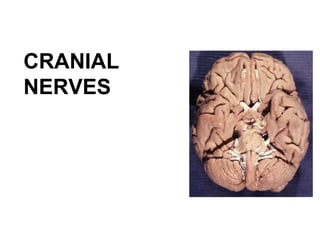
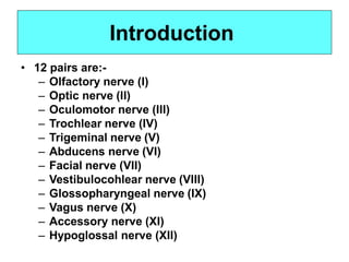
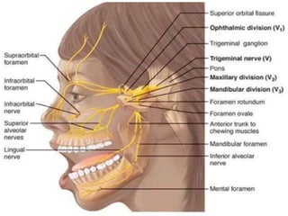
• The 12 pairs of cranial nerves arise
from the brain inside the cranial
cavity and pass through various
foramina in the bones of the
cranium.
• Divides into 3 functions: Sensory
nerves, Motor nerves and Mixed
nerves.
• 12 pairs are:-
– Olfactory nerve (I)
– Optic nerve (II)
– Oculomotor nerve (III)
– Trochlear nerve (IV)
– Trigeminal nerve (V)
– Abducens nerve (VI)
– Facial nerve (VII)
– Vestibulocohlear nerve (VIII)
– Glossopharyngeal nerve (IX)
– Vagus nerve (X)
– Accessory nerve (XI)
– Hypoglossal nerve (XII)
Introduction
CRANIAL NERVES
CRANIAL
NERVEFIBERS
Efferent -
Motor
Afferent -
sensory
Skeletal
muscles
Visceral (smooth)
muscles
Developed from
Somatic
mesoderm
Developed from
Special visceral mesoderm
Branchial arches
General somatic Efferent
GSE
Special Visceral Efferent
SVE
General Visceral Efferent
GVE
General
sensory
Special
sensory
Somatic Visceral
Somatic Visceral
General Visceral Afferent
GVA
general somatic Afferent
GSA
Special Somatic Afferent
SSA
Special Visceral Afferent
SVA
CRANIAL NERVE FIBERS
Cranial nerve supply muscles of branchial arches
( SVE) :
1. Trigeminal nerve 1st arch
2. Facial 2nd arch
3. Glossopharyngeal 3rd arch
4. Vagus 4th & 6th arhes
Cranial nerve carry parasympathetic ( GVE) :
1. Oculomotor nerve
2. Facial
3. Glossopharyngeal
4. Vagus
Cranial nerve carry taste ( SVA) :
1. Facial
2. Glossopharyngeal
3. Vagus
Olfactory Nerve (I)
•
•
Sensory nerve
Contain axons that
conduct impulses for
olfaction, the sense of
smell.
The olfactory
epithelium occupies the
superior part of the
nasal cavity, covering
the inferior surface of
the cribriform plate and
extending down along
the superior nasal
conchae.
•
Olfactory Nerve (I)
• The olfactory receptors
within the olfactory
epithelium are bipolar
neuron.
Each has a single odor
sensitive dendrite projecting
from one side of the cell body
and an unmylinated axons
extending from the other
side.
Bundle of axons of olfactory
receptors extend through
about 20 olfactory foramina
in the cribriform plate of the
•
•
ethmoid bone.
Olfactory Nerve (I)
• Olfactory nerves end in the brain in paired masses of
gray matter called the olfactory bulbs. Two extensions
of the brain that rest on the cribriform plate.
Within the olfactory bulbs, the axon terminals of
olfactory receptor form synapses with the dendrite and
cell bodies of the next neurons in the olfactory
pathway.
The axons of these neuron make up the olfactory tract,
which extend posteriorly from the olfactory bulbs.
Axons in the olfactory tract end in the primary olfactory
area in the temporal lobe of the temporal cerebral
•
•
•
cortex.
Optic Nerve (II)
•
•
Sensory nerve
Contains axons that conduct nerve impulses for
vision.
In the retina, rods and cones initiate visual signals
and relay them to bipolar cells, which transmit the
signals to ganglion cells.
Axons of all ganglion cells in the retina of each eye
join to form an optic nerve, which pass through the
optic foramen.
•
•
•
•
•
• Posterior to the eyeball, the two optic nerves merge
to form the optic chiasm.
Within the chiasm, axons from the medial half of
each eye cross to the opposite side, axons from the
lateral half is remain on the same side.
Posterior to the chiasm, the regrouped axons, some
from each eye, form the optic tracts.
Most axons in the optic tracts end in the lateral
geniculate nucleus of the thalamus.
Optic Nerve (II)
•
•
• There, they synapse with neuron whose axons
extend to the primary visual area in the occipital lobe
of the cerebral cortex.
A few axons pass through the optic chiasm and then
extend to the superior colliculi of the midbrain.
They synapse with motor neurons that control the
extrinsic (move the eyeball) and intrinsic eye
muscles (control light intensity).
Optic Nerve (II)
Oculomotor Nerve (III)
•
•
Motor nerve
Oculomotor nerve extends anteriorly and divides into
superior and inferior branches, both of which pass
through the superior orbital fissure into the orbit.
Axons in the superior branch innervate the superior
rectus (extrinsic eyeball muscle) and the levator
palpebrae superioris (muscles of upper eyelid).
•
•
•
• Axons in the inferior branch supply the medial rectus,
inferior rectus and inferior oblique muscles (all
extrinsic eyeball muscles).
Theses somatic motor neurons control movements of
the eyeball and upper eyelid.
The inferior branch of the oculomtor nerve also
provides parasympathetic innervation to intrinsic
eyeball muscles, which are smooth muscles.
Oculomotor Nerve (III)
• They include the ciliary muscles of the eyeball and the
circular muscles (sphincter pupillae) of the iris.
Parasympatethic impulses propagate from oculomotor
nucleus in the midbrain to the ciliary ganglion, a relay
centre of the autonomic nervous system.
•
• From the ciliary ganglion, parasympathetic axons to
the ciliary muscles, which adjust the lens for near
vision.
• Other parasympathetic axons stimulate the circular
muscles of the iris to contract when bright light
stimulate the eye, causing decrease in the size of the
pupil (constriction).
Oculomotor Nerve (III)
•
•
•
• Proprioceptive sensory axons from the extrinsic
eyeball muscles begin their course towards the brain
in the oculomotor nerve but eventually leave the
nerve to join ophthalmic branch of trigeminal nerve.
They do not return to the brain in the oculomotor
nerve.
The cell bodies of the sensory axons reside in the
trigeminal ganglion, and they enter the midbrain via
trigeminal nerve.
These axons convey nerve impulses for
proprioception, the nonvisual perception of the
movements and position of the body, from extrinsic
eyeball muscles.
Oculomotor Nerve (III)
Trochlear Nerve (IV)
•
•
•
Motor nerve
Smallest cranial nerve
Arises from posterior part of the brainstem.
•
• The motor neurons originate in the trochlear nucleus
in the midbrain, and axons from the nucleus pass
through the superior orbital fissure of orbit.
These somatic motor axons innervate the superior
oblique muscles of the eyeball. (extrinsic eyeball
muscle that control movement of the eyeball)
•
•
• Proprioceptive sensory axons from the superior
oblique muscle begin their course toward the brain in
the trochlear nerve but eventually leave the nerve to
join ophtalmic branch of the trigeminal nerve.
They do not return to the brain in the trochlear nerve.
The cells body of sensory neurons reside in the
trigeminal ganglion, and they enter the midbrain via
trigeminal nerve.
Trochlear Nerve (IV)
• Like those of the oculomotor nerve, these axons
convey nerve impulses for proprioception, the
nonvisual perception of the movements and position
of the body, from extrinsic eyeball muscles.
Trochlear Nerve (IV)
Trigeminal Nerve (V)
•
•
•
Mixed nerve
Largest cranial nerve
2 roots from
venterolateral of the
pons
Have large sensory
root and small motor
root
•
•
• Large sensory root
– Has swelling part –
trigeminal ganglion
– Trigeminal ganglion
located in the fossa inner
surface of petrous
portion.
– The trigeminal ganglion
contain cell bodies of
most of the primary
sensory neurons.
Small motor root
– Originate from nucleusin
the pons
Trigeminal Nerve (V)
• Consists 3
branches
– Ophtalmic
– Maxillary
– Mandibular
Trigeminal Nerve (V)
• Ophthalmic Branch
– Smallest branches of T.N
– Enter orbit through superior orbital fissure
– Contain sensory axon from; (1) skin over upper
eyelid, (2) eyeball, (3) lacrimal gland, (4) upper part
of nasal cavity, (5)side of the nose, forehead,
anterior half of the scalp.
Trigeminal Nerve (V)
• Maxillary Branch
– Intermediate in size
– Enter the foramen rotundum of sphenoid
– Contain sensory axon from; (1)mucosa layer of the
nose, (2) palate, (3) part of the pharynx, (4) upper
teeth, (5) upper lips, (6) lower eyelid.
Trigeminal Nerve (V)
•
•
• Mandibular Branch
– Largest T.N
– Exits through the foramen ovale of sphenoid
– Contain sensory axons from : (1) anterior 2/3 tongue,
(2) cheek and mucosa deep into it, (3) lower teeth, (4)
skin over the mandible and side of the head anterior
to the ear, (5) mucosa of the floor of the mouth.
The sensory axons from 3 branches enter the
trigeminal ganglion and terminate in the nuclei in the
pons.
The trigeminal nerve also contain sensory fiber from
proprioceptors located in the muscles of the
mastication
Trigeminal Nerve (V)
•
•
• Somatic motor axons of the trigeminal nerve are part of
the mandibular nerve and supply muscles of
mastication.
Masseter, temporalis, medial and lateral pterygoid,
anterior belly of digastric, mylohyoid and tensor
tympani.
Important control chewing movements.
Trigeminal Nerve (V)
• Trigeminal Nerve Nuclei: It has four
nucleus:
• (1) Main sensory nucleus.
• (2) Spinal nucleus.
• (3)Mesencephalic nucleus.
• (4) Motor nucleus.
• Main sensory nucleus lies in posterior part of pons
lateral to the motor nucleus.
• Spinal nucleus continuous superiorly with main
sensory nucleus and extends inferiorly through
medulla oblongata and into upper part of
spinal cord as far as second cervical segment.
• Mesencephalic Nucleus composed of unipolar cells
situated in lateral part of gray matter around cerebral
aqueduct.
• It extends inferiorly into pons as far as main
sensory nucleus.
• Motor nucleus is situated in pons medial to main
sensory nucleus.
Abducens Nerve (VI)
•
•
Motor nerve
Origin – abducens nucleus of the pons
• Somatic motor axons
extend from the nucleus to
the lateral rectus muscle of
the eyeball, through the
superior orbital fissure of
the orbit.
Nerve impulses cause
abduction of the eyeball
•
Abducens Nerve (VI)
•
•
•
• Proprioceptive sensory axons from the lateral rectus
muscle begin their course toward the brain in the
abducens nerve but eventually leave the nerve to
join ophtalmic branch of the trigeminal nerve.
They do not return to the brain in the abducens
nerve.
The cells body of sensory neurons reside in the
trigeminal ganglion, and they enter the midbrain via
trigeminal nerve.
These axons convey nerve impulses for
proprioception, the nonvisual perception of the
movements and position of the body, fro extrinsic
eyeball muscles.
Abducens Nerve (VI)
Facial Nerve (VII)
•
•
Mixed nerve
Sensory axons extend from the taste buds of the
tongue (anterior 2/3) through the geniculate ganglion
(a cluster of cell bodies of sensory neuron that lies
beside facial nerve, and end in the pons)
Sensory portion of the facial nerve also contain axons
from proprioceprors in muscles of the face and scalp
and from skin in the ear canal.
•
• Axons of somatic motor
neurons arise from nucleus in
the pons, pass through petrous
portion of temporal and
innervate facial, scalp and neck
muscles.
Innervations this axons cause
contraction of facial
expression muscles, plus
stylohyoid, posteriorbelly of
•
digastric, and stapedius in the
ear.
Facial Nerve (VII)
•
Axons of parasymapthetic
neuron that are part of the
facial nerve end in 2
parasymapthetic ganglia;
pterygopalatine and
submandibular ganglion.
From this 2 ganglia, other
parasympathetic axons
extends to the lacrimal gland,
nasal gland, palatine gland,
sublingual and submandibular
gland.
•
Vestibulocochlear Nerve (VIII)
•
•
•
Acoustic @ Auditory nerve
Sensory nerve
Has 2 branches; vestibular and cochlear branches.
Vestibulocochlear Nerve (VIII)
• Vestibular branch
– Carry impulses fro equibilirium
– Sensory axons in the vestibular branch arise from semicircular
canals, the saccule, and the utricle of the inner ear.
– Then extend to the vestibular ganglion, where the cell bodies
are located.
– And end in the vestibular nuclei in the medulla oblongata.
– Some sensory axons enter the cerebellum via the inferior
cerebellar peduncle.
Vestibulocochlear Nerve (VIII)
• Cochlear Branch
– Carry impulses for hearing
– Sensory axons in the cochlear branch arise in the spiral
organ (Organ of Corti) in the cochlea of the inner ear.
– The cell bodies of cochlear branch sensory neurons are
located in the spiral ganglion of the cochlea.
– From there axons extend to cochlear nuclei in the medulla
oblongata.
Glossopharyngeal Nerve (IX)
•
•
Mix nerve
Sensory axons of GN arise from :
– Taste buds and somatic sensory receptor on the
posterior 1/3 of tongue
– Proprioceptors in swallowing muscles supply by
motor portion
– Baroreceptors in the carotid sinus
– Chemoreseptor in the carotid body
Hermizan Halihanafiah
•
• The cell bodies of these sensory neurons are
located in the superior and inferior ganglia.
From these ganglia, sensory axons pass through
the jugular foramen and end in the medulla
oblongata.
Glossopharyngeal Nerve (IX)
• Axons of motor neurons in GN arise in nuclei of the
MO and exit the skull through the jugular foramen.
Somatic motor neuron innervate the stylopharyngeus
muscle and autonomic motor neurons
(parasympathetic) stimulate the parotid gland to
secrete saliva.
Some of the sell bodies of parasymapthetic motor
•
•
neuron are located in the otic ganglion.
Vagus Nerve (X)
•
•
Mixed nerve
Sensory axon arise from:
– Skin of the external ear
– A few taste bud in the epiglottis and pharynx
– Proprioceptors in muscles of the neck and throat
– Baroreceptor in the arch of aorta
– Chemoreceptor in the aortic bodies
– Visceral sensory receptors in the most organs of thoracic and
abdominal cavities.
• These axons pass
through the jugular
foramen and end in
the MO and pons
The somatic motor
neurons, arise from
nuclei in the MO
and supply muscle
of the pharynx,
larynx, and soft
palate that used in
swallowing and
vocalization.
•
• Axons autonomic motor neuron (parasympathetic) in
the vagus nerve originate in nuclei of MO and end in
the lungs and heart.
Vagal parasympathetic axons also supply gland of GIT
and smooth muscles of respiratory tract, esophagus,
stomach, gallbladder, small intestine and most of the
large intestine.
•
Accessory Nerve (XI)
•
•
Motor nerve
Motor axons arise in the anterior gray of the 1st 5
segments of the cervical portion of the spinal cord.
The axons from the segment exit the spinal cord
laterally and come together, pass through the foramen
magnum and exit through the jugular foramen along
with the vagus nerve.
•
• The AN convey motor impulses to the
sternocleidomastoid and trapezius muscles to
coordinate head movement.
Sesnory axons in the AN originate from proprioceptors
in the muscles supplied by its motor neurons begins
their course toward the brain in the AN but eventually
leave the nerve and to join the cervical plexus.
•
• From cervical plexus, they enetr the spinal cord via the
posterior root of the cervical spinal nerve to pass to
and end in the MO.
The sensory axon do not return to the barin in the AN
and, like all sensory axon, have their cell bodies in
posterior root ganglion.
•
Hypoglossal Nerve (XII)
•
•
Motor nerve
Somatic motor axons originate in the hypoglossal
nuclei in the MO, pass through the hypoglossal canal,
and supply the muscles of the tongue.
These axons conduct impulses for speech and
•
swallowing.
• Sensory axons that originate from proprioceptors in
the tongue muscles begin their course towards the
brain in the hypoglossal nerve.
They leave the nerve and join cervical spinal nerve and
end in the MO, again entering the CNC via posterior
root of cervical spinal nerve.
The sensory axons do not return to the brain in the
•
•
hypoglossal nerve.

CRANIAL NERVES-OVERVIEW.pptx

  • 1.
  • 2.
    Introduction • The 12pairs of cranial nerves arise from the brain inside the cranial cavity and pass through various foramina in the bones of the cranium. • Divides into 3 functions: Sensory nerves, Motor nerves and Mixed nerves.
  • 3.
    • 12 pairsare:- – Olfactory nerve (I) – Optic nerve (II) – Oculomotor nerve (III) – Trochlear nerve (IV) – Trigeminal nerve (V) – Abducens nerve (VI) – Facial nerve (VII) – Vestibulocohlear nerve (VIII) – Glossopharyngeal nerve (IX) – Vagus nerve (X) – Accessory nerve (XI) – Hypoglossal nerve (XII) Introduction
  • 6.
  • 7.
    CRANIAL NERVEFIBERS Efferent - Motor Afferent - sensory Skeletal muscles Visceral(smooth) muscles Developed from Somatic mesoderm Developed from Special visceral mesoderm Branchial arches General somatic Efferent GSE Special Visceral Efferent SVE General Visceral Efferent GVE General sensory Special sensory Somatic Visceral Somatic Visceral General Visceral Afferent GVA general somatic Afferent GSA Special Somatic Afferent SSA Special Visceral Afferent SVA
  • 8.
    CRANIAL NERVE FIBERS Cranialnerve supply muscles of branchial arches ( SVE) : 1. Trigeminal nerve 1st arch 2. Facial 2nd arch 3. Glossopharyngeal 3rd arch 4. Vagus 4th & 6th arhes Cranial nerve carry parasympathetic ( GVE) : 1. Oculomotor nerve 2. Facial 3. Glossopharyngeal 4. Vagus Cranial nerve carry taste ( SVA) : 1. Facial 2. Glossopharyngeal 3. Vagus
  • 9.
    Olfactory Nerve (I) • • Sensorynerve Contain axons that conduct impulses for olfaction, the sense of smell. The olfactory epithelium occupies the superior part of the nasal cavity, covering the inferior surface of the cribriform plate and extending down along the superior nasal conchae. •
  • 11.
    Olfactory Nerve (I) •The olfactory receptors within the olfactory epithelium are bipolar neuron. Each has a single odor sensitive dendrite projecting from one side of the cell body and an unmylinated axons extending from the other side. Bundle of axons of olfactory receptors extend through about 20 olfactory foramina in the cribriform plate of the • • ethmoid bone.
  • 12.
    Olfactory Nerve (I) •Olfactory nerves end in the brain in paired masses of gray matter called the olfactory bulbs. Two extensions of the brain that rest on the cribriform plate. Within the olfactory bulbs, the axon terminals of olfactory receptor form synapses with the dendrite and cell bodies of the next neurons in the olfactory pathway. The axons of these neuron make up the olfactory tract, which extend posteriorly from the olfactory bulbs. Axons in the olfactory tract end in the primary olfactory area in the temporal lobe of the temporal cerebral • • • cortex.
  • 13.
    Optic Nerve (II) • • Sensorynerve Contains axons that conduct nerve impulses for vision. In the retina, rods and cones initiate visual signals and relay them to bipolar cells, which transmit the signals to ganglion cells. Axons of all ganglion cells in the retina of each eye join to form an optic nerve, which pass through the optic foramen. • •
  • 15.
    • • • • Posterior tothe eyeball, the two optic nerves merge to form the optic chiasm. Within the chiasm, axons from the medial half of each eye cross to the opposite side, axons from the lateral half is remain on the same side. Posterior to the chiasm, the regrouped axons, some from each eye, form the optic tracts. Most axons in the optic tracts end in the lateral geniculate nucleus of the thalamus. Optic Nerve (II)
  • 16.
    • • • There, theysynapse with neuron whose axons extend to the primary visual area in the occipital lobe of the cerebral cortex. A few axons pass through the optic chiasm and then extend to the superior colliculi of the midbrain. They synapse with motor neurons that control the extrinsic (move the eyeball) and intrinsic eye muscles (control light intensity). Optic Nerve (II)
  • 18.
    Oculomotor Nerve (III) • • Motornerve Oculomotor nerve extends anteriorly and divides into superior and inferior branches, both of which pass through the superior orbital fissure into the orbit. Axons in the superior branch innervate the superior rectus (extrinsic eyeball muscle) and the levator palpebrae superioris (muscles of upper eyelid). •
  • 19.
    • • • Axons inthe inferior branch supply the medial rectus, inferior rectus and inferior oblique muscles (all extrinsic eyeball muscles). Theses somatic motor neurons control movements of the eyeball and upper eyelid. The inferior branch of the oculomtor nerve also provides parasympathetic innervation to intrinsic eyeball muscles, which are smooth muscles. Oculomotor Nerve (III)
  • 20.
    • They includethe ciliary muscles of the eyeball and the circular muscles (sphincter pupillae) of the iris. Parasympatethic impulses propagate from oculomotor nucleus in the midbrain to the ciliary ganglion, a relay centre of the autonomic nervous system. • • From the ciliary ganglion, parasympathetic axons to the ciliary muscles, which adjust the lens for near vision. • Other parasympathetic axons stimulate the circular muscles of the iris to contract when bright light stimulate the eye, causing decrease in the size of the pupil (constriction). Oculomotor Nerve (III)
  • 21.
    • • • • Proprioceptive sensoryaxons from the extrinsic eyeball muscles begin their course towards the brain in the oculomotor nerve but eventually leave the nerve to join ophthalmic branch of trigeminal nerve. They do not return to the brain in the oculomotor nerve. The cell bodies of the sensory axons reside in the trigeminal ganglion, and they enter the midbrain via trigeminal nerve. These axons convey nerve impulses for proprioception, the nonvisual perception of the movements and position of the body, from extrinsic eyeball muscles. Oculomotor Nerve (III)
  • 22.
    Trochlear Nerve (IV) • • • Motornerve Smallest cranial nerve Arises from posterior part of the brainstem.
  • 23.
    • • The motorneurons originate in the trochlear nucleus in the midbrain, and axons from the nucleus pass through the superior orbital fissure of orbit. These somatic motor axons innervate the superior oblique muscles of the eyeball. (extrinsic eyeball muscle that control movement of the eyeball) • • • Proprioceptive sensory axons from the superior oblique muscle begin their course toward the brain in the trochlear nerve but eventually leave the nerve to join ophtalmic branch of the trigeminal nerve. They do not return to the brain in the trochlear nerve. The cells body of sensory neurons reside in the trigeminal ganglion, and they enter the midbrain via trigeminal nerve. Trochlear Nerve (IV)
  • 24.
    • Like thoseof the oculomotor nerve, these axons convey nerve impulses for proprioception, the nonvisual perception of the movements and position of the body, from extrinsic eyeball muscles. Trochlear Nerve (IV)
  • 25.
    Trigeminal Nerve (V) • • • Mixednerve Largest cranial nerve 2 roots from venterolateral of the pons Have large sensory root and small motor root •
  • 26.
    • • Large sensoryroot – Has swelling part – trigeminal ganglion – Trigeminal ganglion located in the fossa inner surface of petrous portion. – The trigeminal ganglion contain cell bodies of most of the primary sensory neurons. Small motor root – Originate from nucleusin the pons Trigeminal Nerve (V)
  • 27.
    • Consists 3 branches –Ophtalmic – Maxillary – Mandibular Trigeminal Nerve (V)
  • 28.
    • Ophthalmic Branch –Smallest branches of T.N – Enter orbit through superior orbital fissure – Contain sensory axon from; (1) skin over upper eyelid, (2) eyeball, (3) lacrimal gland, (4) upper part of nasal cavity, (5)side of the nose, forehead, anterior half of the scalp. Trigeminal Nerve (V)
  • 29.
    • Maxillary Branch –Intermediate in size – Enter the foramen rotundum of sphenoid – Contain sensory axon from; (1)mucosa layer of the nose, (2) palate, (3) part of the pharynx, (4) upper teeth, (5) upper lips, (6) lower eyelid. Trigeminal Nerve (V)
  • 30.
    • • • Mandibular Branch –Largest T.N – Exits through the foramen ovale of sphenoid – Contain sensory axons from : (1) anterior 2/3 tongue, (2) cheek and mucosa deep into it, (3) lower teeth, (4) skin over the mandible and side of the head anterior to the ear, (5) mucosa of the floor of the mouth. The sensory axons from 3 branches enter the trigeminal ganglion and terminate in the nuclei in the pons. The trigeminal nerve also contain sensory fiber from proprioceptors located in the muscles of the mastication Trigeminal Nerve (V)
  • 31.
    • • • Somatic motoraxons of the trigeminal nerve are part of the mandibular nerve and supply muscles of mastication. Masseter, temporalis, medial and lateral pterygoid, anterior belly of digastric, mylohyoid and tensor tympani. Important control chewing movements. Trigeminal Nerve (V)
  • 32.
    • Trigeminal NerveNuclei: It has four nucleus: • (1) Main sensory nucleus. • (2) Spinal nucleus. • (3)Mesencephalic nucleus. • (4) Motor nucleus. • Main sensory nucleus lies in posterior part of pons lateral to the motor nucleus. • Spinal nucleus continuous superiorly with main sensory nucleus and extends inferiorly through medulla oblongata and into upper part of spinal cord as far as second cervical segment. • Mesencephalic Nucleus composed of unipolar cells situated in lateral part of gray matter around cerebral aqueduct. • It extends inferiorly into pons as far as main sensory nucleus. • Motor nucleus is situated in pons medial to main sensory nucleus.
  • 52.
    Abducens Nerve (VI) • • Motornerve Origin – abducens nucleus of the pons
  • 53.
    • Somatic motoraxons extend from the nucleus to the lateral rectus muscle of the eyeball, through the superior orbital fissure of the orbit. Nerve impulses cause abduction of the eyeball • Abducens Nerve (VI)
  • 54.
    • • • • Proprioceptive sensoryaxons from the lateral rectus muscle begin their course toward the brain in the abducens nerve but eventually leave the nerve to join ophtalmic branch of the trigeminal nerve. They do not return to the brain in the abducens nerve. The cells body of sensory neurons reside in the trigeminal ganglion, and they enter the midbrain via trigeminal nerve. These axons convey nerve impulses for proprioception, the nonvisual perception of the movements and position of the body, fro extrinsic eyeball muscles. Abducens Nerve (VI)
  • 55.
    Facial Nerve (VII) • • Mixednerve Sensory axons extend from the taste buds of the tongue (anterior 2/3) through the geniculate ganglion (a cluster of cell bodies of sensory neuron that lies beside facial nerve, and end in the pons) Sensory portion of the facial nerve also contain axons from proprioceprors in muscles of the face and scalp and from skin in the ear canal. •
  • 56.
    • Axons ofsomatic motor neurons arise from nucleus in the pons, pass through petrous portion of temporal and innervate facial, scalp and neck muscles. Innervations this axons cause contraction of facial expression muscles, plus stylohyoid, posteriorbelly of • digastric, and stapedius in the ear. Facial Nerve (VII)
  • 57.
    • Axons of parasymapthetic neuronthat are part of the facial nerve end in 2 parasymapthetic ganglia; pterygopalatine and submandibular ganglion. From this 2 ganglia, other parasympathetic axons extends to the lacrimal gland, nasal gland, palatine gland, sublingual and submandibular gland. •
  • 65.
    Vestibulocochlear Nerve (VIII) • • • Acoustic@ Auditory nerve Sensory nerve Has 2 branches; vestibular and cochlear branches.
  • 66.
  • 67.
    • Vestibular branch –Carry impulses fro equibilirium – Sensory axons in the vestibular branch arise from semicircular canals, the saccule, and the utricle of the inner ear. – Then extend to the vestibular ganglion, where the cell bodies are located. – And end in the vestibular nuclei in the medulla oblongata. – Some sensory axons enter the cerebellum via the inferior cerebellar peduncle. Vestibulocochlear Nerve (VIII)
  • 68.
    • Cochlear Branch –Carry impulses for hearing – Sensory axons in the cochlear branch arise in the spiral organ (Organ of Corti) in the cochlea of the inner ear. – The cell bodies of cochlear branch sensory neurons are located in the spiral ganglion of the cochlea. – From there axons extend to cochlear nuclei in the medulla oblongata.
  • 69.
    Glossopharyngeal Nerve (IX) • • Mixnerve Sensory axons of GN arise from : – Taste buds and somatic sensory receptor on the posterior 1/3 of tongue – Proprioceptors in swallowing muscles supply by motor portion – Baroreceptors in the carotid sinus – Chemoreseptor in the carotid body
  • 70.
    Hermizan Halihanafiah • • Thecell bodies of these sensory neurons are located in the superior and inferior ganglia. From these ganglia, sensory axons pass through the jugular foramen and end in the medulla oblongata. Glossopharyngeal Nerve (IX)
  • 71.
    • Axons ofmotor neurons in GN arise in nuclei of the MO and exit the skull through the jugular foramen. Somatic motor neuron innervate the stylopharyngeus muscle and autonomic motor neurons (parasympathetic) stimulate the parotid gland to secrete saliva. Some of the sell bodies of parasymapthetic motor • • neuron are located in the otic ganglion.
  • 72.
    Vagus Nerve (X) • • Mixednerve Sensory axon arise from: – Skin of the external ear – A few taste bud in the epiglottis and pharynx – Proprioceptors in muscles of the neck and throat – Baroreceptor in the arch of aorta – Chemoreceptor in the aortic bodies – Visceral sensory receptors in the most organs of thoracic and abdominal cavities.
  • 73.
    • These axonspass through the jugular foramen and end in the MO and pons The somatic motor neurons, arise from nuclei in the MO and supply muscle of the pharynx, larynx, and soft palate that used in swallowing and vocalization. •
  • 74.
    • Axons autonomicmotor neuron (parasympathetic) in the vagus nerve originate in nuclei of MO and end in the lungs and heart. Vagal parasympathetic axons also supply gland of GIT and smooth muscles of respiratory tract, esophagus, stomach, gallbladder, small intestine and most of the large intestine. •
  • 75.
    Accessory Nerve (XI) • • Motornerve Motor axons arise in the anterior gray of the 1st 5 segments of the cervical portion of the spinal cord. The axons from the segment exit the spinal cord laterally and come together, pass through the foramen magnum and exit through the jugular foramen along with the vagus nerve. •
  • 76.
    • The ANconvey motor impulses to the sternocleidomastoid and trapezius muscles to coordinate head movement. Sesnory axons in the AN originate from proprioceptors in the muscles supplied by its motor neurons begins their course toward the brain in the AN but eventually leave the nerve and to join the cervical plexus. •
  • 77.
    • From cervicalplexus, they enetr the spinal cord via the posterior root of the cervical spinal nerve to pass to and end in the MO. The sensory axon do not return to the barin in the AN and, like all sensory axon, have their cell bodies in posterior root ganglion. •
  • 78.
    Hypoglossal Nerve (XII) • • Motornerve Somatic motor axons originate in the hypoglossal nuclei in the MO, pass through the hypoglossal canal, and supply the muscles of the tongue. These axons conduct impulses for speech and • swallowing.
  • 79.
    • Sensory axonsthat originate from proprioceptors in the tongue muscles begin their course towards the brain in the hypoglossal nerve. They leave the nerve and join cervical spinal nerve and end in the MO, again entering the CNC via posterior root of cervical spinal nerve. The sensory axons do not return to the brain in the • • hypoglossal nerve.