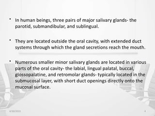
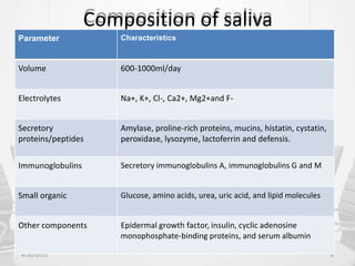
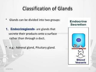
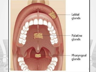
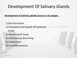
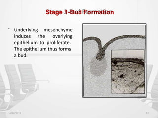
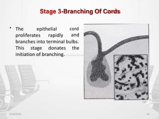
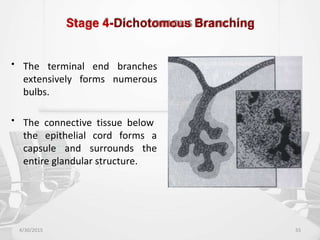
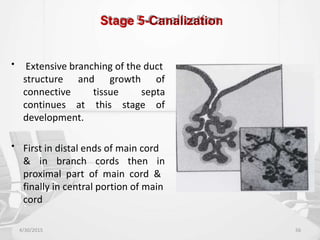
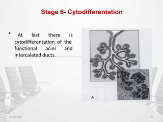
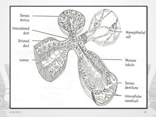
SlideShare a Scribd company logo
1 of 104
Contents
•
•
•
•
•
•
•
•
•
•
•
•
Introduction
Composition of saliva
Functions of saliva
Anatomy of salivary glands
Development
Structure
Formation and secretion of saliva
Ductal modification of saliva
Histology of the major salivary glands
Histology of the minor salivary glands
Age changes
Clinical considerations
4/30/2015 2
Introduction
• The oral cavity is kept moist by a film of fluid called saliva that
coats the teeth and the mucosa.
• Saliva is a complex fluid, produced by the salivary glands, the
most important function of which is to maintain the well-
being of mouth.
• Individuals with a deficiency of salivary secretion experience
difficulty in eating, speaking, and swallowing and become
prone to mucosal infections and rampant caries.
4/30/2015 3
• In human beings, three pairs of major salivary glands- the
parotid, submandibular, and sublingual.
• They are located outside the oral cavity, with extended duct
systems through which the gland secretions reach the mouth.
• Numerous smaller minor salivary glands are located in various
parts of the oral cavity- the labial, lingual palatal, buccal,
glossopalatine, and retromolar glands- typically located in the
submucosal layer, with short duct openings directly onto the
mucosal surface.
4/30/2015 4
Saliva
• Is a clear, slightly acidic, mucoserous secretion.
• The whole saliva is a complex mixture of fluids, with
contributions from major and minor salivary glands and the
gingival crevicular fluid, containing a high population of
normal oral bacteria, desquamated epithelial cells and
transient residues of food or drink, following their ingestion.
4/30/2015 5
Composition of saliva
4/30/2015 6
Parameter Characteristics
Volume 600-1000ml/day
Electrolytes Na+, K+, Cl-, Ca2+, Mg2+and F-
Secretory
proteins/peptides
Amylase, proline-rich proteins, mucins, histatin, cystatin,
peroxidase, lysozyme, lactoferrin and defensis.
Immunoglobulins Secretory immunoglobulins A, immunoglobulins G and M
Small organic Glucose, amino acids, urea, uric acid, and lipid molecules
Other components Epidermal growth factor, insulin, cyclic adenosine
monophosphate-binding proteins, and serum albumin
Functions of saliva
4/30/2015 7
Function Effect Active constituents
Protection Clearance Water
Lubrication Mucins, glycoproteins
Thermal or chemical
insulation
Mucins
Tannin binding Basic proline-rich
proteins, histatins
Buffering pH maintenance Bicarbonate, phosphate,
basic proteins, urea,
ammonia
Neutralization of acids
4/30/2015 8
Function Effect Active constituents
Tooth integrity Enamel maturation,
repair
Calcium, phosphate, fluoride, statherin,
acidic proline-rich proteins
Antimicrobial
activity
Physical barrier Mucins
Immune defense Seretory immunoglobulin A
Non-immune
defense
Peroxidase, lysozyme, lectoferrin,
histatin, mucins, agglutinins, secretory
leukocyte protease inhibitor and
defensins
Tissue repair Wound healing,
epithelial
regeneration
Growth factors, trefoil proteins
Digestion Bolus formation Water, mucins
4/30/2015 9
Function Effect Active constituents
Digestion Starch, triglyceride
digestion
Amylase, lipase
Taste Solution of molecules Water and lipocalins
Maintenance of taste
buds
Epidermal growth factor
and carbonic anhydrase
VI
• Gland- An organized aggregation of cells functioning as a
secretory or excretory organ.
4/30/2015 10
Classification of Glands
• Glands can be divided into two groups:
1. Endocrineglands- are glands that
secrete their products onto a surface
rather than through a duct.
• e.g.- Adrenal gland, Pituitary gland.
4/30/2015 11
2) Exocrine glands
- These glands secrete their products via
a duct. e.g.- Salivary glands. It can be divided into three
groups:-
a) Merocrine gland
e.g.-Mucous and Serous
glands
b) Apocrine gland
e.g.-Sweat glands
c) Holocrine gland
e.g.-Sebaceous glands
4/30/2015 12
Classification Of Salivary Glands
1) ACCORDING TO SIZE:
A) MAJOR B) MINOR
Parotid Glands of Lips,
Submandibular Cheeks , Hard
Sublingual Palate, Soft palate,
Von Ebners and
Blandin & Nuhn.
4/30/2015 13
4/30/2015 44
Major Salivary Glands with Ducts
2) ACCORDING TO BRANCHING OF DUCTS:
A) Compound
- Major glands
B) Simple
- Minor glands
3) ACCORDING TO SECRETION:
SEROUS MUCOUS MIXED
a)Parotid a)Palatine a)Submandibular
b)Von-ebner b)Post. lingual b)Sublingual
c) Glossopalatine c)Buccal
d)Labial.
4/30/2015 14
Anatomy of salivary glands
4/30/2015 15
Parotid gland
• Is the largest
• Weight – 15gms
• Is situated below the External acoustic meatus, b/w the ramus
of the mandible and the sternocleidomastoid and overlaps
them.
• Secretion of parotid gland is serous.
• Anteriorly , the gland overlaps the masseter muscle. A part of
this forward extension is often detached – Accessory Parotid
which lies b/w the zygomatic arch and the parotid duct.
4/30/2015 16
4/30/2015 17
Parotid Capsule
• The investing layer of deep cervical fascia forms the capsule
for the gland.
• The fascia splits to enclose the gland.
• Superficial lamina – thick & adherent to the gland.
• Deep lamina – thin – attached to the styloid process, the
mandible and tympanic plate.
• A portion of deep lamina, extending between the styloid
process and the mandible – thickened to form
Stylomandibular Ligament –separates the Parotid gland from
the Submandibular salivary gland.
4/30/2015 18
External Features
• The glands has four surfaces
a) Superior
b) Superficial
c) Antero-medial
d) Postero-medial
• The surfaces are separated by three borders.
a) Anterior
b) Posterior
c) Medial
4/30/2015 19
Relations
1. Superior surface related to

External acoustic meatus

Superficial temporal vessels

Posterior surface of TMJ

Auriculotemporal nerve
2. Superficial surface (largest)
a) Skin
b) Superficial fascia
i. Great auricular nerve
4/30/2015 20
ii) Pre-auricular lymph node
iii) Posterior fibers of platysma
c) Parotid fascia
d) Deep parotid lymph nodes
3. Anteromedial surface
o Masseter
o Lateral surface of TMJ
o Posterior border of the mandible
o Medial pterygoid
o Terminal branches of facial nerve
4/30/2015 21
4/30/2015 22
3. Posteromedial surface

Mastoid process

Sternocleidomastoid

Posterior belly of the digastric

Styloid process
4/30/2015 23
5. Anterior border :
– Separates the superficial surface from the
Anteromedial surface
– Extends from –anterior part of the superior surface
to the apex
– Structures emerging:

Parotid duct

Terminal branches of the facial nerve

Transverse facial vessels
4/30/2015 24
4/30/2015 25
6. Posterior border :
– Separated the superficial surface from the
posteromedial surface
– Overlaps the sternomastoid.
7. Medial border :
– Separates the Anteromedial surface from the
posteromedial surface.
– Related to the lateral wall of pharynx.
4/30/2015 26
Structures within the Parotid Gland
1. Arteries
• ECA-external carotid
artery
• Maxillary artery
• Superficial temporal
vessels
• Posterior auricular
artery
4/30/2015 27
2. Veins
• Retromandibular vein
divides into:
1. Posterior division drains into
external jugular vein
2. Anterior division drains into
common facial vein
4/30/2015 28
3. Nerves
• Terminal branches of facial
nerve
a) Temporal
b) Zygomatic
c) Buccal
d) Mandibular
e) cervical
• Parotid lymph nodes
4/30/2015 29
Nerve Supply:
• Parasympathetic nerves are secretomotor.
-Reach the gland through the auriculotemporal nerve.
•
•
Sensory – auriculotemporal nerve.
Sympathetic nerves are vasomotor
–Derived from the plexus around the external carotid
artery
Lymphatic Drainage
• Upper deep cervical nodes
Blood Supply :
•
•
ECA
Veins – drain into external jugular vein
4/30/2015 30
Parotid Duct
• Stensen’s duct-5cm long
• Emerges from the substance of the gland to course anteriorly
until it reaches the anterior border of the masseter muscle at
a point of the upper and middle third.
• When it crosses the masseter muscle it receives the duct of
the accessory lobe.
4/30/2015 31
• Around the border of the masseter muscle-turns sharply
medially, often embedded in the furrow of the protruding
buccal fat pad.
• In its medial course ,the duct reaches the outer surface of the
buccinator muscle, where it
direction anteriorly and medially.
perforates in the oblique
• Then runs for a short distance obliquely forward, between the
buccinator and the mucous membrane –opens on the oral
surface of the cheek
4/30/2015 32
Submandibular gland
• Situated in the anterior part of the Digastric triangle
• Size that of a walnut
• Is J-shaped
• Secretion of submandibular gland is both serous and mucous.
• Divided by the posterior border of the Mylohyoid Muscle into
1. Larger part superficial to the muscle
• Covers the inferior surface of the gland
• Attached to the base of the mandible
4/30/2015 33
2. Small part lying deep to the muscle
• Covers the medial surface of the gland
• Attached to the mylohyoid line of the mandible
4/30/2015 34
4/30/2015 35
Superficial part:
– Fills the digastric triangle
– It has
a. Inferior surface-covered by-

Skin

Platysma

Cervical branch of facial nerve

Deep fascia

Facial vein

Submandibular lymph nodes.
4/30/2015 36
b. Lateral surface
• Submandibular fossa on the mandible
• Facial artery
c. Medial surface divided into
a) Anterior part – related to the
– Mylohyoid muscle
– Nerve
– Vessels
4/30/2015 37
b) Middle part-
• Hyoglossus
• Styloglossus
• Lingual nerve
• Hypoglossal nerve
c) Posterior part-
• Styloglossus
• Stylohyoid ligament
• IXth
nerve
• Wall of pharynx
4/30/2015 38
Submandibular Duct
• Called “WHARTONS” duct
• 5 cm long
• Emerges at the anterior end of deep part of the gland
• Runs forwards on the hyoglossus ,between the lingual and
the hypoglossal nerves
• Opens in the floor of the mouth on the summit of the
sublingual papilla, at the side of the frenum of the tongue
4/30/2015 39
4/30/2015 40
BLOOD SUPPLY AND LYMPHATIC DRAINAGE :
• Facial artery
• Veins drain into common facial vein or lingual vein
• Lymph passes into the Submandibular lymph nodes
NERVE SUPPLY :
• Supplied by branches from
• Submandibular ganglion
• These convey
– Secretomotor fibers
– Sensory fibers from lingual nerve
– Vasomotor sympathetic fibers from facial artery.
4/30/2015 41
Sublingual gland
• Is the smallest
• Almond shaped
• 3-4gm weight
• Secretion of sublingual gland is both serous and mucous.
• Lies above the mylohyoid, below the mucosa of the floor of
the mouth
• Medial to the sublingual fossa
4/30/2015 42
• Lateral to genioglossus
• About 15 ducts emerge from
the gland –
Called ducts of rivinus
BLOOD SUPPLY:
• Lingual and submental
arteries.
4/30/2015 43
Minor Salivary Glands
• Scattered & distributed in gaps beneath the epithelium having
small & narrow ducts
• Present almost on all parts of the oral cavity
• Absence of separate capsule on the glands
• They can be divided into:
a)
b)
c)
Labial & buccal glands
Palatine glands
Glands of tongue-anterior glands
-posterior glands
-Von ebners’ glands
4/30/2015 45
4/30/2015 46
LABIAL & BUCCAL GLANDS
• Present under epithelium of lips & cheeks
• Mixed in nature
• Intercalated duct are variable in length.
4/30/2015 47
PALATINE GLANDS
• Present at glandular region of hard palate i.e. posterolateral
part of hard palate.
• Pure mucous gland
4/30/2015 48
GLANDS OF TONGUE
Anterior Gland:
• Near apex of tongue anterior lingual gland are present. These
are glands of Blandin & Nuhn.
• Mucous in nature
• Ducts of these gland open on ventral surface of tongue near
lingual frenum.
4/30/2015 49
Posterior Glands:
• Present on posterior part of tongue near suclcus terminalis
• Mucous in nature
• Ducts open at dorsal surface of tongue
von Ebner’s gland:
• Serous gland
• Opening through vallate papilla.
4/30/2015 50
Development Of Salivary Glands
Development of salivary glands occurs in six stages:
1) Bud Formation
2) Formation And Growth Of Epithelial
Cords
3) Branching Of Cords
4) Dichotomous Branching
5) Canalization
6) Cytodifferentation.
4/30/2015 51
Stage 1-Bud Formation
• Underlying mesenchyme
induces the overlying
epithelium to proliferate.
The epithelium thus forms
a bud.
4/30/2015 52
Stage 2 -Formation And Growth Of
Epithelial Cord
• The epithelial bud proliferates
to form a solid cord of cells.
• The underlying mesenchymal
condensation
proliferates.
also
4/30/2015 53
Stage 3-Branching Of Cords
• The epithelial cord
and
proliferates rapidly
branches into terminal bulbs.
This stage donates the
initiation of branching.
4/30/2015 54
Stage 4-Dichotomous Branching
• The terminal end branches
extensively
bulbs.
forms numerous
• The connective tissue below
the epithelial cord forms a
the
capsule and surrounds
entire glandular structure.
4/30/2015 55
Stage 5-Canalization
• Extensive branching of the duct
structure
connective
continues
and growth
tissue
of
septa
at
development.
this stage of
• First in distal ends of main cord
& in branch cords then in
proximal part of main cord &
finally in central portion of main
cord
4/30/2015 56
Stage 6- Cytodifferentation
• At last there is
cytodifferentation of the
functional acini
intercalated ducts.
and
4/30/2015 57
Development Of Salivary Gland In Intrauterine
Life
• ParotidGland- 4
th
Week of intrauterine life
• SubmandibularGland- 6
th
Week of intrauterine life
• SublingualGland- 8
th
week of intrauterine life
• Minorglands- 12
th
week of intrauterine life.
4/30/2015 58
Molecular analysis
• The mechanism(s) by which tissues interact include
"instructive" interactions in which the changes induced in the
responding tissue are totally new and would not have
occurred unless the specific interaction had taken place.
• The changes associated with the determination (the
commitment to a specific function) of a cell's developmental
pathway are usually induced by an "instructive“ type of
interactive event.
4/30/2015 59
• Other types of heterotypic interactions are "permissive" in
nature in that they facilitate the full expression of tissue
characteristics that have already been determined.
• Thus, "instructive" interactions tend to restrict the cell's
developmental options, while 'permissive'' interactions
regulate or amplify the degree of expression or stabilization of
the remaining developmental potential of the cell.
• Developmental, heterotypic tissue interactions between the
mesenchyme and the epithelium play a significant role in
inducing and controlling both the morphogenesis and the
cytodifferentiation of developing salivary glands.
4/30/2015 60
Epithelial-mesenchymal interactions in salivary gland
morphogenesis
• Epithelial mesenchymal interactions between the epithelium
of the salivary gland anlage and its mesenchymal capsule
directly control the development of the gland's characteristic
branching pattern.
• The mesenchyme appears to exercise control over the
behavior of the salivary epithelium through the selective
production and destruction of specific extracellular matrix
molecules
4/30/2015 61
• These extracellular matrix molecules are deposited in and
near the basement membrane, which serves as the interface
between the epithelial cells and their investing mesenchymal
capsule.
• It consists of a complex mixture of molecules, including
proteoglycans (heparan sulfate, chondroitin sulfate), laminin,
type IV collagen, fibronectin, and entactin .
• The dynamic nature of the molecular composition of the
basement membrane permits the structure to induce and
maintain developmental changes in the encased epithelium.
4/30/2015 62
Role of collagen
• Collagen, produced by the capsular mesenchyme, was the
important molecule in regulating and maintaining
branching pattern of the submandibular gland.
the
• Collagen serves as a stabilizing component that inhibits cell
growth in specific areas of the growing salivary gland
rudiment, while other areas are devoid of collagen and thus
able to grow.
• The synthesis and deposition of both types I and III collagen
appear to be
morphogenesis.
required for salivary gland branching
4/30/2015 63
• Types I and III collagen appear to prevent hyaluronidases and
other enzymes from digesting the basement membrane by
binding with heparan sulfate proteoglycan and other
basement membrane components to stabilize the basal
lamina.
• Deposition of type III collagen at the cleft or branch point
areas seems to play a key role in regulating both assembly of
basal lamina components and stabilizing these points so that
branching may go forward.
4/30/2015 64
Role of fibronectin
• Fibronectin is expressed by the salivary gland epithelium, but
also that it was expressed at much higher levels in cleft
compared to bud epithelium.
• Fibronectin and fibronectin receptor function are needed for
cleft formation, the initial stage of salivary gland branching
morphogenesis.
4/30/2015 65
Other extracellular molecules
• Laminin γ2- it is expressed highly in the epithelial end bud
regions of salivary glands.
• It might regulate the development of embryonic epithelium in
various organs by stimulating cell motility ; in the salivary
gland, it might be involved in outward bud expansion.
• Extracellular matrix proteins undergo degradation
turnover by matrix metalloproteinases (MMPs).
and
4/30/2015 66
Structure of Salivary Glands
• Is architecturally TUBULOACINAR
• Consists mainly of:
1.Terminal secretory units- Acini
2. Ductal system
3. Myoepithelial cells
4/30/2015 67
4/30/2015 68
• General structure of salivary
gland is compared with bunch
of grapes with grapes
represent the secretory acini
while stalk represent ductal
system.
• Acini
• The basic functional unit of salivary gland is the terminal
secretory unit called Acini.
• The cells in the acini rest on basement membrane and are
arranged in single layer.
• The central lumen of each acini may have a star shaped
morphology .
• The secretory terminal unit in serous acini is generally made
up of 8-12 serous surrounding a central lumen.
4/30/2015 69
4/30/2015 70
• Serous cells
• Cells are pyramidal in shape.
• Base resting on basal lamina & apex towards lumen
• Nucleus spherical in shape & located basal third of pyramid.
• Accumulation of secretory granules in apical cytoplasm
• That have many protein granules in their cytoplasm which
secrete polysaccharides.
4/30/2015 71
4/30/2015 72
• Plasma membrane
specialization.
of serous cells exhibit several
• Luminal Surface : contain intercellular canaliculi with few
microvilli.
• Lateral surface: occasional folds
• Basal surface : regular folds .5µm deep( this folding increases
surface area of cells)
• Basal cytoplasm contain cisternae of rough endoplasmic
reticulum, golgi complex
variable size & density.
forming secretory granules of
4/30/2015 73
4/30/2015 74
• These granules increase in density as their content condensed
& forming mature secretory granules.
• This cells joined by each
intercellular junction such as:
other or mucous gland by
1. Tight junctions (zonula occludens)-->fusion of outer cell
layer
2. Intermediate junction (zonula adherens)-->intercellular
communication
3. Desmosomes-->firm adhesion
4/30/2015 75
4/30/2015 76
• Mucous cells
• Specialized for synthesis, storage, and secretion of secretory
proteins. Mucous secretion differs from secretion of serous in
two respects:
a) have little or no enzymatic activity.
b) ratio of carbohydrate to protein is greater.
• In cross section these tubules appear as round profile with
mucous cell surrounding central lumen
• In electron microscope mucous secretory appear swollen,
their membrane are disrupted & they often fused with one
another.
4/30/2015 77
4/30/2015 78
• Mucous end pieces in major salivary glands & some minor
glands have serous cell associated with them in form of
crescent covering the mucosa cells at end of tubules.
• Accumulation of
product
apical cytoplasm of large
compress
amount
nucleus
of
&
secretory (mucous) which
endoplasmic reticulum against basal cell membrane.
• Have large golgi complex located mainly basal to mass of
secretory granules.
4/30/2015 79
4/30/2015 80
4/30/2015 81
• Ductal system
• Comprises a varied network of ducts characterized by
progressively narrow membranes.
• The network contains 3 classes of ducts:
a. Intercalated ducts
b. Striated ducts
c. Terminal excretory ducts
4/30/2015 82
INTERCALATED DUCTS
– The secretion of the terminal end piece first pass
through the intercalated ducts.
– Are of small diameter
– Lined by short cuboidal cells with centrally placed nucleus
and little cytoplasm
– Contains RER which is situated basally and golgi complex
situated apically
4/30/2015 83
• Secretory granules are occasionally found.
• Prominent in glands with watery secretion
• Hence occur in parotid gland
4/30/2015 84
4/30/2015 85
• Striated Ducts
• Receive saliva from intercalated ducts.
• The intercalated ducts pass into the striated ducts.
• Lined by columnar cells
• Have centrally placed nucleus and intense eosinophilic
cytoplasm.
• Characteristic features –
Prominent striations at the basal ends of the cell
4/30/2015 86
• Around the nucleus a few RER and golgi complex are found.
• Luminal surface is characterized by short, stubby microvilli
• Adjacent cells are united by junctional complexes and
desmosomal attachments
• Are always surrounded by no. of longitudinally oriented blood
vessels.
4/30/2015 87
4/30/2015 88
• Excretory ducts
• The salivary fluid is secreted into oral cavity through the
terminal excretory ducts.
• Near the striated ducts it is lined by a pseudo stratified
epithelium consisting on tall, columnar cells.
• After passing through the striated ducts ,the salivary fluid is
secreted through the terminal excretory ducts.
4/30/2015 89
• As the terminal secretory ducts approach the oral cavity, their
epithelium changes to true stratified epithelium merging with
that of the oral cavity.
4/30/2015 90
• Myoepethelial cells
• Stellate or spider-like
perinuclear cytoplasm.
with flattened nucleus, scanty
• Their appearance is reminiscent of a basket cradling the
secretory unit, hence the term BASKET CELL.
• These cells are contractile in nature like muscle. These cells
contract causing pulsation & release of secretory product
from end cell.
• These are also end piece cells but non secretory in nature.
4/30/2015 91
• These cells have octopus like structure having cell body
present in intercalated duct region with processes extending
onto parts of secretory end piece.
• In body of cell small flattened nucleus is present.
• From cell body 4 to 8 processes radiate.
• b/w myoepithelial
attachment present.
cells & secretary cells desmosomal
4/30/2015 92
• Cytoplasmic organism present in perinuclear region.
• Cells act as support for secretory cells by preventing their over
distention, because secretory product accumulate within their
cytoplasm.
• These cells also contract & widen the diameter of intercalated
ducts. Thus they lower or increase their resistance to flow.
4/30/2015 93
4/30/2015 94
• Primary saliva
– Serous and mucous cells
– Intercalated ducts
• Modified saliva
– Striated and terminal ducts
4/30/2015 95
• Production And Secretion Of Saliva
• Saliva is formed in the secretory end pieces and consists of 2
components:
a) Macromolecular components:
Derived from the Secretory and synthetic activity
of acinar cells.
b) Fluid component:
derived from blood
4/30/2015 96
REGULATION OF SALIVA
2 varieties of salivary secretion:
1. Spontaneous : Occurs all the time, without any known
stimulus this keeps mouth moist all the time.
2. Stimulated : Occurs of known stimulus may be
1. Psychological
2. Visual
3. Taste
4. Others (during vomiting)
4/30/2015 104
Secretory Unit (salivon)
• The basic unit “salivon” consists of:
• Acinus -initial secretory process
• Intercalated duct -initial portion of duct
• Striated duct -modification of secretory product
• Myoepithelial cells surround acinus and intercalated duct
contraction of which moves saliva and prevents development
of back pressure
4/30/2015 105
Age changes
• Generalized loss of salivary gland parenchymal tissues occurs.
• Lost salivary cells often replaced by adipose tissues.
• Increase in fibrous connective tissue and vascular cells
• Gradual reduction in proportional acinar volume in major
salivary glands.
• Decreased production of saliva
4/30/2015 106
Chemical Biology of Saliva in Disease
4/30/2015 114
Saliva in dental caries
• Decreased saliva flow rate, decreased buffer capacity,
increased number of S. mutans and Lactobacilli in saliva are
usually associated with increased caries prevalence.
• Similarly, decreased level of certain salivary proteins such as
proline-rich proteins (PRP1, PRP3), histatin 1, and statherin is
associated with signifcantly higher caries-susceptibility
4/30/2015 115
Saliva and periodontal disorders
• Several marker proteins, saliva may be used for periodontal
disorder screening.
• The levels of proteolytic granulocyte enzyme elastase,
protease inhibitor alpha1-antitrypsin,, and elastase inhibitor
alpha2-macroglobulin may increase considerably under
gingivitis and/or periodontitis.
4/30/2015 116
Saliva and xerostomia
• In xerostomia usually resting unstimulated whole saliva flow
rate is less than 0.1.0.2 mL/min and stimulated flow rate is
less than 0.4.0.7 mL/min.
• In serious cases, saliva demonstrates low pH and buffer
capacity, increased total protein albumin and sodium
concentration, decreased amylase/protein ratio, and high
lactobacillus and yeast concentration.
4/30/2015 117
• Moreover, the level of alpha2-macroglobulin is also a good
indicator of an individual’s periodontal status).
• Salivary level of 3-hydroxy-fatty acids (lipid constituent of
lipopolysacharide endotoxin of several anaerobic bacteria) are
also good indicators of chronic periodontitis.
• Albumin may also correlate with gingival inflammation.
4/30/2015 118
4/30/2015 121
The most important salivary changes in several systemic
conditions
Condition Usual but not speciÞc changes in the saliva
Anxiety Decrease of flow rate
Depression Decrease of flow rate
Acute stress Decrease of sIgA,
Increase of amylase, salivary chaperon Hsp70, stress
hormones
Prompt changes of mucins. adhesive properties
Sj¨ogren.s syndrome Decrease of flow rate, phosphate
Increase of sodium, chloride and several salivary proteins
Cystic Þbrosis Increase of electrolytes, lipids, and prostaglandin E2
Unusual form of EGF

More Related Content

Similar to SALIVARY GLANDS.pptx

Anatomy of Submandibular Gland
Anatomy of Submandibular GlandAnatomy of Submandibular Gland
Anatomy of Submandibular Gland
Fuad Ridha Mahabot
 

Similar to SALIVARY GLANDS.pptx (20)

SALIVARY (1) (1).pptx
SALIVARY (1) (1).pptxSALIVARY (1) (1).pptx
SALIVARY (1) (1).pptx
 
Surgical anatomy of major salivary glands
Surgical anatomy of major salivary glandsSurgical anatomy of major salivary glands
Surgical anatomy of major salivary glands
 
Salivary glands
Salivary glandsSalivary glands
Salivary glands
 
Saliva and salivary glands by Dr.Nilesh Vaidya
Saliva and salivary glands by Dr.Nilesh VaidyaSaliva and salivary glands by Dr.Nilesh Vaidya
Saliva and salivary glands by Dr.Nilesh Vaidya
 
saliva - body's mirror
saliva - body's mirror saliva - body's mirror
saliva - body's mirror
 
Salivary Glands AR.pptx
Salivary Glands AR.pptxSalivary Glands AR.pptx
Salivary Glands AR.pptx
 
Salivary glands.pptx
Salivary glands.pptxSalivary glands.pptx
Salivary glands.pptx
 
Digestive System ppt.pdf
Digestive System ppt.pdfDigestive System ppt.pdf
Digestive System ppt.pdf
 
DIGESTIVE SYSTEM
DIGESTIVE SYSTEMDIGESTIVE SYSTEM
DIGESTIVE SYSTEM
 
Salivary glands Disorders and management.
Salivary glands Disorders and management.Salivary glands Disorders and management.
Salivary glands Disorders and management.
 
Salivary glands
Salivary glandsSalivary glands
Salivary glands
 
Salivary glands anatomy & applied aspects
Salivary glands anatomy & applied aspectsSalivary glands anatomy & applied aspects
Salivary glands anatomy & applied aspects
 
Anatomy and physiology of salivary gland
Anatomy and physiology of salivary glandAnatomy and physiology of salivary gland
Anatomy and physiology of salivary gland
 
Parotid glands by dr parthsarthi gautam, MDS
Parotid glands by dr parthsarthi gautam, MDSParotid glands by dr parthsarthi gautam, MDS
Parotid glands by dr parthsarthi gautam, MDS
 
Surgical anatomy of salivary gland
Surgical anatomy of salivary gland Surgical anatomy of salivary gland
Surgical anatomy of salivary gland
 
Saliva ppt
Saliva  pptSaliva  ppt
Saliva ppt
 
Anatomy of Submandibular Gland
Anatomy of Submandibular GlandAnatomy of Submandibular Gland
Anatomy of Submandibular Gland
 
Major salivary gland by Dr.Hardik Rupareliya
 Major salivary gland by Dr.Hardik Rupareliya Major salivary gland by Dr.Hardik Rupareliya
Major salivary gland by Dr.Hardik Rupareliya
 
Anatomy of maxilla and mandible
Anatomy of maxilla and mandibleAnatomy of maxilla and mandible
Anatomy of maxilla and mandible
 
anatomyofmaxillaandmandible-
anatomyofmaxillaandmandible-anatomyofmaxillaandmandible-
anatomyofmaxillaandmandible-
 

More from DrRashidKaziMahbubur

More from DrRashidKaziMahbubur (20)

ANATOMICAL DETAILS OF THE ETHMOID BONE.pptx
ANATOMICAL DETAILS OF  THE ETHMOID BONE.pptxANATOMICAL DETAILS OF  THE ETHMOID BONE.pptx
ANATOMICAL DETAILS OF THE ETHMOID BONE.pptx
 
BRAINSTEM-ANATOMICAL DETAILS WITH EXPLANATIOON.pptx
BRAINSTEM-ANATOMICAL DETAILS WITH EXPLANATIOON.pptxBRAINSTEM-ANATOMICAL DETAILS WITH EXPLANATIOON.pptx
BRAINSTEM-ANATOMICAL DETAILS WITH EXPLANATIOON.pptx
 
ANATOMICAL AND FUNCTIONAL VARIATIONS OF DUODENUM.ppt
ANATOMICAL AND FUNCTIONAL VARIATIONS OF DUODENUM.pptANATOMICAL AND FUNCTIONAL VARIATIONS OF DUODENUM.ppt
ANATOMICAL AND FUNCTIONAL VARIATIONS OF DUODENUM.ppt
 
DETAILED ANATOMICAL FEATURES OF DUODENUM.ppt
DETAILED ANATOMICAL FEATURES OF DUODENUM.pptDETAILED ANATOMICAL FEATURES OF DUODENUM.ppt
DETAILED ANATOMICAL FEATURES OF DUODENUM.ppt
 
NEUROLOGICAL VESSELS. ELABORATED BY ANATOMY
NEUROLOGICAL VESSELS. ELABORATED BY ANATOMYNEUROLOGICAL VESSELS. ELABORATED BY ANATOMY
NEUROLOGICAL VESSELS. ELABORATED BY ANATOMY
 
PERITONEUM.pptx
PERITONEUM.pptxPERITONEUM.pptx
PERITONEUM.pptx
 
BRAINSTEM-SUMMARY.pptx
BRAINSTEM-SUMMARY.pptxBRAINSTEM-SUMMARY.pptx
BRAINSTEM-SUMMARY.pptx
 
TEMPORAL BONE.pptx
TEMPORAL BONE.pptxTEMPORAL BONE.pptx
TEMPORAL BONE.pptx
 
EMBRYOLOGY.pptx
EMBRYOLOGY.pptxEMBRYOLOGY.pptx
EMBRYOLOGY.pptx
 
CRANIAL NERVES-SUMMARY.pptx
CRANIAL NERVES-SUMMARY.pptxCRANIAL NERVES-SUMMARY.pptx
CRANIAL NERVES-SUMMARY.pptx
 
CRANIAL NERVES-OVERVIEW.pptx
CRANIAL NERVES-OVERVIEW.pptxCRANIAL NERVES-OVERVIEW.pptx
CRANIAL NERVES-OVERVIEW.pptx
 
DERIVATIVES OF GERM LAYERS-3.ppt
DERIVATIVES OF GERM LAYERS-3.pptDERIVATIVES OF GERM LAYERS-3.ppt
DERIVATIVES OF GERM LAYERS-3.ppt
 
BRAINSTEM-DETAILED.pptx
BRAINSTEM-DETAILED.pptxBRAINSTEM-DETAILED.pptx
BRAINSTEM-DETAILED.pptx
 
thebrainstem-170219034215.pdf
thebrainstem-170219034215.pdfthebrainstem-170219034215.pdf
thebrainstem-170219034215.pdf
 
CEREBRUM-SUMMARY_1.pptx
CEREBRUM-SUMMARY_1.pptxCEREBRUM-SUMMARY_1.pptx
CEREBRUM-SUMMARY_1.pptx
 
MUSCLE TISSUE.pptx
MUSCLE TISSUE.pptxMUSCLE TISSUE.pptx
MUSCLE TISSUE.pptx
 
X-RAY PNS.pptx
X-RAY PNS.pptxX-RAY PNS.pptx
X-RAY PNS.pptx
 
THYROID.pptx
THYROID.pptxTHYROID.pptx
THYROID.pptx
 
TRIANGLES OF THE NECK
TRIANGLES OF THE NECKTRIANGLES OF THE NECK
TRIANGLES OF THE NECK
 
GENERAL OSTEOLOGY.pptx
GENERAL OSTEOLOGY.pptxGENERAL OSTEOLOGY.pptx
GENERAL OSTEOLOGY.pptx
 

Recently uploaded

Call Girls Bhubaneswar Just Call 9907093804 Top Class Call Girl Service Avail...
Call Girls Bhubaneswar Just Call 9907093804 Top Class Call Girl Service Avail...Call Girls Bhubaneswar Just Call 9907093804 Top Class Call Girl Service Avail...
Call Girls Bhubaneswar Just Call 9907093804 Top Class Call Girl Service Avail...
Dipal Arora
 
College Call Girls in Haridwar 9667172968 Short 4000 Night 10000 Best call gi...
College Call Girls in Haridwar 9667172968 Short 4000 Night 10000 Best call gi...College Call Girls in Haridwar 9667172968 Short 4000 Night 10000 Best call gi...
College Call Girls in Haridwar 9667172968 Short 4000 Night 10000 Best call gi...
perfect solution
 
Call Girls in Gagan Vihar (delhi) call me [🔝 9953056974 🔝] escort service 24X7
Call Girls in Gagan Vihar (delhi) call me [🔝  9953056974 🔝] escort service 24X7Call Girls in Gagan Vihar (delhi) call me [🔝  9953056974 🔝] escort service 24X7
Call Girls in Gagan Vihar (delhi) call me [🔝 9953056974 🔝] escort service 24X7
9953056974 Low Rate Call Girls In Saket, Delhi NCR
 

Recently uploaded (20)

Best Rate (Patna ) Call Girls Patna ⟟ 8617370543 ⟟ High Class Call Girl In 5 ...
Best Rate (Patna ) Call Girls Patna ⟟ 8617370543 ⟟ High Class Call Girl In 5 ...Best Rate (Patna ) Call Girls Patna ⟟ 8617370543 ⟟ High Class Call Girl In 5 ...
Best Rate (Patna ) Call Girls Patna ⟟ 8617370543 ⟟ High Class Call Girl In 5 ...
 
Night 7k to 12k Chennai City Center Call Girls 👉👉 7427069034⭐⭐ 100% Genuine E...
Night 7k to 12k Chennai City Center Call Girls 👉👉 7427069034⭐⭐ 100% Genuine E...Night 7k to 12k Chennai City Center Call Girls 👉👉 7427069034⭐⭐ 100% Genuine E...
Night 7k to 12k Chennai City Center Call Girls 👉👉 7427069034⭐⭐ 100% Genuine E...
 
Russian Call Girls Service Jaipur {8445551418} ❤️PALLAVI VIP Jaipur Call Gir...
Russian Call Girls Service  Jaipur {8445551418} ❤️PALLAVI VIP Jaipur Call Gir...Russian Call Girls Service  Jaipur {8445551418} ❤️PALLAVI VIP Jaipur Call Gir...
Russian Call Girls Service Jaipur {8445551418} ❤️PALLAVI VIP Jaipur Call Gir...
 
Night 7k to 12k Navi Mumbai Call Girl Photo 👉 BOOK NOW 9833363713 👈 ♀️ night ...
Night 7k to 12k Navi Mumbai Call Girl Photo 👉 BOOK NOW 9833363713 👈 ♀️ night ...Night 7k to 12k Navi Mumbai Call Girl Photo 👉 BOOK NOW 9833363713 👈 ♀️ night ...
Night 7k to 12k Navi Mumbai Call Girl Photo 👉 BOOK NOW 9833363713 👈 ♀️ night ...
 
Call Girls Haridwar Just Call 8250077686 Top Class Call Girl Service Available
Call Girls Haridwar Just Call 8250077686 Top Class Call Girl Service AvailableCall Girls Haridwar Just Call 8250077686 Top Class Call Girl Service Available
Call Girls Haridwar Just Call 8250077686 Top Class Call Girl Service Available
 
Call Girls Bhubaneswar Just Call 9907093804 Top Class Call Girl Service Avail...
Call Girls Bhubaneswar Just Call 9907093804 Top Class Call Girl Service Avail...Call Girls Bhubaneswar Just Call 9907093804 Top Class Call Girl Service Avail...
Call Girls Bhubaneswar Just Call 9907093804 Top Class Call Girl Service Avail...
 
College Call Girls in Haridwar 9667172968 Short 4000 Night 10000 Best call gi...
College Call Girls in Haridwar 9667172968 Short 4000 Night 10000 Best call gi...College Call Girls in Haridwar 9667172968 Short 4000 Night 10000 Best call gi...
College Call Girls in Haridwar 9667172968 Short 4000 Night 10000 Best call gi...
 
O898O367676 Call Girls In Ahmedabad Escort Service Available 24×7 In Ahmedabad
O898O367676 Call Girls In Ahmedabad Escort Service Available 24×7 In AhmedabadO898O367676 Call Girls In Ahmedabad Escort Service Available 24×7 In Ahmedabad
O898O367676 Call Girls In Ahmedabad Escort Service Available 24×7 In Ahmedabad
 
Call Girls Jabalpur Just Call 8250077686 Top Class Call Girl Service Available
Call Girls Jabalpur Just Call 8250077686 Top Class Call Girl Service AvailableCall Girls Jabalpur Just Call 8250077686 Top Class Call Girl Service Available
Call Girls Jabalpur Just Call 8250077686 Top Class Call Girl Service Available
 
Call Girls Gwalior Just Call 8617370543 Top Class Call Girl Service Available
Call Girls Gwalior Just Call 8617370543 Top Class Call Girl Service AvailableCall Girls Gwalior Just Call 8617370543 Top Class Call Girl Service Available
Call Girls Gwalior Just Call 8617370543 Top Class Call Girl Service Available
 
Premium Call Girls Cottonpet Whatsapp 7001035870 Independent Escort Service
Premium Call Girls Cottonpet Whatsapp 7001035870 Independent Escort ServicePremium Call Girls Cottonpet Whatsapp 7001035870 Independent Escort Service
Premium Call Girls Cottonpet Whatsapp 7001035870 Independent Escort Service
 
(Low Rate RASHMI ) Rate Of Call Girls Jaipur ❣ 8445551418 ❣ Elite Models & Ce...
(Low Rate RASHMI ) Rate Of Call Girls Jaipur ❣ 8445551418 ❣ Elite Models & Ce...(Low Rate RASHMI ) Rate Of Call Girls Jaipur ❣ 8445551418 ❣ Elite Models & Ce...
(Low Rate RASHMI ) Rate Of Call Girls Jaipur ❣ 8445551418 ❣ Elite Models & Ce...
 
Call Girls Tirupati Just Call 8250077686 Top Class Call Girl Service Available
Call Girls Tirupati Just Call 8250077686 Top Class Call Girl Service AvailableCall Girls Tirupati Just Call 8250077686 Top Class Call Girl Service Available
Call Girls Tirupati Just Call 8250077686 Top Class Call Girl Service Available
 
Call Girls in Gagan Vihar (delhi) call me [🔝 9953056974 🔝] escort service 24X7
Call Girls in Gagan Vihar (delhi) call me [🔝  9953056974 🔝] escort service 24X7Call Girls in Gagan Vihar (delhi) call me [🔝  9953056974 🔝] escort service 24X7
Call Girls in Gagan Vihar (delhi) call me [🔝 9953056974 🔝] escort service 24X7
 
Pondicherry Call Girls Book Now 9630942363 Top Class Pondicherry Escort Servi...
Pondicherry Call Girls Book Now 9630942363 Top Class Pondicherry Escort Servi...Pondicherry Call Girls Book Now 9630942363 Top Class Pondicherry Escort Servi...
Pondicherry Call Girls Book Now 9630942363 Top Class Pondicherry Escort Servi...
 
VIP Service Call Girls Sindhi Colony 📳 7877925207 For 18+ VIP Call Girl At Th...
VIP Service Call Girls Sindhi Colony 📳 7877925207 For 18+ VIP Call Girl At Th...VIP Service Call Girls Sindhi Colony 📳 7877925207 For 18+ VIP Call Girl At Th...
VIP Service Call Girls Sindhi Colony 📳 7877925207 For 18+ VIP Call Girl At Th...
 
The Most Attractive Hyderabad Call Girls Kothapet 𖠋 9332606886 𖠋 Will You Mis...
The Most Attractive Hyderabad Call Girls Kothapet 𖠋 9332606886 𖠋 Will You Mis...The Most Attractive Hyderabad Call Girls Kothapet 𖠋 9332606886 𖠋 Will You Mis...
The Most Attractive Hyderabad Call Girls Kothapet 𖠋 9332606886 𖠋 Will You Mis...
 
Call Girls Coimbatore Just Call 9907093804 Top Class Call Girl Service Available
Call Girls Coimbatore Just Call 9907093804 Top Class Call Girl Service AvailableCall Girls Coimbatore Just Call 9907093804 Top Class Call Girl Service Available
Call Girls Coimbatore Just Call 9907093804 Top Class Call Girl Service Available
 
(👑VVIP ISHAAN ) Russian Call Girls Service Navi Mumbai🖕9920874524🖕Independent...
(👑VVIP ISHAAN ) Russian Call Girls Service Navi Mumbai🖕9920874524🖕Independent...(👑VVIP ISHAAN ) Russian Call Girls Service Navi Mumbai🖕9920874524🖕Independent...
(👑VVIP ISHAAN ) Russian Call Girls Service Navi Mumbai🖕9920874524🖕Independent...
 
Best Rate (Guwahati ) Call Girls Guwahati ⟟ 8617370543 ⟟ High Class Call Girl...
Best Rate (Guwahati ) Call Girls Guwahati ⟟ 8617370543 ⟟ High Class Call Girl...Best Rate (Guwahati ) Call Girls Guwahati ⟟ 8617370543 ⟟ High Class Call Girl...
Best Rate (Guwahati ) Call Girls Guwahati ⟟ 8617370543 ⟟ High Class Call Girl...
 

SALIVARY GLANDS.pptx

  • 1. Contents • • • • • • • • • • • • Introduction Composition of saliva Functions of saliva Anatomy of salivary glands Development Structure Formation and secretion of saliva Ductal modification of saliva Histology of the major salivary glands Histology of the minor salivary glands Age changes Clinical considerations 4/30/2015 2
  • 2. Introduction • The oral cavity is kept moist by a film of fluid called saliva that coats the teeth and the mucosa. • Saliva is a complex fluid, produced by the salivary glands, the most important function of which is to maintain the well- being of mouth. • Individuals with a deficiency of salivary secretion experience difficulty in eating, speaking, and swallowing and become prone to mucosal infections and rampant caries. 4/30/2015 3
  • 3. • In human beings, three pairs of major salivary glands- the parotid, submandibular, and sublingual. • They are located outside the oral cavity, with extended duct systems through which the gland secretions reach the mouth. • Numerous smaller minor salivary glands are located in various parts of the oral cavity- the labial, lingual palatal, buccal, glossopalatine, and retromolar glands- typically located in the submucosal layer, with short duct openings directly onto the mucosal surface. 4/30/2015 4
  • 4. Saliva • Is a clear, slightly acidic, mucoserous secretion. • The whole saliva is a complex mixture of fluids, with contributions from major and minor salivary glands and the gingival crevicular fluid, containing a high population of normal oral bacteria, desquamated epithelial cells and transient residues of food or drink, following their ingestion. 4/30/2015 5
  • 5. Composition of saliva 4/30/2015 6 Parameter Characteristics Volume 600-1000ml/day Electrolytes Na+, K+, Cl-, Ca2+, Mg2+and F- Secretory proteins/peptides Amylase, proline-rich proteins, mucins, histatin, cystatin, peroxidase, lysozyme, lactoferrin and defensis. Immunoglobulins Secretory immunoglobulins A, immunoglobulins G and M Small organic Glucose, amino acids, urea, uric acid, and lipid molecules Other components Epidermal growth factor, insulin, cyclic adenosine monophosphate-binding proteins, and serum albumin
  • 6. Functions of saliva 4/30/2015 7 Function Effect Active constituents Protection Clearance Water Lubrication Mucins, glycoproteins Thermal or chemical insulation Mucins Tannin binding Basic proline-rich proteins, histatins Buffering pH maintenance Bicarbonate, phosphate, basic proteins, urea, ammonia Neutralization of acids
  • 7. 4/30/2015 8 Function Effect Active constituents Tooth integrity Enamel maturation, repair Calcium, phosphate, fluoride, statherin, acidic proline-rich proteins Antimicrobial activity Physical barrier Mucins Immune defense Seretory immunoglobulin A Non-immune defense Peroxidase, lysozyme, lectoferrin, histatin, mucins, agglutinins, secretory leukocyte protease inhibitor and defensins Tissue repair Wound healing, epithelial regeneration Growth factors, trefoil proteins Digestion Bolus formation Water, mucins
  • 8. 4/30/2015 9 Function Effect Active constituents Digestion Starch, triglyceride digestion Amylase, lipase Taste Solution of molecules Water and lipocalins Maintenance of taste buds Epidermal growth factor and carbonic anhydrase VI
  • 9. • Gland- An organized aggregation of cells functioning as a secretory or excretory organ. 4/30/2015 10
  • 10. Classification of Glands • Glands can be divided into two groups: 1. Endocrineglands- are glands that secrete their products onto a surface rather than through a duct. • e.g.- Adrenal gland, Pituitary gland. 4/30/2015 11
  • 11. 2) Exocrine glands - These glands secrete their products via a duct. e.g.- Salivary glands. It can be divided into three groups:- a) Merocrine gland e.g.-Mucous and Serous glands b) Apocrine gland e.g.-Sweat glands c) Holocrine gland e.g.-Sebaceous glands 4/30/2015 12
  • 12. Classification Of Salivary Glands 1) ACCORDING TO SIZE: A) MAJOR B) MINOR Parotid Glands of Lips, Submandibular Cheeks , Hard Sublingual Palate, Soft palate, Von Ebners and Blandin & Nuhn. 4/30/2015 13
  • 13. 4/30/2015 44 Major Salivary Glands with Ducts
  • 14. 2) ACCORDING TO BRANCHING OF DUCTS: A) Compound - Major glands B) Simple - Minor glands 3) ACCORDING TO SECRETION: SEROUS MUCOUS MIXED a)Parotid a)Palatine a)Submandibular b)Von-ebner b)Post. lingual b)Sublingual c) Glossopalatine c)Buccal d)Labial. 4/30/2015 14
  • 15. Anatomy of salivary glands 4/30/2015 15
  • 16. Parotid gland • Is the largest • Weight – 15gms • Is situated below the External acoustic meatus, b/w the ramus of the mandible and the sternocleidomastoid and overlaps them. • Secretion of parotid gland is serous. • Anteriorly , the gland overlaps the masseter muscle. A part of this forward extension is often detached – Accessory Parotid which lies b/w the zygomatic arch and the parotid duct. 4/30/2015 16
  • 18. Parotid Capsule • The investing layer of deep cervical fascia forms the capsule for the gland. • The fascia splits to enclose the gland. • Superficial lamina – thick & adherent to the gland. • Deep lamina – thin – attached to the styloid process, the mandible and tympanic plate. • A portion of deep lamina, extending between the styloid process and the mandible – thickened to form Stylomandibular Ligament –separates the Parotid gland from the Submandibular salivary gland. 4/30/2015 18
  • 19. External Features • The glands has four surfaces a) Superior b) Superficial c) Antero-medial d) Postero-medial • The surfaces are separated by three borders. a) Anterior b) Posterior c) Medial 4/30/2015 19
  • 20. Relations 1. Superior surface related to  External acoustic meatus  Superficial temporal vessels  Posterior surface of TMJ  Auriculotemporal nerve 2. Superficial surface (largest) a) Skin b) Superficial fascia i. Great auricular nerve 4/30/2015 20
  • 21. ii) Pre-auricular lymph node iii) Posterior fibers of platysma c) Parotid fascia d) Deep parotid lymph nodes 3. Anteromedial surface o Masseter o Lateral surface of TMJ o Posterior border of the mandible o Medial pterygoid o Terminal branches of facial nerve 4/30/2015 21
  • 23. 3. Posteromedial surface  Mastoid process  Sternocleidomastoid  Posterior belly of the digastric  Styloid process 4/30/2015 23
  • 24. 5. Anterior border : – Separates the superficial surface from the Anteromedial surface – Extends from –anterior part of the superior surface to the apex – Structures emerging:  Parotid duct  Terminal branches of the facial nerve  Transverse facial vessels 4/30/2015 24
  • 26. 6. Posterior border : – Separated the superficial surface from the posteromedial surface – Overlaps the sternomastoid. 7. Medial border : – Separates the Anteromedial surface from the posteromedial surface. – Related to the lateral wall of pharynx. 4/30/2015 26
  • 27. Structures within the Parotid Gland 1. Arteries • ECA-external carotid artery • Maxillary artery • Superficial temporal vessels • Posterior auricular artery 4/30/2015 27
  • 28. 2. Veins • Retromandibular vein divides into: 1. Posterior division drains into external jugular vein 2. Anterior division drains into common facial vein 4/30/2015 28
  • 29. 3. Nerves • Terminal branches of facial nerve a) Temporal b) Zygomatic c) Buccal d) Mandibular e) cervical • Parotid lymph nodes 4/30/2015 29
  • 30. Nerve Supply: • Parasympathetic nerves are secretomotor. -Reach the gland through the auriculotemporal nerve. • • Sensory – auriculotemporal nerve. Sympathetic nerves are vasomotor –Derived from the plexus around the external carotid artery Lymphatic Drainage • Upper deep cervical nodes Blood Supply : • • ECA Veins – drain into external jugular vein 4/30/2015 30
  • 31. Parotid Duct • Stensen’s duct-5cm long • Emerges from the substance of the gland to course anteriorly until it reaches the anterior border of the masseter muscle at a point of the upper and middle third. • When it crosses the masseter muscle it receives the duct of the accessory lobe. 4/30/2015 31
  • 32. • Around the border of the masseter muscle-turns sharply medially, often embedded in the furrow of the protruding buccal fat pad. • In its medial course ,the duct reaches the outer surface of the buccinator muscle, where it direction anteriorly and medially. perforates in the oblique • Then runs for a short distance obliquely forward, between the buccinator and the mucous membrane –opens on the oral surface of the cheek 4/30/2015 32
  • 33. Submandibular gland • Situated in the anterior part of the Digastric triangle • Size that of a walnut • Is J-shaped • Secretion of submandibular gland is both serous and mucous. • Divided by the posterior border of the Mylohyoid Muscle into 1. Larger part superficial to the muscle • Covers the inferior surface of the gland • Attached to the base of the mandible 4/30/2015 33
  • 34. 2. Small part lying deep to the muscle • Covers the medial surface of the gland • Attached to the mylohyoid line of the mandible 4/30/2015 34
  • 36. Superficial part: – Fills the digastric triangle – It has a. Inferior surface-covered by-  Skin  Platysma  Cervical branch of facial nerve  Deep fascia  Facial vein  Submandibular lymph nodes. 4/30/2015 36
  • 37. b. Lateral surface • Submandibular fossa on the mandible • Facial artery c. Medial surface divided into a) Anterior part – related to the – Mylohyoid muscle – Nerve – Vessels 4/30/2015 37
  • 38. b) Middle part- • Hyoglossus • Styloglossus • Lingual nerve • Hypoglossal nerve c) Posterior part- • Styloglossus • Stylohyoid ligament • IXth nerve • Wall of pharynx 4/30/2015 38
  • 39. Submandibular Duct • Called “WHARTONS” duct • 5 cm long • Emerges at the anterior end of deep part of the gland • Runs forwards on the hyoglossus ,between the lingual and the hypoglossal nerves • Opens in the floor of the mouth on the summit of the sublingual papilla, at the side of the frenum of the tongue 4/30/2015 39
  • 41. BLOOD SUPPLY AND LYMPHATIC DRAINAGE : • Facial artery • Veins drain into common facial vein or lingual vein • Lymph passes into the Submandibular lymph nodes NERVE SUPPLY : • Supplied by branches from • Submandibular ganglion • These convey – Secretomotor fibers – Sensory fibers from lingual nerve – Vasomotor sympathetic fibers from facial artery. 4/30/2015 41
  • 42. Sublingual gland • Is the smallest • Almond shaped • 3-4gm weight • Secretion of sublingual gland is both serous and mucous. • Lies above the mylohyoid, below the mucosa of the floor of the mouth • Medial to the sublingual fossa 4/30/2015 42
  • 43. • Lateral to genioglossus • About 15 ducts emerge from the gland – Called ducts of rivinus BLOOD SUPPLY: • Lingual and submental arteries. 4/30/2015 43
  • 44. Minor Salivary Glands • Scattered & distributed in gaps beneath the epithelium having small & narrow ducts • Present almost on all parts of the oral cavity • Absence of separate capsule on the glands • They can be divided into: a) b) c) Labial & buccal glands Palatine glands Glands of tongue-anterior glands -posterior glands -Von ebners’ glands 4/30/2015 45
  • 46. LABIAL & BUCCAL GLANDS • Present under epithelium of lips & cheeks • Mixed in nature • Intercalated duct are variable in length. 4/30/2015 47
  • 47. PALATINE GLANDS • Present at glandular region of hard palate i.e. posterolateral part of hard palate. • Pure mucous gland 4/30/2015 48
  • 48. GLANDS OF TONGUE Anterior Gland: • Near apex of tongue anterior lingual gland are present. These are glands of Blandin & Nuhn. • Mucous in nature • Ducts of these gland open on ventral surface of tongue near lingual frenum. 4/30/2015 49
  • 49. Posterior Glands: • Present on posterior part of tongue near suclcus terminalis • Mucous in nature • Ducts open at dorsal surface of tongue von Ebner’s gland: • Serous gland • Opening through vallate papilla. 4/30/2015 50
  • 50. Development Of Salivary Glands Development of salivary glands occurs in six stages: 1) Bud Formation 2) Formation And Growth Of Epithelial Cords 3) Branching Of Cords 4) Dichotomous Branching 5) Canalization 6) Cytodifferentation. 4/30/2015 51
  • 51. Stage 1-Bud Formation • Underlying mesenchyme induces the overlying epithelium to proliferate. The epithelium thus forms a bud. 4/30/2015 52
  • 52. Stage 2 -Formation And Growth Of Epithelial Cord • The epithelial bud proliferates to form a solid cord of cells. • The underlying mesenchymal condensation proliferates. also 4/30/2015 53
  • 53. Stage 3-Branching Of Cords • The epithelial cord and proliferates rapidly branches into terminal bulbs. This stage donates the initiation of branching. 4/30/2015 54
  • 54. Stage 4-Dichotomous Branching • The terminal end branches extensively bulbs. forms numerous • The connective tissue below the epithelial cord forms a the capsule and surrounds entire glandular structure. 4/30/2015 55
  • 55. Stage 5-Canalization • Extensive branching of the duct structure connective continues and growth tissue of septa at development. this stage of • First in distal ends of main cord & in branch cords then in proximal part of main cord & finally in central portion of main cord 4/30/2015 56
  • 56. Stage 6- Cytodifferentation • At last there is cytodifferentation of the functional acini intercalated ducts. and 4/30/2015 57
  • 57. Development Of Salivary Gland In Intrauterine Life • ParotidGland- 4 th Week of intrauterine life • SubmandibularGland- 6 th Week of intrauterine life • SublingualGland- 8 th week of intrauterine life • Minorglands- 12 th week of intrauterine life. 4/30/2015 58
  • 58. Molecular analysis • The mechanism(s) by which tissues interact include "instructive" interactions in which the changes induced in the responding tissue are totally new and would not have occurred unless the specific interaction had taken place. • The changes associated with the determination (the commitment to a specific function) of a cell's developmental pathway are usually induced by an "instructive“ type of interactive event. 4/30/2015 59
  • 59. • Other types of heterotypic interactions are "permissive" in nature in that they facilitate the full expression of tissue characteristics that have already been determined. • Thus, "instructive" interactions tend to restrict the cell's developmental options, while 'permissive'' interactions regulate or amplify the degree of expression or stabilization of the remaining developmental potential of the cell. • Developmental, heterotypic tissue interactions between the mesenchyme and the epithelium play a significant role in inducing and controlling both the morphogenesis and the cytodifferentiation of developing salivary glands. 4/30/2015 60
  • 60. Epithelial-mesenchymal interactions in salivary gland morphogenesis • Epithelial mesenchymal interactions between the epithelium of the salivary gland anlage and its mesenchymal capsule directly control the development of the gland's characteristic branching pattern. • The mesenchyme appears to exercise control over the behavior of the salivary epithelium through the selective production and destruction of specific extracellular matrix molecules 4/30/2015 61
  • 61. • These extracellular matrix molecules are deposited in and near the basement membrane, which serves as the interface between the epithelial cells and their investing mesenchymal capsule. • It consists of a complex mixture of molecules, including proteoglycans (heparan sulfate, chondroitin sulfate), laminin, type IV collagen, fibronectin, and entactin . • The dynamic nature of the molecular composition of the basement membrane permits the structure to induce and maintain developmental changes in the encased epithelium. 4/30/2015 62
  • 62. Role of collagen • Collagen, produced by the capsular mesenchyme, was the important molecule in regulating and maintaining branching pattern of the submandibular gland. the • Collagen serves as a stabilizing component that inhibits cell growth in specific areas of the growing salivary gland rudiment, while other areas are devoid of collagen and thus able to grow. • The synthesis and deposition of both types I and III collagen appear to be morphogenesis. required for salivary gland branching 4/30/2015 63
  • 63. • Types I and III collagen appear to prevent hyaluronidases and other enzymes from digesting the basement membrane by binding with heparan sulfate proteoglycan and other basement membrane components to stabilize the basal lamina. • Deposition of type III collagen at the cleft or branch point areas seems to play a key role in regulating both assembly of basal lamina components and stabilizing these points so that branching may go forward. 4/30/2015 64
  • 64. Role of fibronectin • Fibronectin is expressed by the salivary gland epithelium, but also that it was expressed at much higher levels in cleft compared to bud epithelium. • Fibronectin and fibronectin receptor function are needed for cleft formation, the initial stage of salivary gland branching morphogenesis. 4/30/2015 65
  • 65. Other extracellular molecules • Laminin γ2- it is expressed highly in the epithelial end bud regions of salivary glands. • It might regulate the development of embryonic epithelium in various organs by stimulating cell motility ; in the salivary gland, it might be involved in outward bud expansion. • Extracellular matrix proteins undergo degradation turnover by matrix metalloproteinases (MMPs). and 4/30/2015 66
  • 66. Structure of Salivary Glands • Is architecturally TUBULOACINAR • Consists mainly of: 1.Terminal secretory units- Acini 2. Ductal system 3. Myoepithelial cells 4/30/2015 67
  • 67. 4/30/2015 68 • General structure of salivary gland is compared with bunch of grapes with grapes represent the secretory acini while stalk represent ductal system.
  • 68. • Acini • The basic functional unit of salivary gland is the terminal secretory unit called Acini. • The cells in the acini rest on basement membrane and are arranged in single layer. • The central lumen of each acini may have a star shaped morphology . • The secretory terminal unit in serous acini is generally made up of 8-12 serous surrounding a central lumen. 4/30/2015 69
  • 70. • Serous cells • Cells are pyramidal in shape. • Base resting on basal lamina & apex towards lumen • Nucleus spherical in shape & located basal third of pyramid. • Accumulation of secretory granules in apical cytoplasm • That have many protein granules in their cytoplasm which secrete polysaccharides. 4/30/2015 71
  • 72. • Plasma membrane specialization. of serous cells exhibit several • Luminal Surface : contain intercellular canaliculi with few microvilli. • Lateral surface: occasional folds • Basal surface : regular folds .5µm deep( this folding increases surface area of cells) • Basal cytoplasm contain cisternae of rough endoplasmic reticulum, golgi complex variable size & density. forming secretory granules of 4/30/2015 73
  • 74. • These granules increase in density as their content condensed & forming mature secretory granules. • This cells joined by each intercellular junction such as: other or mucous gland by 1. Tight junctions (zonula occludens)-->fusion of outer cell layer 2. Intermediate junction (zonula adherens)-->intercellular communication 3. Desmosomes-->firm adhesion 4/30/2015 75
  • 76. • Mucous cells • Specialized for synthesis, storage, and secretion of secretory proteins. Mucous secretion differs from secretion of serous in two respects: a) have little or no enzymatic activity. b) ratio of carbohydrate to protein is greater. • In cross section these tubules appear as round profile with mucous cell surrounding central lumen • In electron microscope mucous secretory appear swollen, their membrane are disrupted & they often fused with one another. 4/30/2015 77
  • 78. • Mucous end pieces in major salivary glands & some minor glands have serous cell associated with them in form of crescent covering the mucosa cells at end of tubules. • Accumulation of product apical cytoplasm of large compress amount nucleus of & secretory (mucous) which endoplasmic reticulum against basal cell membrane. • Have large golgi complex located mainly basal to mass of secretory granules. 4/30/2015 79
  • 81. • Ductal system • Comprises a varied network of ducts characterized by progressively narrow membranes. • The network contains 3 classes of ducts: a. Intercalated ducts b. Striated ducts c. Terminal excretory ducts 4/30/2015 82
  • 82. INTERCALATED DUCTS – The secretion of the terminal end piece first pass through the intercalated ducts. – Are of small diameter – Lined by short cuboidal cells with centrally placed nucleus and little cytoplasm – Contains RER which is situated basally and golgi complex situated apically 4/30/2015 83
  • 83. • Secretory granules are occasionally found. • Prominent in glands with watery secretion • Hence occur in parotid gland 4/30/2015 84
  • 85. • Striated Ducts • Receive saliva from intercalated ducts. • The intercalated ducts pass into the striated ducts. • Lined by columnar cells • Have centrally placed nucleus and intense eosinophilic cytoplasm. • Characteristic features – Prominent striations at the basal ends of the cell 4/30/2015 86
  • 86. • Around the nucleus a few RER and golgi complex are found. • Luminal surface is characterized by short, stubby microvilli • Adjacent cells are united by junctional complexes and desmosomal attachments • Are always surrounded by no. of longitudinally oriented blood vessels. 4/30/2015 87
  • 88. • Excretory ducts • The salivary fluid is secreted into oral cavity through the terminal excretory ducts. • Near the striated ducts it is lined by a pseudo stratified epithelium consisting on tall, columnar cells. • After passing through the striated ducts ,the salivary fluid is secreted through the terminal excretory ducts. 4/30/2015 89
  • 89. • As the terminal secretory ducts approach the oral cavity, their epithelium changes to true stratified epithelium merging with that of the oral cavity. 4/30/2015 90
  • 90. • Myoepethelial cells • Stellate or spider-like perinuclear cytoplasm. with flattened nucleus, scanty • Their appearance is reminiscent of a basket cradling the secretory unit, hence the term BASKET CELL. • These cells are contractile in nature like muscle. These cells contract causing pulsation & release of secretory product from end cell. • These are also end piece cells but non secretory in nature. 4/30/2015 91
  • 91. • These cells have octopus like structure having cell body present in intercalated duct region with processes extending onto parts of secretory end piece. • In body of cell small flattened nucleus is present. • From cell body 4 to 8 processes radiate. • b/w myoepithelial attachment present. cells & secretary cells desmosomal 4/30/2015 92
  • 92. • Cytoplasmic organism present in perinuclear region. • Cells act as support for secretory cells by preventing their over distention, because secretory product accumulate within their cytoplasm. • These cells also contract & widen the diameter of intercalated ducts. Thus they lower or increase their resistance to flow. 4/30/2015 93
  • 94. • Primary saliva – Serous and mucous cells – Intercalated ducts • Modified saliva – Striated and terminal ducts 4/30/2015 95 • Production And Secretion Of Saliva
  • 95. • Saliva is formed in the secretory end pieces and consists of 2 components: a) Macromolecular components: Derived from the Secretory and synthetic activity of acinar cells. b) Fluid component: derived from blood 4/30/2015 96
  • 96. REGULATION OF SALIVA 2 varieties of salivary secretion: 1. Spontaneous : Occurs all the time, without any known stimulus this keeps mouth moist all the time. 2. Stimulated : Occurs of known stimulus may be 1. Psychological 2. Visual 3. Taste 4. Others (during vomiting) 4/30/2015 104
  • 97. Secretory Unit (salivon) • The basic unit “salivon” consists of: • Acinus -initial secretory process • Intercalated duct -initial portion of duct • Striated duct -modification of secretory product • Myoepithelial cells surround acinus and intercalated duct contraction of which moves saliva and prevents development of back pressure 4/30/2015 105
  • 98. Age changes • Generalized loss of salivary gland parenchymal tissues occurs. • Lost salivary cells often replaced by adipose tissues. • Increase in fibrous connective tissue and vascular cells • Gradual reduction in proportional acinar volume in major salivary glands. • Decreased production of saliva 4/30/2015 106
  • 99. Chemical Biology of Saliva in Disease 4/30/2015 114
  • 100. Saliva in dental caries • Decreased saliva flow rate, decreased buffer capacity, increased number of S. mutans and Lactobacilli in saliva are usually associated with increased caries prevalence. • Similarly, decreased level of certain salivary proteins such as proline-rich proteins (PRP1, PRP3), histatin 1, and statherin is associated with signifcantly higher caries-susceptibility 4/30/2015 115
  • 101. Saliva and periodontal disorders • Several marker proteins, saliva may be used for periodontal disorder screening. • The levels of proteolytic granulocyte enzyme elastase, protease inhibitor alpha1-antitrypsin,, and elastase inhibitor alpha2-macroglobulin may increase considerably under gingivitis and/or periodontitis. 4/30/2015 116
  • 102. Saliva and xerostomia • In xerostomia usually resting unstimulated whole saliva flow rate is less than 0.1.0.2 mL/min and stimulated flow rate is less than 0.4.0.7 mL/min. • In serious cases, saliva demonstrates low pH and buffer capacity, increased total protein albumin and sodium concentration, decreased amylase/protein ratio, and high lactobacillus and yeast concentration. 4/30/2015 117
  • 103. • Moreover, the level of alpha2-macroglobulin is also a good indicator of an individual’s periodontal status). • Salivary level of 3-hydroxy-fatty acids (lipid constituent of lipopolysacharide endotoxin of several anaerobic bacteria) are also good indicators of chronic periodontitis. • Albumin may also correlate with gingival inflammation. 4/30/2015 118
  • 104. 4/30/2015 121 The most important salivary changes in several systemic conditions Condition Usual but not speciÞc changes in the saliva Anxiety Decrease of flow rate Depression Decrease of flow rate Acute stress Decrease of sIgA, Increase of amylase, salivary chaperon Hsp70, stress hormones Prompt changes of mucins. adhesive properties Sj¨ogren.s syndrome Decrease of flow rate, phosphate Increase of sodium, chloride and several salivary proteins Cystic Þbrosis Increase of electrolytes, lipids, and prostaglandin E2 Unusual form of EGF