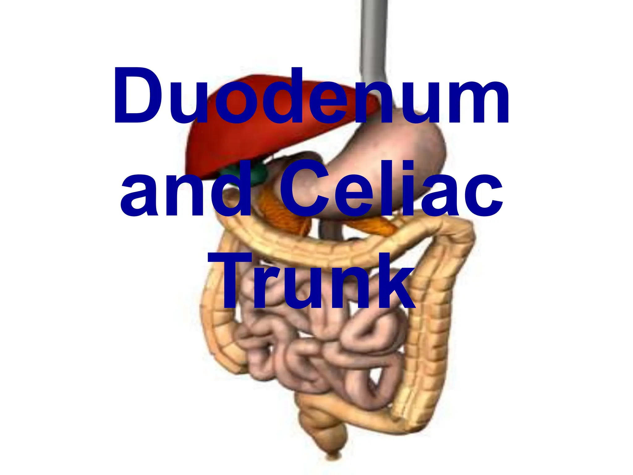
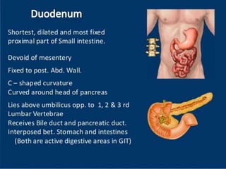
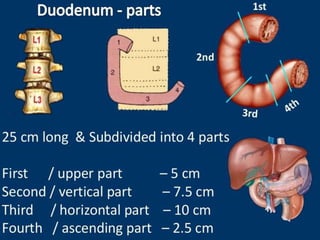
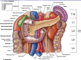
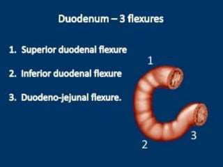
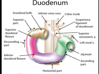
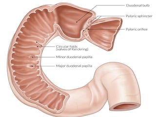
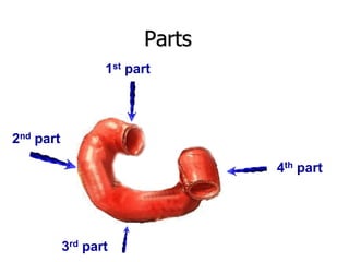
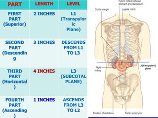
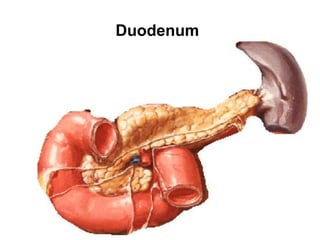
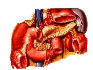
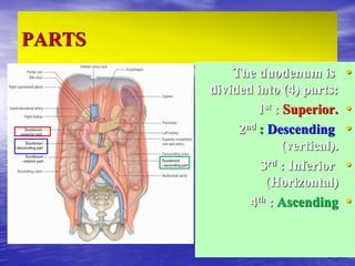
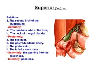
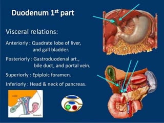
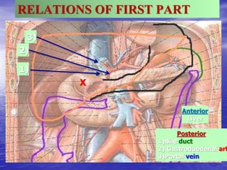
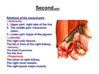
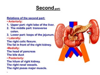
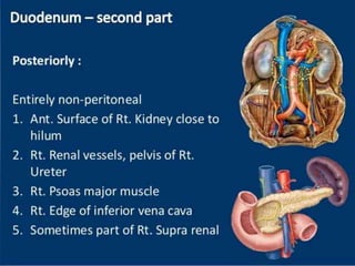
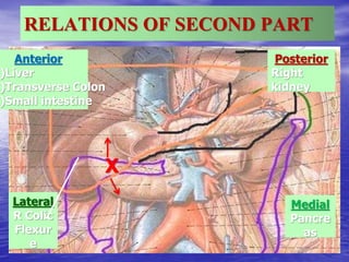
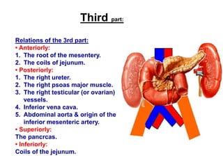
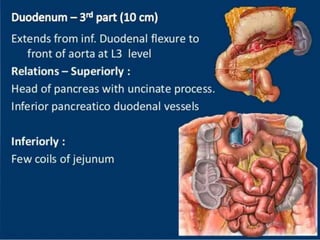
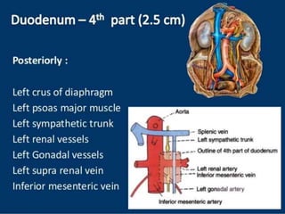
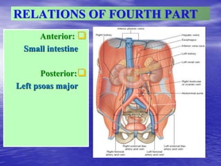
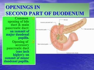
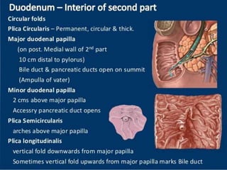
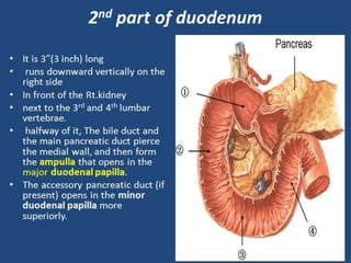
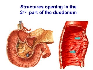
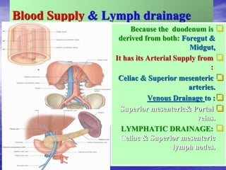
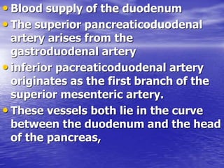
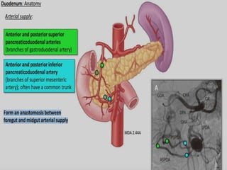
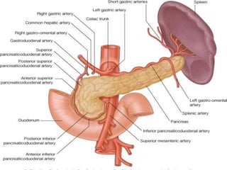
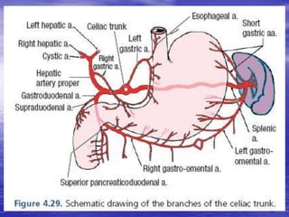
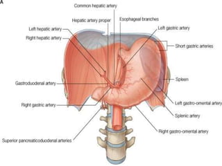
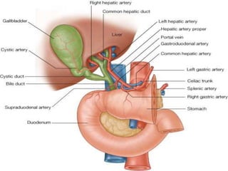
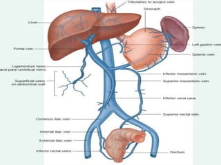
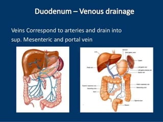
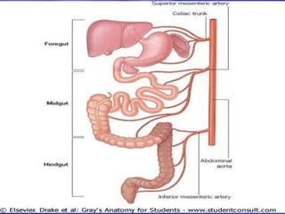
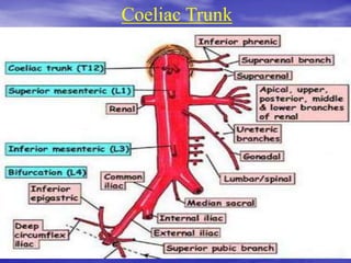
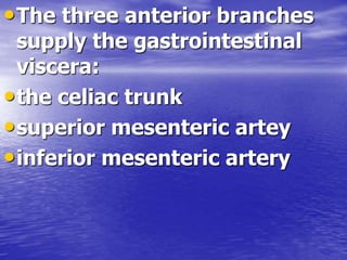
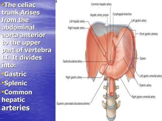
The document discusses the duodenum and its blood supply. It is divided into 4 parts, with the first part located superiorly and the fourth part ascending. Each part has specific relations to surrounding structures. The second part of the duodenum contains openings for the bile duct and pancreatic ducts. The duodenum receives its blood supply from the celiac trunk and superior mesenteric artery and drains into the portal vein and superior mesenteric vein.

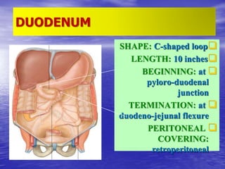
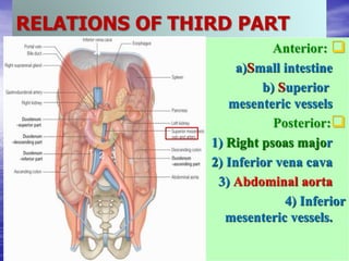
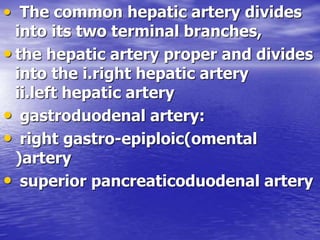
![Fourth part:
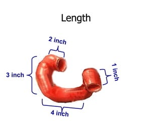
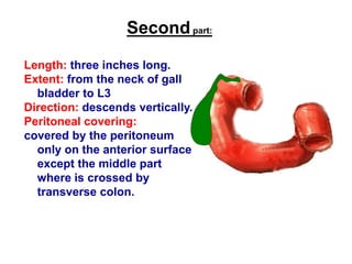
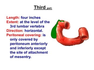
Length: one inch long.
Extent: from the leve] of the
3rd to the level of the 2nd
lumbar vertebrae.
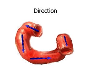
Direction: ascends to end by
forming the duodenojejunal
flexure.
Peritoneal covering: is
covered by the peritoneum
anteriorly and to the left.](https://image.slidesharecdn.com/anatomicalvariationsofduodenum-240228054248-efa6f4dd/85/ANATOMICAL-AND-FUNCTIONAL-VARIATIONS-OF-DUODENUM-ppt-41-320.jpg)
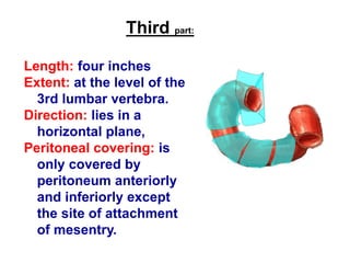
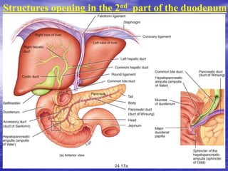
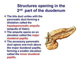
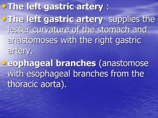
![Fourth part:
Length: one inch long.
Extent: from the leve] of the
3rd to the level of the 2nd
lumbar vertebrae.
Direction: ascends to end by
forming the duodenojejunal
flexure.
Peritoneal covering: is
covered by the peritoneum
anteriorly and to the left.](https://image.slidesharecdn.com/anatomicalvariationsofduodenum-240228054248-efa6f4dd/85/ANATOMICAL-AND-FUNCTIONAL-VARIATIONS-OF-DUODENUM-ppt-42-320.jpg)


![CTEV [ clubfoot] DR ARUN LAL ,DR MOHAMED ASHRAF travancore medical college k...](https://cdn.slidesharecdn.com/ss_thumbnails/ctevclubfootdrarunlaldrmohamedashraftravancoremedicalcollegekollamkeralaindia-260208063247-18fc466c-thumbnail.jpg?width=640&height=640&fit=bounds)

