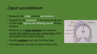
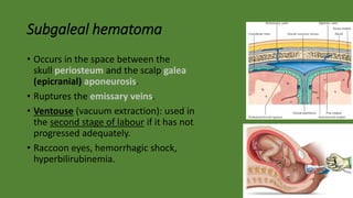
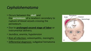
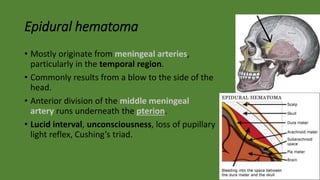
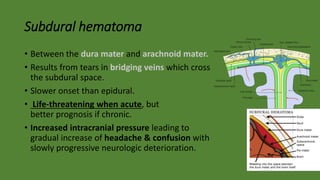
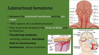
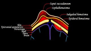
Cranial Hematomas
Hematoma
• Localized collection of blood outside the blood vessels, &
then coagulate and solidify before blood is reabsorbed.
Disease: sickle cell disease, infection, tumors
Trauma: injury or surgery
• Hemorrhage is active or ongoing bleeding.
• Hematoma is a pathologic collection of blood in body tissues, outside
of blood vessels.
Classification
Loaction
• Cranial hematoma
• Subdermal hematoma
• Breast hematoma
• Perichondral hematoma
• Perianal hematoma
• Subungual hematoma
Size
• Petechia < 2 mm
• Purpura 2 mm – 1 cm
• Bruise (ecchymosis, contusion) > 1 cm
Cranial hematomas
• Intra-axial (cerebral) hematoma:
• Intraparenchymal
• Intraventricular
• Extra-axial hematoma:
• Epidural
• Subdural
• Subarachnoid
Caput succedaneum
• Between the scalp and the periosteum.
• Caused by the pressure of the presenting part
of the scalp against the dilating cervix during
delivery.
• Presents as a scalp swelling that extends
across the midline and over suture lines and is
associated with head molding.
• Usually resolves over the first few days.
• Management consists of observation only.
Subgaleal hematoma
• Occurs in the space between the
skull periosteum and the scalp galea
(epicranial) aponeurosis.
• Ruptures the emissary veins.
• Ventouse (vacuum extraction): used in
the second stage of labour if it has not
progressed adequately.
• Raccoon eyes, hemorrhagic shock,
hyperbilirubinemia.
Cephalohematoma
• Occurs between the skull and
the periosteum of a newborn secondary to
rupture of blood vessels crossing the
periosteum.
• Due to prolonged second stage of labor or
instrumental delivery.
• Jaundice, anemia, hypotension
• Risk of infection: osteomyelitis, meningitis
• Differential diagnosis: subgaleal hematoma
Epidural hematoma
• Mostly originate from meningeal arteries,
particularly in the temporal region.
• Commonly results from a blow to the side of the
head.
• Anterior division of the middle meningeal
artery runs underneath the pterion.
• Lucid interval, unconsciousness, loss of pupillary
light reflex, Cushing’s triad.
Subdural hematoma
• Between the dura mater and arachnoid mater.
• Results from tears in bridging veins which cross
the subdural space.
• Slower onset than epidural.
• Life-threatening when acute, but
better prognosis if chronic.
• Increased intracranial pressure leading to
gradual increase of headache & confusion with
slowly progressive neurologic deterioration.
Subarachnoid hematoma
• Between the arachnoid membrane and the pia
mater.
• ~85% rupture of a cerebral aneurysm.
• They tend to be located in the Circle of Willis and
its branches.
• Thunderclap headache,
vomiting, seizures, decreased
level of consciousness,
hemiparesis, Terson syndrome.
Cranial hematomas (injury)

Cranial hematomas (injury)

  • 1.
  • 2.
    Hematoma • Localized collectionof blood outside the blood vessels, & then coagulate and solidify before blood is reabsorbed. Disease: sickle cell disease, infection, tumors Trauma: injury or surgery • Hemorrhage is active or ongoing bleeding. • Hematoma is a pathologic collection of blood in body tissues, outside of blood vessels.
  • 3.
    Classification Loaction • Cranial hematoma •Subdermal hematoma • Breast hematoma • Perichondral hematoma • Perianal hematoma • Subungual hematoma Size • Petechia < 2 mm • Purpura 2 mm – 1 cm • Bruise (ecchymosis, contusion) > 1 cm
  • 4.
    Cranial hematomas • Intra-axial(cerebral) hematoma: • Intraparenchymal • Intraventricular • Extra-axial hematoma: • Epidural • Subdural • Subarachnoid
  • 5.
    Caput succedaneum • Betweenthe scalp and the periosteum. • Caused by the pressure of the presenting part of the scalp against the dilating cervix during delivery. • Presents as a scalp swelling that extends across the midline and over suture lines and is associated with head molding. • Usually resolves over the first few days. • Management consists of observation only.
  • 6.
    Subgaleal hematoma • Occursin the space between the skull periosteum and the scalp galea (epicranial) aponeurosis. • Ruptures the emissary veins. • Ventouse (vacuum extraction): used in the second stage of labour if it has not progressed adequately. • Raccoon eyes, hemorrhagic shock, hyperbilirubinemia.
  • 7.
    Cephalohematoma • Occurs betweenthe skull and the periosteum of a newborn secondary to rupture of blood vessels crossing the periosteum. • Due to prolonged second stage of labor or instrumental delivery. • Jaundice, anemia, hypotension • Risk of infection: osteomyelitis, meningitis • Differential diagnosis: subgaleal hematoma
  • 8.
    Epidural hematoma • Mostlyoriginate from meningeal arteries, particularly in the temporal region. • Commonly results from a blow to the side of the head. • Anterior division of the middle meningeal artery runs underneath the pterion. • Lucid interval, unconsciousness, loss of pupillary light reflex, Cushing’s triad.
  • 9.
    Subdural hematoma • Betweenthe dura mater and arachnoid mater. • Results from tears in bridging veins which cross the subdural space. • Slower onset than epidural. • Life-threatening when acute, but better prognosis if chronic. • Increased intracranial pressure leading to gradual increase of headache & confusion with slowly progressive neurologic deterioration.
  • 10.
    Subarachnoid hematoma • Betweenthe arachnoid membrane and the pia mater. • ~85% rupture of a cerebral aneurysm. • They tend to be located in the Circle of Willis and its branches. • Thunderclap headache, vomiting, seizures, decreased level of consciousness, hemiparesis, Terson syndrome.

Editor's Notes

  • #7 Raccoon eyes due to facial fractures Hyperbilirubinemia due to increased breakdown of RBCs
  • #9 The pterion is the region where the frontal, parietal, temporal, and sphenoid join together. The pterion may also be fractured indirectly by blows to the top or back of the head that place sufficient force on the skull to fracture the pterion. Epidural hematoma can cause herniation of cerebellar tonsils in the posterior cranial fossa (which contains brainstem & cerebellum) causing loss of pupillary light reflex & Cushing’s triad (hypertension, bradycardia, irregular respiration.
  • #10 Subdural hematoma have slower onset of symptoms than epidural, because the lower pressure veins (subdural) bleed more slowly than arteries (epidural).
  • #11 Thunderclap headache: described as “like being kicked in the head” or “the worst ever”, often pulsates towards the occiput (back of the head). Terson's syndrome is the occurrence of a vitreous hemorrhage of the eye in association with subarachnoid hemorrhage.