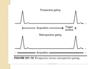
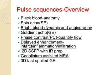
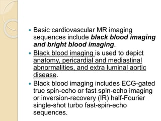
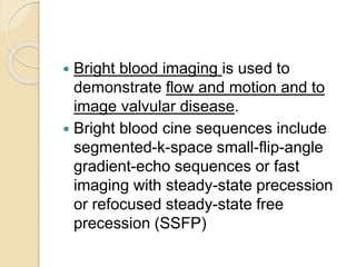
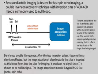
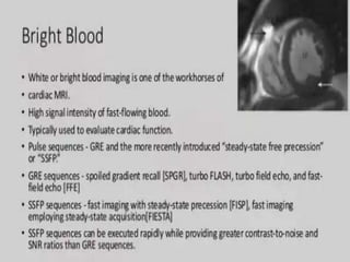
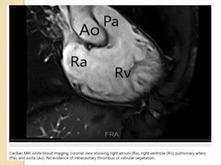
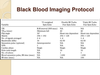
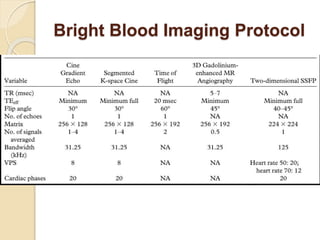
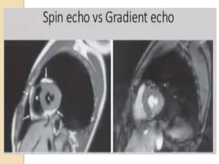
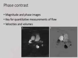
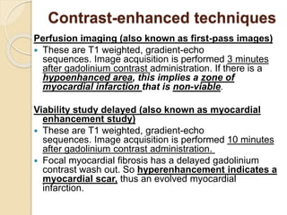
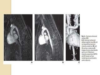
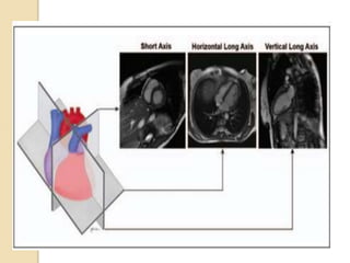
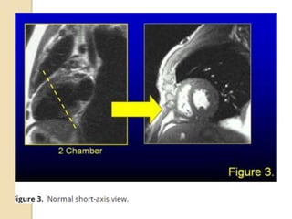
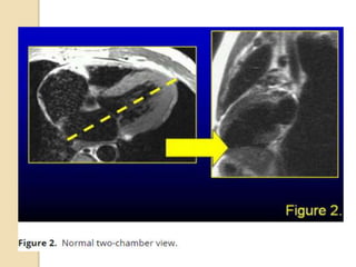
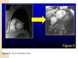
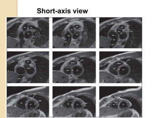
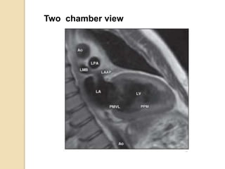
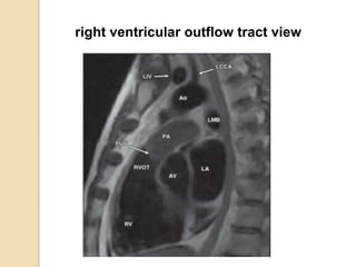
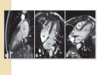
SlideShare a Scribd company logo
Cardiac MRI-Basics
By
Dr.Shaik Asma
Post graduate student
 Cardiac MRI consists of using MRI to
study heart anatomy, physiology, and
pathology.
Advantages
Cardiac MRI offers:
 improved soft tissue definition
 protocol can be tailored to likely
differential diagnoses
◦ a large number of sequences are available
◦ dynamic imaging provides functional
assessment
 no ionizing radiation
◦ MRI safety still requires consideration
Technique
Cardiac gating
MR imaging techniques
 Blood techniques
Dark blood
Bright blood
 Imaging sequences
 Imaging planes
Pulse sequences-Overview
 Black blood-anatomy
Spin echo(SE)
 Bright blood-dynamic and angiography
Gradient echo(GE)
 Phase contrast(PC)-quantify flow
 Delayed enhancement-
infarct/inflammation/infiltration
 2D SSFP with IR prep
 Gadolinium assisted MRA
3D fast spoiled GE
 Basic cardiovascular MR imaging
sequences include black blood imaging
and bright blood imaging.
 Black blood imaging is used to depict
anatomy, pericardial and mediastinal
abnormalities, and extra luminal aortic
disease.
 Black blood imaging includes ECG-gated
true spin-echo or fast spin-echo imaging
or inversion-recovery (IR) half-Fourier
single-shot turbo fast-spin-echo
sequences.
 Bright blood imaging is used to
demonstrate flow and motion and to
image valvular disease.
 Bright blood cine sequences include
segmented-k-space small-flip-angle
gradient-echo sequences or fast
imaging with steady-state precession
or refocused steady-state free
precession (SSFP)
Advantages
 Since it uses the residual transverse
magnetization instead of wasting
it,there is increased signal to noise
ratio.
 Since transverse magnetization is
added back to longitudinal
magnetization,the steady state is
reached quicker,in as little as single
TR.
 The TRs are extremely
 The images have both T1and T2
weighted.(The molecules with long T2
and short T1 will have bright
signal(both fat and water)
 Thus bright signal in steady state
white blood images is attributable to
T1 and T2 signal from the blood,not
flow related.
 Gating of white blood images allows
evaluation of dynamic cardiac function
and physiology throughout cardiac
cycle.Examples include motion of
myocardium and valve leaflets.
 Gating in white blood images serve to
time image acquisition during diastolic
phase of cardiac cycle,thereby limiting
cardiac motion artifact.
Black Blood Imaging Protocol
Bright Blood Imaging Protocol
Cine imaging
Cine imaging or cine MRI, are a type of MRI
sequence acquired to capture motion
 For the heart, this is achieved by acquisition at
multiple time points throughout the cardiac
cycle, after synchronisation with the ECG has
been achieved.
 Separate k-spaces are assigned to different
phases or segments of the cardiac cycle.
 Images are reconstructed from each k-
space and shown in a movie.
 This type of imaging technique requires fast
imaging techniques using very short repetition
and echo times such as balanced steady-state
free precession or spoiled gradient
Contrast-enhanced techniques
Perfusion imaging (also known as first-pass images)
 These are T1 weighted, gradient-echo
sequences. Image acquisition is performed 3 minutes
after gadolinium contrast administration. If there is a
hypoenhanced area, this implies a zone of
myocardial infarction that is non-viable.
Viability study delayed (also known as myocardial
enhancement study)
 These are T1 weighted, gradient-echo
sequences. Image acquisition is performed 10 minutes
after gadolinium contrast administration.
 Focal myocardial fibrosis has a delayed gadolinium
contrast wash out. So hyperenhancement indicates a
myocardial scar, thus an evolved myocardial
infarction.
MR planes of imaging
 The two main planes used for cardiac
MRI include the body (scanner) planes
and the cardiac planes.
 Body Planes
Body planes are oriented orthogonal to
the long axis of the body and consist
of axial, sagittal, and coronal planes .
 The axial plane can depict the four
chambers of the heart and the
pericardium simultaneously.
 The sagittal plane can show the great
vessels arising in continuity from the
ventricles.
 The coronal plane can be used to
assess the left ventricular outflow
tract, the left atrium, and the
pulmonary veins.
Cardiac planes
 The standard cardiac planes are
established using the scout images
and include
short axis view
horizontal long axis (four-chamber view)
vertical long axis (two-chamber view)
 These planes are prescribed along a line
extending from the cardiac apex to the
center of the mitral valve (long axis of the
heart) using the axial body plane images.
 The short-axis plane extends perpendicular
to this true long axis of the heart at the level
of the mid left ventricle.
 The horizontal long(4 chamber) axis is
generated by selecting the horizontal plane
that is perpendicular to the short axis
 The vertical long (2 chamber)axis is
prescribed along a vertical plane orthogonal
to the short-axis plane
Short axis view
 The short-axis plane extends
perpendicular to this true long axis of
the heart at the level of the mid left
ventricle
Two chamber view
 To achieve a two-chamber scout view,
a true axial view through the left
ventricle should be obtained, and then
an oblique coronal scout view should
be positioned parallel to the
interventricular septum
4 chamber view
 The horizontal long axis(4 chamber
view) is generated by selecting the
horizontal plane that is perpendicular
to the short axis
Left ventricular outflow view
 To obtain a long-axis left ventricular
view, an imaging plane should be
positioned from the left ventricular
apex through the mitral valve by using
a two-chamber view depicting the
mitral valve
Planning and acquisition of
standard cardiac views
Short-axis view
Horizontal long axis view
Two chamber view
right ventricular outflow tract view
left ventricular outflow tract view
Indications of cardiac MRI
Conclusion
THANK YOU

More Related Content

What's hot

Coronary CT
Coronary CTCoronary CT
Coronary CT
Dr.Suhas Basavaiah
 
Coronary artery calcium scoring IDoR 2018
Coronary artery calcium scoring IDoR 2018Coronary artery calcium scoring IDoR 2018
Coronary artery calcium scoring IDoR 2018
Shawnm Nasih Dawood
 
Cardiac MRI principle
Cardiac MRI principleCardiac MRI principle
Cardiac MRI principle
Thaiwat Tatsanawiwat
 
Ct angio in cardiology
Ct angio in cardiologyCt angio in cardiology
Ct angio in cardiology
Amit Verma
 
CT Angiography Lower Limb
CT Angiography Lower LimbCT Angiography Lower Limb
CT Angiography Lower Limb
Dr. Yash Kumar Achantani
 
BASIC MRI SEQUENCES
BASIC MRI SEQUENCESBASIC MRI SEQUENCES
BASIC MRI SEQUENCES
Ganesan Yogananthem
 
magnetic resonance angiography
magnetic resonance angiographymagnetic resonance angiography
magnetic resonance angiography
qavi786
 
Ct coronary angiography edited 1st
Ct   coronary  angiography edited 1stCt   coronary  angiography edited 1st
Ct coronary angiography edited 1st
Yashawant Yadav
 
Cardiac CT
Cardiac CTCardiac CT
CAROTID DOPPLER STUDY
CAROTID DOPPLER STUDYCAROTID DOPPLER STUDY
CAROTID DOPPLER STUDY
Dr I Gurubharath .
 
Tissue harmonic imaging
Tissue harmonic imaging Tissue harmonic imaging
Tissue harmonic imaging
MAMTA PANDA
 
CT Coronary angiography
CT Coronary angiography CT Coronary angiography
CT Coronary angiography
Dr-Girish Gunari
 
Coronary CT- angiogram: physics
Coronary CT- angiogram:  physics Coronary CT- angiogram:  physics
Coronary CT- angiogram: physics
REMIX MAHARJAN
 
Role of ct angiography in diagnosis of coronary anomalies
Role of ct angiography in diagnosis of coronary anomalies Role of ct angiography in diagnosis of coronary anomalies
Role of ct angiography in diagnosis of coronary anomalies
GhadaSheta
 
Myocardial perfusion scintigraphy overview
Myocardial perfusion scintigraphy overviewMyocardial perfusion scintigraphy overview
Myocardial perfusion scintigraphy overview
Mostafa Sayed
 
Coronary CT
Coronary CTCoronary CT
Coronary CT
lokesh213
 
CT Angiography presentation
CT Angiography presentation CT Angiography presentation
CT Angiography presentation
Shatha M
 
Coronary ct angiography
Coronary ct angiographyCoronary ct angiography
Coronary ct angiography
SGPGI, lucknow
 
Magnetic resonance angiography
Magnetic resonance angiographyMagnetic resonance angiography
Magnetic resonance angiography
Rahman Ud Din
 
Ct angio ppt
Ct angio pptCt angio ppt
Ct angio ppt
drksp
 

What's hot (20)

Coronary CT
Coronary CTCoronary CT
Coronary CT
 
Coronary artery calcium scoring IDoR 2018
Coronary artery calcium scoring IDoR 2018Coronary artery calcium scoring IDoR 2018
Coronary artery calcium scoring IDoR 2018
 
Cardiac MRI principle
Cardiac MRI principleCardiac MRI principle
Cardiac MRI principle
 
Ct angio in cardiology
Ct angio in cardiologyCt angio in cardiology
Ct angio in cardiology
 
CT Angiography Lower Limb
CT Angiography Lower LimbCT Angiography Lower Limb
CT Angiography Lower Limb
 
BASIC MRI SEQUENCES
BASIC MRI SEQUENCESBASIC MRI SEQUENCES
BASIC MRI SEQUENCES
 
magnetic resonance angiography
magnetic resonance angiographymagnetic resonance angiography
magnetic resonance angiography
 
Ct coronary angiography edited 1st
Ct   coronary  angiography edited 1stCt   coronary  angiography edited 1st
Ct coronary angiography edited 1st
 
Cardiac CT
Cardiac CTCardiac CT
Cardiac CT
 
CAROTID DOPPLER STUDY
CAROTID DOPPLER STUDYCAROTID DOPPLER STUDY
CAROTID DOPPLER STUDY
 
Tissue harmonic imaging
Tissue harmonic imaging Tissue harmonic imaging
Tissue harmonic imaging
 
CT Coronary angiography
CT Coronary angiography CT Coronary angiography
CT Coronary angiography
 
Coronary CT- angiogram: physics
Coronary CT- angiogram:  physics Coronary CT- angiogram:  physics
Coronary CT- angiogram: physics
 
Role of ct angiography in diagnosis of coronary anomalies
Role of ct angiography in diagnosis of coronary anomalies Role of ct angiography in diagnosis of coronary anomalies
Role of ct angiography in diagnosis of coronary anomalies
 
Myocardial perfusion scintigraphy overview
Myocardial perfusion scintigraphy overviewMyocardial perfusion scintigraphy overview
Myocardial perfusion scintigraphy overview
 
Coronary CT
Coronary CTCoronary CT
Coronary CT
 
CT Angiography presentation
CT Angiography presentation CT Angiography presentation
CT Angiography presentation
 
Coronary ct angiography
Coronary ct angiographyCoronary ct angiography
Coronary ct angiography
 
Magnetic resonance angiography
Magnetic resonance angiographyMagnetic resonance angiography
Magnetic resonance angiography
 
Ct angio ppt
Ct angio pptCt angio ppt
Ct angio ppt
 

Similar to Cardiac MRI basics

CT Coronary Angiography.pptx, eart muscle) Acute angina (type of chest pain) ...
CT Coronary Angiography.pptx, eart muscle) Acute angina (type of chest pain) ...CT Coronary Angiography.pptx, eart muscle) Acute angina (type of chest pain) ...
CT Coronary Angiography.pptx, eart muscle) Acute angina (type of chest pain) ...
RukamaneeYadav
 
Nuclear medicine for cardiothoracic surgeons
Nuclear medicine for cardiothoracic surgeonsNuclear medicine for cardiothoracic surgeons
Nuclear medicine for cardiothoracic surgeons
SrikanthK120
 
ECHO WINDOWS AND VIEWS.pptx
ECHO WINDOWS AND VIEWS.pptxECHO WINDOWS AND VIEWS.pptx
ECHO WINDOWS AND VIEWS.pptx
RutviThaker
 
Angiography of Head and Neck
Angiography of Head and NeckAngiography of Head and Neck
Angiography of Head and Neck
SharmaRajan4
 
Diagnostic Procedures for Cardiovascular system
Diagnostic Procedures for Cardiovascular systemDiagnostic Procedures for Cardiovascular system
Diagnostic Procedures for Cardiovascular systemrangeles5
 
ECHO in Cardiac Failure
ECHO in Cardiac Failure ECHO in Cardiac Failure
ECHO in Cardiac Failure
MANULALVS
 
Echo parameters in crt patients selection
Echo parameters in crt patients selectionEcho parameters in crt patients selection
Echo parameters in crt patients selection
Jai Babu
 
Echocardiography an introduction
Echocardiography an introductionEchocardiography an introduction
Echocardiography an introduction
LPS Institute of Cardiology Kanpur UP India
 
intto to echo.pdf
intto to echo.pdfintto to echo.pdf
intto to echo.pdf
MariyaQazi
 
1804.06812
1804.068121804.06812
1804.06812
maicu1
 
Radiology 5th year, 12th lecture (Dr. Abeer)
Radiology 5th year, 12th lecture (Dr. Abeer)Radiology 5th year, 12th lecture (Dr. Abeer)
Radiology 5th year, 12th lecture (Dr. Abeer)
College of Medicine, Sulaymaniyah
 
radiology.CVS 1st lecture.(dr.abeer)
radiology.CVS 1st lecture.(dr.abeer)radiology.CVS 1st lecture.(dr.abeer)
radiology.CVS 1st lecture.(dr.abeer)student
 
Cardiac diagnostics and laboratory tests
Cardiac diagnostics and laboratory tests Cardiac diagnostics and laboratory tests
Cardiac diagnostics and laboratory tests
Dr. Rima Das
 
CSF Flow Study In MRI
CSF Flow Study In MRICSF Flow Study In MRI
CSF Flow Study In MRI
Mohitdeswal13
 
7-190517175000.pdf
7-190517175000.pdf7-190517175000.pdf
7-190517175000.pdf
mdmedicine2012
 
invasive non invasive procedures.pdf for bsc nursing students
invasive non invasive procedures.pdf for bsc nursing studentsinvasive non invasive procedures.pdf for bsc nursing students
invasive non invasive procedures.pdf for bsc nursing students
shanmukhadevi
 
basicechocardiography-170913182344 (2).pdf
basicechocardiography-170913182344 (2).pdfbasicechocardiography-170913182344 (2).pdf
basicechocardiography-170913182344 (2).pdf
littlealphonsa
 

Similar to Cardiac MRI basics (20)

Cardiac MRI
Cardiac MRICardiac MRI
Cardiac MRI
 
introduction
introductionintroduction
introduction
 
4D Flow MRI
4D Flow MRI4D Flow MRI
4D Flow MRI
 
CT Coronary Angiography.pptx, eart muscle) Acute angina (type of chest pain) ...
CT Coronary Angiography.pptx, eart muscle) Acute angina (type of chest pain) ...CT Coronary Angiography.pptx, eart muscle) Acute angina (type of chest pain) ...
CT Coronary Angiography.pptx, eart muscle) Acute angina (type of chest pain) ...
 
Nuclear medicine for cardiothoracic surgeons
Nuclear medicine for cardiothoracic surgeonsNuclear medicine for cardiothoracic surgeons
Nuclear medicine for cardiothoracic surgeons
 
ECHO WINDOWS AND VIEWS.pptx
ECHO WINDOWS AND VIEWS.pptxECHO WINDOWS AND VIEWS.pptx
ECHO WINDOWS AND VIEWS.pptx
 
Angiography of Head and Neck
Angiography of Head and NeckAngiography of Head and Neck
Angiography of Head and Neck
 
Diagnostic Procedures for Cardiovascular system
Diagnostic Procedures for Cardiovascular systemDiagnostic Procedures for Cardiovascular system
Diagnostic Procedures for Cardiovascular system
 
ECHO in Cardiac Failure
ECHO in Cardiac Failure ECHO in Cardiac Failure
ECHO in Cardiac Failure
 
Echo parameters in crt patients selection
Echo parameters in crt patients selectionEcho parameters in crt patients selection
Echo parameters in crt patients selection
 
Echocardiography an introduction
Echocardiography an introductionEchocardiography an introduction
Echocardiography an introduction
 
intto to echo.pdf
intto to echo.pdfintto to echo.pdf
intto to echo.pdf
 
1804.06812
1804.068121804.06812
1804.06812
 
Radiology 5th year, 12th lecture (Dr. Abeer)
Radiology 5th year, 12th lecture (Dr. Abeer)Radiology 5th year, 12th lecture (Dr. Abeer)
Radiology 5th year, 12th lecture (Dr. Abeer)
 
radiology.CVS 1st lecture.(dr.abeer)
radiology.CVS 1st lecture.(dr.abeer)radiology.CVS 1st lecture.(dr.abeer)
radiology.CVS 1st lecture.(dr.abeer)
 
Cardiac diagnostics and laboratory tests
Cardiac diagnostics and laboratory tests Cardiac diagnostics and laboratory tests
Cardiac diagnostics and laboratory tests
 
CSF Flow Study In MRI
CSF Flow Study In MRICSF Flow Study In MRI
CSF Flow Study In MRI
 
7-190517175000.pdf
7-190517175000.pdf7-190517175000.pdf
7-190517175000.pdf
 
invasive non invasive procedures.pdf for bsc nursing students
invasive non invasive procedures.pdf for bsc nursing studentsinvasive non invasive procedures.pdf for bsc nursing students
invasive non invasive procedures.pdf for bsc nursing students
 
basicechocardiography-170913182344 (2).pdf
basicechocardiography-170913182344 (2).pdfbasicechocardiography-170913182344 (2).pdf
basicechocardiography-170913182344 (2).pdf
 

Recently uploaded

basicmodesofventilation2022-220313203758.pdf
basicmodesofventilation2022-220313203758.pdfbasicmodesofventilation2022-220313203758.pdf
basicmodesofventilation2022-220313203758.pdf
aljamhori teaching hospital
 
Non-respiratory Functions of the Lungs.pdf
Non-respiratory Functions of the Lungs.pdfNon-respiratory Functions of the Lungs.pdf
Non-respiratory Functions of the Lungs.pdf
MedicoseAcademics
 
263778731218 Abortion Clinic /Pills In Harare ,
263778731218 Abortion Clinic /Pills In Harare ,263778731218 Abortion Clinic /Pills In Harare ,
263778731218 Abortion Clinic /Pills In Harare ,
sisternakatoto
 
ARTHROLOGY PPT NCISM SYLLABUS AYURVEDA STUDENTS
ARTHROLOGY PPT NCISM SYLLABUS AYURVEDA STUDENTSARTHROLOGY PPT NCISM SYLLABUS AYURVEDA STUDENTS
ARTHROLOGY PPT NCISM SYLLABUS AYURVEDA STUDENTS
Dr. Vinay Pareek
 
Hemodialysis: Chapter 4, Dialysate Circuit - Dr.Gawad
Hemodialysis: Chapter 4, Dialysate Circuit - Dr.GawadHemodialysis: Chapter 4, Dialysate Circuit - Dr.Gawad
Hemodialysis: Chapter 4, Dialysate Circuit - Dr.Gawad
NephroTube - Dr.Gawad
 
How STIs Influence the Development of Pelvic Inflammatory Disease.pptx
How STIs Influence the Development of Pelvic Inflammatory Disease.pptxHow STIs Influence the Development of Pelvic Inflammatory Disease.pptx
How STIs Influence the Development of Pelvic Inflammatory Disease.pptx
FFragrant
 
Ozempic: Preoperative Management of Patients on GLP-1 Receptor Agonists
Ozempic: Preoperative Management of Patients on GLP-1 Receptor Agonists  Ozempic: Preoperative Management of Patients on GLP-1 Receptor Agonists
Ozempic: Preoperative Management of Patients on GLP-1 Receptor Agonists
Saeid Safari
 
ANATOMY AND PHYSIOLOGY OF URINARY SYSTEM.pptx
ANATOMY AND PHYSIOLOGY OF URINARY SYSTEM.pptxANATOMY AND PHYSIOLOGY OF URINARY SYSTEM.pptx
ANATOMY AND PHYSIOLOGY OF URINARY SYSTEM.pptx
Swetaba Besh
 
Knee anatomy and clinical tests 2024.pdf
Knee anatomy and clinical tests 2024.pdfKnee anatomy and clinical tests 2024.pdf
Knee anatomy and clinical tests 2024.pdf
vimalpl1234
 
Light House Retreats: Plant Medicine Retreat Europe
Light House Retreats: Plant Medicine Retreat EuropeLight House Retreats: Plant Medicine Retreat Europe
Light House Retreats: Plant Medicine Retreat Europe
Lighthouse Retreat
 
Top-Vitamin-Supplement-Brands-in-India List
Top-Vitamin-Supplement-Brands-in-India ListTop-Vitamin-Supplement-Brands-in-India List
Top-Vitamin-Supplement-Brands-in-India List
SwisschemDerma
 
Basavarajeeyam - Ayurvedic heritage book of Andhra pradesh
Basavarajeeyam - Ayurvedic heritage book of Andhra pradeshBasavarajeeyam - Ayurvedic heritage book of Andhra pradesh
Basavarajeeyam - Ayurvedic heritage book of Andhra pradesh
Dr. Madduru Muni Haritha
 
Journal Article Review on Rasamanikya
Journal Article Review on RasamanikyaJournal Article Review on Rasamanikya
Journal Article Review on Rasamanikya
Dr. Jyothirmai Paindla
 
The Electrocardiogram - Physiologic Principles
The Electrocardiogram - Physiologic PrinciplesThe Electrocardiogram - Physiologic Principles
The Electrocardiogram - Physiologic Principles
MedicoseAcademics
 
Adv. biopharm. APPLICATION OF PHARMACOKINETICS : TARGETED DRUG DELIVERY SYSTEMS
Adv. biopharm. APPLICATION OF PHARMACOKINETICS : TARGETED DRUG DELIVERY SYSTEMSAdv. biopharm. APPLICATION OF PHARMACOKINETICS : TARGETED DRUG DELIVERY SYSTEMS
Adv. biopharm. APPLICATION OF PHARMACOKINETICS : TARGETED DRUG DELIVERY SYSTEMS
AkankshaAshtankar
 
micro teaching on communication m.sc nursing.pdf
micro teaching on communication m.sc nursing.pdfmicro teaching on communication m.sc nursing.pdf
micro teaching on communication m.sc nursing.pdf
Anurag Sharma
 
Chapter 11 Nutrition and Chronic Diseases.pptx
Chapter 11 Nutrition and Chronic Diseases.pptxChapter 11 Nutrition and Chronic Diseases.pptx
Chapter 11 Nutrition and Chronic Diseases.pptx
Earlene McNair
 
Integrating Ayurveda into Parkinson’s Management: A Holistic Approach
Integrating Ayurveda into Parkinson’s Management: A Holistic ApproachIntegrating Ayurveda into Parkinson’s Management: A Holistic Approach
Integrating Ayurveda into Parkinson’s Management: A Holistic Approach
Ayurveda ForAll
 
Maxilla, Mandible & Hyoid Bone & Clinical Correlations by Dr. RIG.pptx
Maxilla, Mandible & Hyoid Bone & Clinical Correlations by Dr. RIG.pptxMaxilla, Mandible & Hyoid Bone & Clinical Correlations by Dr. RIG.pptx
Maxilla, Mandible & Hyoid Bone & Clinical Correlations by Dr. RIG.pptx
Dr. Rabia Inam Gandapore
 
Local Advanced Lung Cancer: Artificial Intelligence, Synergetics, Complex Sys...
Local Advanced Lung Cancer: Artificial Intelligence, Synergetics, Complex Sys...Local Advanced Lung Cancer: Artificial Intelligence, Synergetics, Complex Sys...
Local Advanced Lung Cancer: Artificial Intelligence, Synergetics, Complex Sys...
Oleg Kshivets
 

Recently uploaded (20)

basicmodesofventilation2022-220313203758.pdf
basicmodesofventilation2022-220313203758.pdfbasicmodesofventilation2022-220313203758.pdf
basicmodesofventilation2022-220313203758.pdf
 
Non-respiratory Functions of the Lungs.pdf
Non-respiratory Functions of the Lungs.pdfNon-respiratory Functions of the Lungs.pdf
Non-respiratory Functions of the Lungs.pdf
 
263778731218 Abortion Clinic /Pills In Harare ,
263778731218 Abortion Clinic /Pills In Harare ,263778731218 Abortion Clinic /Pills In Harare ,
263778731218 Abortion Clinic /Pills In Harare ,
 
ARTHROLOGY PPT NCISM SYLLABUS AYURVEDA STUDENTS
ARTHROLOGY PPT NCISM SYLLABUS AYURVEDA STUDENTSARTHROLOGY PPT NCISM SYLLABUS AYURVEDA STUDENTS
ARTHROLOGY PPT NCISM SYLLABUS AYURVEDA STUDENTS
 
Hemodialysis: Chapter 4, Dialysate Circuit - Dr.Gawad
Hemodialysis: Chapter 4, Dialysate Circuit - Dr.GawadHemodialysis: Chapter 4, Dialysate Circuit - Dr.Gawad
Hemodialysis: Chapter 4, Dialysate Circuit - Dr.Gawad
 
How STIs Influence the Development of Pelvic Inflammatory Disease.pptx
How STIs Influence the Development of Pelvic Inflammatory Disease.pptxHow STIs Influence the Development of Pelvic Inflammatory Disease.pptx
How STIs Influence the Development of Pelvic Inflammatory Disease.pptx
 
Ozempic: Preoperative Management of Patients on GLP-1 Receptor Agonists
Ozempic: Preoperative Management of Patients on GLP-1 Receptor Agonists  Ozempic: Preoperative Management of Patients on GLP-1 Receptor Agonists
Ozempic: Preoperative Management of Patients on GLP-1 Receptor Agonists
 
ANATOMY AND PHYSIOLOGY OF URINARY SYSTEM.pptx
ANATOMY AND PHYSIOLOGY OF URINARY SYSTEM.pptxANATOMY AND PHYSIOLOGY OF URINARY SYSTEM.pptx
ANATOMY AND PHYSIOLOGY OF URINARY SYSTEM.pptx
 
Knee anatomy and clinical tests 2024.pdf
Knee anatomy and clinical tests 2024.pdfKnee anatomy and clinical tests 2024.pdf
Knee anatomy and clinical tests 2024.pdf
 
Light House Retreats: Plant Medicine Retreat Europe
Light House Retreats: Plant Medicine Retreat EuropeLight House Retreats: Plant Medicine Retreat Europe
Light House Retreats: Plant Medicine Retreat Europe
 
Top-Vitamin-Supplement-Brands-in-India List
Top-Vitamin-Supplement-Brands-in-India ListTop-Vitamin-Supplement-Brands-in-India List
Top-Vitamin-Supplement-Brands-in-India List
 
Basavarajeeyam - Ayurvedic heritage book of Andhra pradesh
Basavarajeeyam - Ayurvedic heritage book of Andhra pradeshBasavarajeeyam - Ayurvedic heritage book of Andhra pradesh
Basavarajeeyam - Ayurvedic heritage book of Andhra pradesh
 
Journal Article Review on Rasamanikya
Journal Article Review on RasamanikyaJournal Article Review on Rasamanikya
Journal Article Review on Rasamanikya
 
The Electrocardiogram - Physiologic Principles
The Electrocardiogram - Physiologic PrinciplesThe Electrocardiogram - Physiologic Principles
The Electrocardiogram - Physiologic Principles
 
Adv. biopharm. APPLICATION OF PHARMACOKINETICS : TARGETED DRUG DELIVERY SYSTEMS
Adv. biopharm. APPLICATION OF PHARMACOKINETICS : TARGETED DRUG DELIVERY SYSTEMSAdv. biopharm. APPLICATION OF PHARMACOKINETICS : TARGETED DRUG DELIVERY SYSTEMS
Adv. biopharm. APPLICATION OF PHARMACOKINETICS : TARGETED DRUG DELIVERY SYSTEMS
 
micro teaching on communication m.sc nursing.pdf
micro teaching on communication m.sc nursing.pdfmicro teaching on communication m.sc nursing.pdf
micro teaching on communication m.sc nursing.pdf
 
Chapter 11 Nutrition and Chronic Diseases.pptx
Chapter 11 Nutrition and Chronic Diseases.pptxChapter 11 Nutrition and Chronic Diseases.pptx
Chapter 11 Nutrition and Chronic Diseases.pptx
 
Integrating Ayurveda into Parkinson’s Management: A Holistic Approach
Integrating Ayurveda into Parkinson’s Management: A Holistic ApproachIntegrating Ayurveda into Parkinson’s Management: A Holistic Approach
Integrating Ayurveda into Parkinson’s Management: A Holistic Approach
 
Maxilla, Mandible & Hyoid Bone & Clinical Correlations by Dr. RIG.pptx
Maxilla, Mandible & Hyoid Bone & Clinical Correlations by Dr. RIG.pptxMaxilla, Mandible & Hyoid Bone & Clinical Correlations by Dr. RIG.pptx
Maxilla, Mandible & Hyoid Bone & Clinical Correlations by Dr. RIG.pptx
 
Local Advanced Lung Cancer: Artificial Intelligence, Synergetics, Complex Sys...
Local Advanced Lung Cancer: Artificial Intelligence, Synergetics, Complex Sys...Local Advanced Lung Cancer: Artificial Intelligence, Synergetics, Complex Sys...
Local Advanced Lung Cancer: Artificial Intelligence, Synergetics, Complex Sys...
 

Cardiac MRI basics

  • 2.  Cardiac MRI consists of using MRI to study heart anatomy, physiology, and pathology.
  • 3. Advantages Cardiac MRI offers:  improved soft tissue definition  protocol can be tailored to likely differential diagnoses ◦ a large number of sequences are available ◦ dynamic imaging provides functional assessment  no ionizing radiation ◦ MRI safety still requires consideration
  • 5.
  • 6.
  • 7.
  • 8.
  • 9.
  • 10.
  • 12.
  • 13. MR imaging techniques  Blood techniques Dark blood Bright blood  Imaging sequences  Imaging planes
  • 14. Pulse sequences-Overview  Black blood-anatomy Spin echo(SE)  Bright blood-dynamic and angiography Gradient echo(GE)  Phase contrast(PC)-quantify flow  Delayed enhancement- infarct/inflammation/infiltration  2D SSFP with IR prep  Gadolinium assisted MRA 3D fast spoiled GE
  • 15.  Basic cardiovascular MR imaging sequences include black blood imaging and bright blood imaging.  Black blood imaging is used to depict anatomy, pericardial and mediastinal abnormalities, and extra luminal aortic disease.  Black blood imaging includes ECG-gated true spin-echo or fast spin-echo imaging or inversion-recovery (IR) half-Fourier single-shot turbo fast-spin-echo sequences.
  • 16.  Bright blood imaging is used to demonstrate flow and motion and to image valvular disease.  Bright blood cine sequences include segmented-k-space small-flip-angle gradient-echo sequences or fast imaging with steady-state precession or refocused steady-state free precession (SSFP)
  • 17.
  • 18.
  • 19.
  • 20.
  • 21.
  • 22. Advantages  Since it uses the residual transverse magnetization instead of wasting it,there is increased signal to noise ratio.  Since transverse magnetization is added back to longitudinal magnetization,the steady state is reached quicker,in as little as single TR.  The TRs are extremely
  • 23.  The images have both T1and T2 weighted.(The molecules with long T2 and short T1 will have bright signal(both fat and water)  Thus bright signal in steady state white blood images is attributable to T1 and T2 signal from the blood,not flow related.
  • 24.  Gating of white blood images allows evaluation of dynamic cardiac function and physiology throughout cardiac cycle.Examples include motion of myocardium and valve leaflets.  Gating in white blood images serve to time image acquisition during diastolic phase of cardiac cycle,thereby limiting cardiac motion artifact.
  • 25.
  • 28. Cine imaging Cine imaging or cine MRI, are a type of MRI sequence acquired to capture motion  For the heart, this is achieved by acquisition at multiple time points throughout the cardiac cycle, after synchronisation with the ECG has been achieved.  Separate k-spaces are assigned to different phases or segments of the cardiac cycle.  Images are reconstructed from each k- space and shown in a movie.  This type of imaging technique requires fast imaging techniques using very short repetition and echo times such as balanced steady-state free precession or spoiled gradient
  • 29.
  • 30.
  • 31.
  • 32. Contrast-enhanced techniques Perfusion imaging (also known as first-pass images)  These are T1 weighted, gradient-echo sequences. Image acquisition is performed 3 minutes after gadolinium contrast administration. If there is a hypoenhanced area, this implies a zone of myocardial infarction that is non-viable. Viability study delayed (also known as myocardial enhancement study)  These are T1 weighted, gradient-echo sequences. Image acquisition is performed 10 minutes after gadolinium contrast administration.  Focal myocardial fibrosis has a delayed gadolinium contrast wash out. So hyperenhancement indicates a myocardial scar, thus an evolved myocardial infarction.
  • 33.
  • 34.
  • 35. MR planes of imaging
  • 36.  The two main planes used for cardiac MRI include the body (scanner) planes and the cardiac planes.  Body Planes Body planes are oriented orthogonal to the long axis of the body and consist of axial, sagittal, and coronal planes .
  • 37.  The axial plane can depict the four chambers of the heart and the pericardium simultaneously.  The sagittal plane can show the great vessels arising in continuity from the ventricles.  The coronal plane can be used to assess the left ventricular outflow tract, the left atrium, and the pulmonary veins.
  • 38.
  • 39. Cardiac planes  The standard cardiac planes are established using the scout images and include short axis view horizontal long axis (four-chamber view) vertical long axis (two-chamber view)
  • 40.  These planes are prescribed along a line extending from the cardiac apex to the center of the mitral valve (long axis of the heart) using the axial body plane images.  The short-axis plane extends perpendicular to this true long axis of the heart at the level of the mid left ventricle.  The horizontal long(4 chamber) axis is generated by selecting the horizontal plane that is perpendicular to the short axis  The vertical long (2 chamber)axis is prescribed along a vertical plane orthogonal to the short-axis plane
  • 41.
  • 42. Short axis view  The short-axis plane extends perpendicular to this true long axis of the heart at the level of the mid left ventricle
  • 43.
  • 44. Two chamber view  To achieve a two-chamber scout view, a true axial view through the left ventricle should be obtained, and then an oblique coronal scout view should be positioned parallel to the interventricular septum
  • 45.
  • 46. 4 chamber view  The horizontal long axis(4 chamber view) is generated by selecting the horizontal plane that is perpendicular to the short axis
  • 47.
  • 48. Left ventricular outflow view  To obtain a long-axis left ventricular view, an imaging plane should be positioned from the left ventricular apex through the mitral valve by using a two-chamber view depicting the mitral valve
  • 49.
  • 50.
  • 51. Planning and acquisition of standard cardiac views
  • 57.
  • 59.