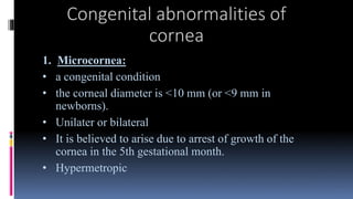
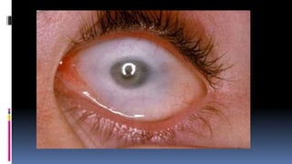
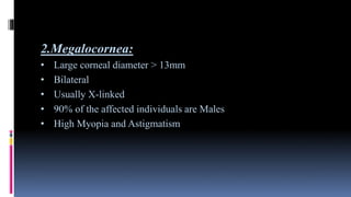
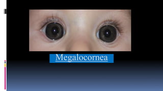
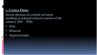
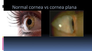
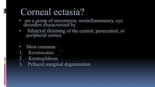
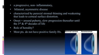
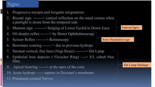
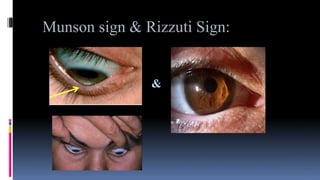
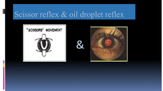
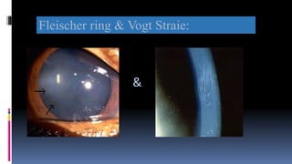
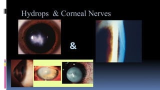
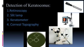
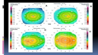
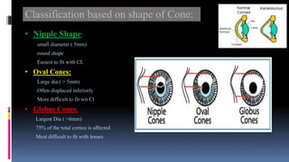
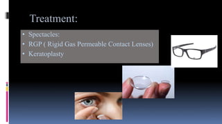
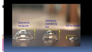
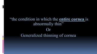
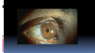
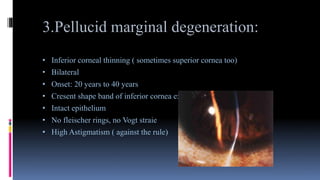
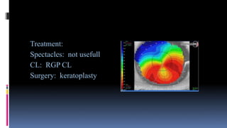
Corneal Disease
Lecturer:
Miss.Yumna Tariq
M.Phil. Scholar ( Optom)
Congenital abnormalities of
cornea
1. Microcornea:
• a congenital condition
• the corneal diameter is <10 mm (or <9 mm in
newborns).
• Unilater or bilateral
• It is believed to arise due to arrest of growth of the
cornea in the 5th gestational month.
• Hypermetropic
2.Megalocornea:
• Large corneal diameter > 13mm
• Bilateral
• Usually X-linked
• 90% of the affected individuals are Males
• High Myopia and Astigmatism
Megalocornea
c. Cornea Plana:
Severe decrease in corneal curvature
resulting in reduced refractive power of the
cornea ( 20D – 30D)
• Rare
• Bilateral
• Hypermetropia
Normal cornea vs cornea plana
Corneal ectasias:
Ectasia:
• an enlargement, dilation, or ballooning effect
Corneal ectasia?
• are a group of uncommon, noninflammatory, eye
disorders characterised by
• bilateral thinning of the central, paracentral, or
peripheral cornea
• Most common
1. Keratoconus
2. Keratoglubous
3. Pellucid marginal degeneration
1.Keratoconus:
• a progressive, non- inflammatory,
• bilateral, asymmetric disease
• characterized by paraxial stromal thinning and weakening
that leads to corneal surface distortion.
• Onset = around puberty, slow progression thereafter until
the 3rd & 4th decades of life
• Role of heredity?
• Most pts. do not have positive family Hx.
Presentation:
1. Reduce visual Acuity
2. Frequent changes in spectacle prescription
3. Glare
4. Ghost images
5. Monocular Diplopia
Signs:
1. Progressive myopia and irregular astigmatism
2. Rizzuti sign -------> conical reflection on the nasal cornea when
a penlight is shone from the temporal side
3. Munson sign --------> bulging of Lower Eyelid in Down Gaze
4. Oil droplet reflex -------> by Direct Ophthalmoscopy
5. Scissor Reflex --------> Retinoscopy
6. Bowmans scarring ------- > due to previous hydrops
7. Stromal vertical, fine lines (Vogt Straie) --------> Slit Lamp
8. Epithelial Iron deposits ( Fleischer Ring) ----> S/L cobalt blue
filter
9. Apical Scarring ------> at the apex of the cone
10. Acute hydrops ----> rapture in Descmet’s membrane
11. Prominent corneal Nerves
External Signs:
Slit Lamp findings:
Retro illumination sign
Munson sign & Rizzuti Sign:
&
Scissor reflex & oil droplet reflex
&
Fleischer ring & Vogt Straie:
&
Hydrops & Corneal Nerves
&
Detection of Keratoconus:
1.Retinoscopy
2. Slit lamp
3. Keratometer
4. Corneal Topography
CLASSIFICATION OF KERATOCONUS
Classification based on shape of Cone:
• Nipple Shape:
small diameter ( 5mm)
round shape
Easiest to fit with CL
• Oval Cones:
Large dia ( > 5mm)
Often displaced inferiorly
More difficult to fit wit Cl
• Globus Cones:
Largest Dia ( >6mm)
75% of the total cornea is affected
Most difficult to fit with lenses
ON THE BASIS OF Keratometer
Reading:
Grades of Keratoconus:
• Mild K ( < 48 D)
• Moderate K ( 48D – 54D)
• Severe K ( > 54D )
Associations:
Systemic:
• Downs Syndrome
• Marfan’s Syndrome
• Turner syndrome
• Ehlers- Danlos Syndrom etc
Ocular :
• Persistent Eye rubbing
• Aniridia
• Retinitis pigmentosa
Treatment:
• Spectacles:
• RGP ( Rigid Gas Permeable Contact Lenses)
• Keratoplasty
2. Keratoglobus:
“the condition in which the entire cornea is
abnormally thin”
Or
Generalized thinning of cornea
Onset: at birth
Signs: ectasia is Generalized
acute hydrops occur less commonly
Cornea is more prone to rupture on trauma
Management:
• CL ------ scleral CL
• Surgery ------poor results
3.Pellucid marginal degeneration:
• Inferior corneal thinning ( sometimes superior cornea too)
• Bilateral
• Onset: 20 years to 40 years
• Cresent shape band of inferior cornea extending from 4 – 8 o’clock
• Intact epithelium
• No fleischer rings, no Vogt straie
• High Astigmatism ( against the rule)
Treatment:
Spectacles: not usefull
CL: RGP CL
Surgery: keratoplasty
Thanks.

Corneal diseases

  • 1.
  • 2.
    Congenital abnormalities of cornea 1.Microcornea: • a congenital condition • the corneal diameter is <10 mm (or <9 mm in newborns). • Unilater or bilateral • It is believed to arise due to arrest of growth of the cornea in the 5th gestational month. • Hypermetropic
  • 4.
    2.Megalocornea: • Large cornealdiameter > 13mm • Bilateral • Usually X-linked • 90% of the affected individuals are Males • High Myopia and Astigmatism
  • 5.
  • 6.
    c. Cornea Plana: Severedecrease in corneal curvature resulting in reduced refractive power of the cornea ( 20D – 30D) • Rare • Bilateral • Hypermetropia
  • 7.
    Normal cornea vscornea plana
  • 8.
    Corneal ectasias: Ectasia: • anenlargement, dilation, or ballooning effect
  • 9.
    Corneal ectasia? • area group of uncommon, noninflammatory, eye disorders characterised by • bilateral thinning of the central, paracentral, or peripheral cornea • Most common 1. Keratoconus 2. Keratoglubous 3. Pellucid marginal degeneration
  • 10.
  • 11.
    • a progressive,non- inflammatory, • bilateral, asymmetric disease • characterized by paraxial stromal thinning and weakening that leads to corneal surface distortion. • Onset = around puberty, slow progression thereafter until the 3rd & 4th decades of life • Role of heredity? • Most pts. do not have positive family Hx.
  • 12.
    Presentation: 1. Reduce visualAcuity 2. Frequent changes in spectacle prescription 3. Glare 4. Ghost images 5. Monocular Diplopia
  • 13.
    Signs: 1. Progressive myopiaand irregular astigmatism 2. Rizzuti sign -------> conical reflection on the nasal cornea when a penlight is shone from the temporal side 3. Munson sign --------> bulging of Lower Eyelid in Down Gaze 4. Oil droplet reflex -------> by Direct Ophthalmoscopy 5. Scissor Reflex --------> Retinoscopy 6. Bowmans scarring ------- > due to previous hydrops 7. Stromal vertical, fine lines (Vogt Straie) --------> Slit Lamp 8. Epithelial Iron deposits ( Fleischer Ring) ----> S/L cobalt blue filter 9. Apical Scarring ------> at the apex of the cone 10. Acute hydrops ----> rapture in Descmet’s membrane 11. Prominent corneal Nerves External Signs: Slit Lamp findings: Retro illumination sign
  • 14.
    Munson sign &Rizzuti Sign: &
  • 15.
    Scissor reflex &oil droplet reflex &
  • 16.
    Fleischer ring &Vogt Straie: &
  • 17.
  • 18.
    Detection of Keratoconus: 1.Retinoscopy 2.Slit lamp 3. Keratometer 4. Corneal Topography
  • 20.
  • 21.
    Classification based onshape of Cone: • Nipple Shape: small diameter ( 5mm) round shape Easiest to fit with CL • Oval Cones: Large dia ( > 5mm) Often displaced inferiorly More difficult to fit wit Cl • Globus Cones: Largest Dia ( >6mm) 75% of the total cornea is affected Most difficult to fit with lenses
  • 22.
    ON THE BASISOF Keratometer Reading: Grades of Keratoconus: • Mild K ( < 48 D) • Moderate K ( 48D – 54D) • Severe K ( > 54D )
  • 23.
    Associations: Systemic: • Downs Syndrome •Marfan’s Syndrome • Turner syndrome • Ehlers- Danlos Syndrom etc Ocular : • Persistent Eye rubbing • Aniridia • Retinitis pigmentosa
  • 24.
    Treatment: • Spectacles: • RGP( Rigid Gas Permeable Contact Lenses) • Keratoplasty
  • 26.
  • 27.
    “the condition inwhich the entire cornea is abnormally thin” Or Generalized thinning of cornea
  • 29.
    Onset: at birth Signs:ectasia is Generalized acute hydrops occur less commonly Cornea is more prone to rupture on trauma Management: • CL ------ scleral CL • Surgery ------poor results
  • 30.
    3.Pellucid marginal degeneration: •Inferior corneal thinning ( sometimes superior cornea too) • Bilateral • Onset: 20 years to 40 years • Cresent shape band of inferior cornea extending from 4 – 8 o’clock • Intact epithelium • No fleischer rings, no Vogt straie • High Astigmatism ( against the rule)
  • 31.
    Treatment: Spectacles: not usefull CL:RGP CL Surgery: keratoplasty
  • 32.