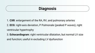
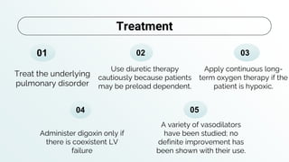
Cor Pulmonale
Ghadeer Ismail Eideh
Supervised by Dr. Ashraf Zughair
19-sep-2022
From: step up to medicine
Definition and
causes
Clinical features
Diagnosis Treatment
01
03
02
04
Outlines
1. is defined as right ventricular hypertrophy with
eventual RV failure resulting from pulmonary
HTN, secondary to pulmonary disease.
2. The definition does not encompass any of the
causes of pulmonary HTN due to left-sided heart
disease (such as mitral stenosis or left-to-right
shunts).
Definition
1. It is most commonly secondary to
COPD.
2. Other causes include recurrent
PE, ILD, asthma, CF, sleep apnea,
and pneumoconioses.
Causes:
Clinical features
1 .Decrease in exercise tolerance
2. Cyanosis and digital clubbing
3. Signs of right ventricular failure: hepatomegaly, edema, JVD
4. Parasternal lift
1. CXR: enlargement of the RA, RV, and pulmonary arteries
2. ECG: right-axis deviation, P Pulmonale (peaked P waves), right
ventricular hypertrophy
3. Echocardiogram: right ventricular dilatation, but normal LV size
and function; useful in excluding LV dysfunction
Diagnosis
Apply continuous long-
term oxygen therapy if the
patient is hypoxic.
Treat the underlying
pulmonary disorder
Use diuretic therapy
cautiously because patients
may be preload dependent.
Administer digoxin only if
there is coexistent LV
failure
01 02 03
04
Treatment
05
A variety of vasodilators
have been studied; no
definite improvement has
been shown with their use.
Thank you.

Cor pulmonale..pptx

  • 1.
    Cor Pulmonale Ghadeer IsmailEideh Supervised by Dr. Ashraf Zughair 19-sep-2022 From: step up to medicine
  • 2.
  • 3.
    1. is definedas right ventricular hypertrophy with eventual RV failure resulting from pulmonary HTN, secondary to pulmonary disease. 2. The definition does not encompass any of the causes of pulmonary HTN due to left-sided heart disease (such as mitral stenosis or left-to-right shunts). Definition
  • 4.
    1. It ismost commonly secondary to COPD. 2. Other causes include recurrent PE, ILD, asthma, CF, sleep apnea, and pneumoconioses. Causes:
  • 6.
    Clinical features 1 .Decreasein exercise tolerance 2. Cyanosis and digital clubbing 3. Signs of right ventricular failure: hepatomegaly, edema, JVD 4. Parasternal lift
  • 7.
    1. CXR: enlargementof the RA, RV, and pulmonary arteries 2. ECG: right-axis deviation, P Pulmonale (peaked P waves), right ventricular hypertrophy 3. Echocardiogram: right ventricular dilatation, but normal LV size and function; useful in excluding LV dysfunction Diagnosis
  • 8.
    Apply continuous long- termoxygen therapy if the patient is hypoxic. Treat the underlying pulmonary disorder Use diuretic therapy cautiously because patients may be preload dependent. Administer digoxin only if there is coexistent LV failure 01 02 03 04 Treatment 05 A variety of vasodilators have been studied; no definite improvement has been shown with their use.
  • 9.