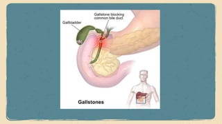
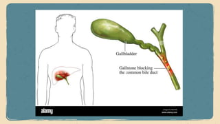
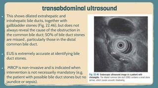
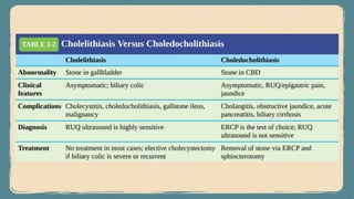
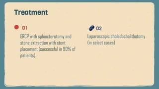
Choledocholithiasis
Ghadeer Ismail Eideh
Supervised by Dr. Aref Rajabi
From: step up to medicine
Outlines
Introduction
01 02 Clinical features
03 Diagnosis
04 Treatment
INTRODUCTION
choledocholithiasis
Stones in the common bile duct,
occur in 10–15% of patients with
gallstones, which have usually
migrated from the gallbladder.
Primary VS secondary stones
• Primary stones originate in the CBD (usually
pigmented stones).
• They are rare but can develop within the
common bile duct many years after a
cholecystectomy, and are sometimes related
to biliary sludge arising from dysfunction of
the sphincter of Oddi.
• In Far Eastern countries, are thought to follow
bacterial infection secondary to parasitic
infections with Clonorchis sinensis, Ascaris
lumbricoides or Fasciola hepatica
Primary stones
Secondary stones originate in the
gallbladder and then pass into the
CBD (usually cholesterol or mixed
stones). These account for 95% of
all cases.
Secondary stones
Clinical features
Choledocholithiasis may be
asymptomatic, may be
found incidentally by
operative cholangiography
at cholecystectomy, or may
manifest as recurrent
abdominal pain with or
without jaundice.
Clinical features
The pain is usually in the
right upper quadrant, and
fever, pruritus and dark
urine may be present.
Physical examination
may show the scar of a
previous
cholecystectomy; if the
gallbladder is present, it
is usually small, fibrotic
and impalpable.
Diagnosis
Diagnosis
Laboratory tests
ERCP
RUQ ultrasound
ERCP
Is the gold standard (sensitivity and
specificity of 95%) and should
follow ultrasound. ERCP is
diagnostic and therapeutic
Lab-test
Total and direct bilirubin
levels are elevated, as well
as ALK-P.
PTC is an
alternative to ERCP
RUQ ultrasound
Is usually the initial study, but is not a
sensitive study for choledocholithiasis.
It detects CBD in only 50% of cases.
transabdominal ultrasound
â—Ź This shows dilated extrahepatic and
intrahepatic bile ducts, together with
gallbladder stones (Fig. 22.46), but does not
always reveal the cause of the obstruction in
the common bile duct; 50% of bile duct stones
are missed , particularly those in the distal
common bile duct.
â—Ź EUS is extremely accurate at identifying bile
duct stones.
â—Ź MRCP is non-invasive and is indicated when
intervention is not necessarily mandatory (e.g.
the patient with possible bile duct stones but no
jaundice or sepsis).
Complications
Complications of choledocholithiasis
Biliary colic Biliary cirrhosis
Cholangitis Obstructive
jaundice
Acute
pancreatitis
Cholelithiasis Versus
Choledocholithiasis
Treatment
Treatment
01
ERCP with sphincterotomy and
stone extraction with stent
placement (successful in 90% of
patients).
02
Laparoscopic choledocholithotomy
(in select cases)
Thank you

Choledocholithiasis.pptx

  • 1.
    Choledocholithiasis Ghadeer Ismail Eideh Supervisedby Dr. Aref Rajabi From: step up to medicine
  • 2.
    Outlines Introduction 01 02 Clinicalfeatures 03 Diagnosis 04 Treatment
  • 3.
    INTRODUCTION choledocholithiasis Stones in thecommon bile duct, occur in 10–15% of patients with gallstones, which have usually migrated from the gallbladder.
  • 6.
  • 7.
    • Primary stonesoriginate in the CBD (usually pigmented stones). • They are rare but can develop within the common bile duct many years after a cholecystectomy, and are sometimes related to biliary sludge arising from dysfunction of the sphincter of Oddi. • In Far Eastern countries, are thought to follow bacterial infection secondary to parasitic infections with Clonorchis sinensis, Ascaris lumbricoides or Fasciola hepatica Primary stones Secondary stones originate in the gallbladder and then pass into the CBD (usually cholesterol or mixed stones). These account for 95% of all cases. Secondary stones
  • 8.
  • 9.
    Choledocholithiasis may be asymptomatic,may be found incidentally by operative cholangiography at cholecystectomy, or may manifest as recurrent abdominal pain with or without jaundice. Clinical features The pain is usually in the right upper quadrant, and fever, pruritus and dark urine may be present. Physical examination may show the scar of a previous cholecystectomy; if the gallbladder is present, it is usually small, fibrotic and impalpable.
  • 10.
  • 11.
    Diagnosis Laboratory tests ERCP RUQ ultrasound ERCP Isthe gold standard (sensitivity and specificity of 95%) and should follow ultrasound. ERCP is diagnostic and therapeutic Lab-test Total and direct bilirubin levels are elevated, as well as ALK-P. PTC is an alternative to ERCP RUQ ultrasound Is usually the initial study, but is not a sensitive study for choledocholithiasis. It detects CBD in only 50% of cases.
  • 12.
    transabdominal ultrasound â—Ź Thisshows dilated extrahepatic and intrahepatic bile ducts, together with gallbladder stones (Fig. 22.46), but does not always reveal the cause of the obstruction in the common bile duct; 50% of bile duct stones are missed , particularly those in the distal common bile duct. â—Ź EUS is extremely accurate at identifying bile duct stones. â—Ź MRCP is non-invasive and is indicated when intervention is not necessarily mandatory (e.g. the patient with possible bile duct stones but no jaundice or sepsis).
  • 13.
  • 14.
    Complications of choledocholithiasis Biliarycolic Biliary cirrhosis Cholangitis Obstructive jaundice Acute pancreatitis
  • 15.
  • 17.
  • 18.
    Treatment 01 ERCP with sphincterotomyand stone extraction with stent placement (successful in 90% of patients). 02 Laparoscopic choledocholithotomy (in select cases)
  • 19.

Editor's Notes

  • #2 1. Table of contents 2. Introduction 3. Identifying information 4. Patient medical history 5. Review of systems 6. Physical examination 7. Big picture 8. Findings 9. Discussion 10. Discussion summary 11. Comparison 12. Diagnosis 13. Treatment 14. Patient monitoring 15. Contraindications and indications 16. Post-prevention 17. Case timeline 18. Conclusions 19. References 20. Our team