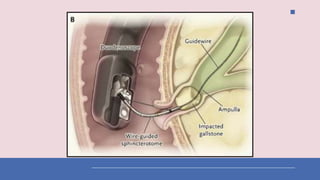
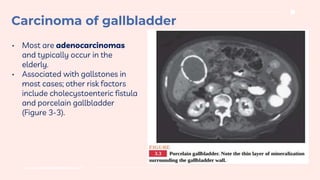
Cholangitis
Ghadeer Ismail Eideh
Supervised by Dr. Aref Rajabi
From: step up to medicine
03
01 02
04
Outlines
Introduction Clinical features
Diagnosis Treatment
Introduction
01
INTRODUCTION
• Infection of biliary tract secondary to obstruction,
which leads to biliary stasis and bacterial
overgrowth.
• Choledocholithiasis accounts for 60% of cases.
• Other causes include pancreatic and biliary
neoplasm, postoperative strictures, invasive
procedures such as ERCP or PTC, and choledochal
cysts.
• Cholangitis is potentially life threatening and
requires emergency treatment.
Clinical features
02
RUQ pain
This classic triad is present in only 50% to 70% of cases.
Jaundice
Charcot triad
Fever
Raynolds pentad =
Septic shock Altered mental status
Charcot traid
Reynolds pentad is a highly toxic state that requires
emergency treatment. It can be rapidly fatal.
Diagnosis
03
a. This is the definitive test, but it should not be performed during the acute phase of
illness. Once cholangitis resolves, proceed with PTC or ERCP to identify the
underlying problem and plan treatment.
b. Perform PTC when the duct system is dilated (per ultrasound) and ERCP when the
duct system is normal.
RUQ ultrasound
Is the initial study, very accurate in detecting gallstones and biliary
tract dilatation, but not very accurate in detecting CBD stones.
Laboratory findings
Hyperbilirubinemia, leukocytosis, mild elevation in serum transaminases.
Cholangiography (PTC or ERCP).
Treatment
04
Treatment
1. IV antibiotics and IV fluids
a. Close monitoring of hemodynamics, BP, and urine
output is important.
b. Most patients respond rapidly. Once the patient has
been afebrile for 48 hours, cholangiography (PTC or
ERCP) can be performed for evaluation of the underlying
condition.
2. Decompress CBD via PTC (catheter drainage); ERCP
(sphincterotomy), or laparotomy (T-tube insertion) once
the patient is stabilized, or emergently if the condition
does not respond to antibiotics.
Thank you
Carcinoma of
the gallbladder
www.traditionalmedicine.com
• Most are adenocarcinomas
and typically occur in the
elderly.
• Associated with gallstones in
most cases; other risk factors
include cholecystoenteric fistula
and porcelain gallbladder
(Figure 3-3).
Carcinoma of gallbladder
• Clinical features are nonspecific and suggest
extrahepatic bile duct obstruction: jaundice, biliary
colic, weight loss, anorexia, and RUQ mass.
Palpable gallbladder is a sign of advanced disease.
• Difficult to remove with surgery: cholecystectomy
versus radical cholecystectomy (with wedge
resection of liver and lymph node dissection)
depending on depth of invasion.
• Prognosis is dismal—more than 90% of patients
die of advanced disease within 1 year of diagnosis.
Disease often goes undetected until it is advanced.
Thank you

cholangitis.pptx

  • 1.
    Cholangitis Ghadeer Ismail Eideh Supervisedby Dr. Aref Rajabi From: step up to medicine
  • 2.
    03 01 02 04 Outlines Introduction Clinicalfeatures Diagnosis Treatment
  • 3.
  • 4.
    INTRODUCTION • Infection ofbiliary tract secondary to obstruction, which leads to biliary stasis and bacterial overgrowth. • Choledocholithiasis accounts for 60% of cases. • Other causes include pancreatic and biliary neoplasm, postoperative strictures, invasive procedures such as ERCP or PTC, and choledochal cysts. • Cholangitis is potentially life threatening and requires emergency treatment.
  • 5.
  • 6.
    RUQ pain This classictriad is present in only 50% to 70% of cases. Jaundice Charcot triad Fever
  • 7.
    Raynolds pentad = Septicshock Altered mental status Charcot traid Reynolds pentad is a highly toxic state that requires emergency treatment. It can be rapidly fatal.
  • 8.
  • 9.
    a. This isthe definitive test, but it should not be performed during the acute phase of illness. Once cholangitis resolves, proceed with PTC or ERCP to identify the underlying problem and plan treatment. b. Perform PTC when the duct system is dilated (per ultrasound) and ERCP when the duct system is normal. RUQ ultrasound Is the initial study, very accurate in detecting gallstones and biliary tract dilatation, but not very accurate in detecting CBD stones. Laboratory findings Hyperbilirubinemia, leukocytosis, mild elevation in serum transaminases. Cholangiography (PTC or ERCP).
  • 10.
  • 11.
    Treatment 1. IV antibioticsand IV fluids a. Close monitoring of hemodynamics, BP, and urine output is important. b. Most patients respond rapidly. Once the patient has been afebrile for 48 hours, cholangiography (PTC or ERCP) can be performed for evaluation of the underlying condition. 2. Decompress CBD via PTC (catheter drainage); ERCP (sphincterotomy), or laparotomy (T-tube insertion) once the patient is stabilized, or emergently if the condition does not respond to antibiotics.
  • 15.
  • 16.
  • 17.
    www.traditionalmedicine.com • Most areadenocarcinomas and typically occur in the elderly. • Associated with gallstones in most cases; other risk factors include cholecystoenteric fistula and porcelain gallbladder (Figure 3-3). Carcinoma of gallbladder
  • 18.
    Carcinoma of gallbladder •Clinical features are nonspecific and suggest extrahepatic bile duct obstruction: jaundice, biliary colic, weight loss, anorexia, and RUQ mass. Palpable gallbladder is a sign of advanced disease. • Difficult to remove with surgery: cholecystectomy versus radical cholecystectomy (with wedge resection of liver and lymph node dissection) depending on depth of invasion. • Prognosis is dismal—more than 90% of patients die of advanced disease within 1 year of diagnosis. Disease often goes undetected until it is advanced.
  • 19.