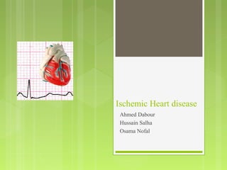
The document discusses ischemic heart disease (IHD), including its causes, symptoms, types (such as stable angina, unstable angina, and myocardial infarction), risk factors, diagnosis, and management. IHD is caused by reduced blood flow to the heart muscle, usually due to coronary artery disease. It presents with chest pain and other symptoms and is diagnosed through electrocardiograms, exercise tolerance tests, echocardiograms, isotope scans, and coronary angiography. Investigation aims to determine the severity and location of arterial blockages for guiding revascularization procedures.
Chronic Stable Angina- Diagnosis & management
By Dr Awadhesh Kumar Sharma
Dr. Awadhesh kumar sharma is a young, diligent and dynamic interventional cardiologist. He did his graduation from GSVM Medical College Kanpur and MD in Internal Medicine from MLB Medical college jhansi. Then he did his superspecilisation degree DM in Cardiology from PGIMER & DR Ram Manoher Lohia Hospital Delhi. He had excellent academic record with Gold medal in MBBS,MD and first class in DM.He was also awarded chief ministers medal in 2009 for his academic excellence by former chief minister of UP Smt Mayawati in 2009.He is also receiver of GEMS international award.He had many national & international publications.He is also in editorial board of international journal- Journal of clinical medicine & research(JCMR).He is also active member of reviewer board of many journals.He is also trainee fellow of American college of cardiology. He is currently working in NABH Approved Gracian Superspeciality Hospital Mohali as Consultant Cardiologist.
definition of heart failure, classification of heart failure, risk factors for heart failure, clinical features, general physical examination findings in heart failure
CARDIAC TAMPONADE ( Cardiac emergency) • Cardiac Tamponade is a life threatening complication caused by excessive accumulation of fluid in the pericardium. Or • Compression of all cardiac chambers due to excessive accumulation of pericardial fluid leading to compromised cardiac out put.
Definition of arrhythmia - background on cardiac physiology including conduction in heart - action potential - pathogensis of arrhythmia - causes and risk factors for arrhythmia- diagnosis of arrhythmia - symptoms of tachyarrhythmias and bradyarrhythmias - investigations for arrhythmia - treatment of arrhythmia - pharmacological and other modalities of therapy for arrhythmia - managment of different types of arrhythmias
Kindly leave your comment if you found this helpful ;)
Some of the slides, i hide it from my real presentations for my own reference. Download to see all of them.
The Advanced Cardiovascular Life Support (ACLS) algorithm is a systematic, evidence-based approach designed to guide healthcare providers in the urgent treatment of: Cardiac arrest. Arrhythmias. Stroke. Other life-threatening cardiovascular emergencies.
The human heart is a muscular organ with four chambers The size of the heart is the size of about a clenched fist. The function of the heart is to maintain a constant flow of blood throughout the body. This replenishes oxygen and circulates nutrients among the cells and tissues.
Several conditions impair the heart’s function. In Medical Terminology we use the term "heart disease". A list of Some Heart diseases is as follows:-
1(a). Disorders of heart rate, rhythm, and conduction
1.1 Sinus Arrhythmia -
Phasic alteration of heart rate during respiration may be due to activity in the parasympathetic system. can be two types:-
sinus bradycardia - Sinus rate is less than 60/min, Like normally present in Athletes.
Pathological Causes -Myocardial Infarction, Sinus Node Disease, Hypothermia, Hypothyroidism, Cholestatic jaundice, Raised Intracranial pressure, drugs like beta-blockers or verapamil.
Sinus Tachycardia - Heart rate of more than 100/min, it may be associated with exercise, pregnancy, and emotion.
After that Pathological Causes of Anxiety, Fever, Anemia, Heart Failure, Thyrotoxicosis, Phaeochromocytoma, and Drugs like bronchodilators.
1.2 Atrial tachyarrhythmias
Heart Disease having Atrial tachyarrhythmias are irregular fast heartbeat in the upper chambers of the heart(atria)
1.3 Atrial ectopic beats
Ectopic heartbeats are extra heartbeats that occur just before a regular beat. Ectopic beats are normal but can give the sensation of a missed beat.
1.4 Atrial tachycardia
It is a type of Heart Disease in which arrhythmia(an irregular heart rhythm) causes the upper chambers(atria) of the heart to beat faster than normal. This condition has several possible causes but is usually not dangerous. It is often curable or manageable with medication.
1.5 Atrial flutter
It Is one of the abnormal heart rhythms characterized by the right atrium beating quickly and encircling the tricuspid annulus.
1.6 Atrial fibrillation
In AF the upper chambers of the heart (the atria) beat irregularly instead of beating effectively to move blood into the ventricles. It is characterized by the presence of multiple, interacting re-entry circuits looping around the area. if untreated atrial fibrillation doubles the risk of heart-related deaths and associated serious conditions like stroke.
common causes may be coronary artery disease, valvular heart disease, hypertension, sinoatrial disease, hyperthyroidism, alcohol, cardiomyopathy, chest infection, congenital heart disease, pericardial disease, and pulmonary embolism.
Acute scrotum is a general term referring to an emergency condition affecting the contents or the wall of the scrotum.
There are a number of conditions that present acutely, predominantly with pain and/or swelling
A careful and detailed history and examination, and in some cases, investigations allow differentiation between these diagnoses. A prompt diagnosis is essential as the patient may require urgent surgical intervention
Testicular torsion refers to twisting of the spermatic cord, causing ischaemia of the testicle.
Testicular torsion results from inadequate fixation of the testis to the tunica vaginalis producing ischemia from reduced arterial inflow and venous outflow obstruction.
The prevalence of testicular torsion in adult patients hospitalized with acute scrotal pain is approximately 25 to 50 percent
NVBDCP.pptx Nation vector borne disease control programSapna Thakur
NVBDCP was launched in 2003-2004 . Vector-Borne Disease: Disease that results from an infection transmitted to humans and other animals by blood-feeding arthropods, such as mosquitoes, ticks, and fleas. Examples of vector-borne diseases include Dengue fever, West Nile Virus, Lyme disease, and malaria.
263778731218 Abortion Clinic /Pills In Harare ,sisternakatoto
263778731218 Abortion Clinic /Pills In Harare ,ABORTION WOMEN’S CLINIC +27730423979 IN women clinic we believe that every woman should be able to make choices in her pregnancy. Our job is to provide compassionate care, safety,affordable and confidential services. That’s why we have won the trust from all generations of women all over the world. we use non surgical method(Abortion pills) to terminate…Dr.LISA +27730423979women Clinic is committed to providing the highest quality of obstetrical and gynecological care to women of all ages. Our dedicated staff aim to treat each patient and her health concerns with compassion and respect.Our dedicated group ABORTION WOMEN’S CLINIC +27730423979 IN women clinic we believe that every woman should be able to make choices in her pregnancy. Our job is to provide compassionate care, safety,affordable and confidential services. That’s why we have won the trust from all generations of women all over the world. we use non surgical method(Abortion pills) to terminate…Dr.LISA +27730423979women Clinic is committed to providing the highest quality of obstetrical and gynecological care to women of all ages. Our dedicated staff aim to treat each patient and her health concerns with compassion and respect.Our dedicated group of receptionists, nurses, and physicians have worked together as a teamof receptionists, nurses, and physicians have worked together as a team wwww.lisywomensclinic.co.za/
Prix Galien International 2024 Forum ProgramLevi Shapiro
June 20, 2024, Prix Galien International and Jerusalem Ethics Forum in ROME. Detailed agenda including panels:
- ADVANCES IN CARDIOLOGY: A NEW PARADIGM IS COMING
- WOMEN’S HEALTH: FERTILITY PRESERVATION
- WHAT’S NEW IN THE TREATMENT OF INFECTIOUS,
ONCOLOGICAL AND INFLAMMATORY SKIN DISEASES?
- ARTIFICIAL INTELLIGENCE AND ETHICS
- GENE THERAPY
- BEYOND BORDERS: GLOBAL INITIATIVES FOR DEMOCRATIZING LIFE SCIENCE TECHNOLOGIES AND PROMOTING ACCESS TO HEALTHCARE
- ETHICAL CHALLENGES IN LIFE SCIENCES
- Prix Galien International Awards Ceremony
Title: Sense of Smell
Presenter: Dr. Faiza, Assistant Professor of Physiology
Qualifications:
MBBS (Best Graduate, AIMC Lahore)
FCPS Physiology
ICMT, CHPE, DHPE (STMU)
MPH (GC University, Faisalabad)
MBA (Virtual University of Pakistan)
Learning Objectives:
Describe the primary categories of smells and the concept of odor blindness.
Explain the structure and location of the olfactory membrane and mucosa, including the types and roles of cells involved in olfaction.
Describe the pathway and mechanisms of olfactory signal transmission from the olfactory receptors to the brain.
Illustrate the biochemical cascade triggered by odorant binding to olfactory receptors, including the role of G-proteins and second messengers in generating an action potential.
Identify different types of olfactory disorders such as anosmia, hyposmia, hyperosmia, and dysosmia, including their potential causes.
Key Topics:
Olfactory Genes:
3% of the human genome accounts for olfactory genes.
400 genes for odorant receptors.
Olfactory Membrane:
Located in the superior part of the nasal cavity.
Medially: Folds downward along the superior septum.
Laterally: Folds over the superior turbinate and upper surface of the middle turbinate.
Total surface area: 5-10 square centimeters.
Olfactory Mucosa:
Olfactory Cells: Bipolar nerve cells derived from the CNS (100 million), with 4-25 olfactory cilia per cell.
Sustentacular Cells: Produce mucus and maintain ionic and molecular environment.
Basal Cells: Replace worn-out olfactory cells with an average lifespan of 1-2 months.
Bowman’s Gland: Secretes mucus.
Stimulation of Olfactory Cells:
Odorant dissolves in mucus and attaches to receptors on olfactory cilia.
Involves a cascade effect through G-proteins and second messengers, leading to depolarization and action potential generation in the olfactory nerve.
Quality of a Good Odorant:
Small (3-20 Carbon atoms), volatile, water-soluble, and lipid-soluble.
Facilitated by odorant-binding proteins in mucus.
Membrane Potential and Action Potential:
Resting membrane potential: -55mV.
Action potential frequency in the olfactory nerve increases with odorant strength.
Adaptation Towards the Sense of Smell:
Rapid adaptation within the first second, with further slow adaptation.
Psychological adaptation greater than receptor adaptation, involving feedback inhibition from the central nervous system.
Primary Sensations of Smell:
Camphoraceous, Musky, Floral, Pepperminty, Ethereal, Pungent, Putrid.
Odor Detection Threshold:
Examples: Hydrogen sulfide (0.0005 ppm), Methyl-mercaptan (0.002 ppm).
Some toxic substances are odorless at lethal concentrations.
Characteristics of Smell:
Odor blindness for single substances due to lack of appropriate receptor protein.
Behavioral and emotional influences of smell.
Transmission of Olfactory Signals:
From olfactory cells to glomeruli in the olfactory bulb, involving lateral inhibition.
Primitive, less old, and new olfactory systems with different path
Couples presenting to the infertility clinic- Do they really have infertility...Sujoy Dasgupta
Dr Sujoy Dasgupta presented the study on "Couples presenting to the infertility clinic- Do they really have infertility? – The unexplored stories of non-consummation" in the 13th Congress of the Asia Pacific Initiative on Reproduction (ASPIRE 2024) at Manila on 24 May, 2024.
Flu Vaccine Alert in Bangalore Karnatakaaddon Scans
As flu season approaches, health officials in Bangalore, Karnataka, are urging residents to get their flu vaccinations. The seasonal flu, while common, can lead to severe health complications, particularly for vulnerable populations such as young children, the elderly, and those with underlying health conditions.
Dr. Vidisha Kumari, a leading epidemiologist in Bangalore, emphasizes the importance of getting vaccinated. "The flu vaccine is our best defense against the influenza virus. It not only protects individuals but also helps prevent the spread of the virus in our communities," he says.
This year, the flu season is expected to coincide with a potential increase in other respiratory illnesses. The Karnataka Health Department has launched an awareness campaign highlighting the significance of flu vaccinations. They have set up multiple vaccination centers across Bangalore, making it convenient for residents to receive their shots.
To encourage widespread vaccination, the government is also collaborating with local schools, workplaces, and community centers to facilitate vaccination drives. Special attention is being given to ensuring that the vaccine is accessible to all, including marginalized communities who may have limited access to healthcare.
Residents are reminded that the flu vaccine is safe and effective. Common side effects are mild and may include soreness at the injection site, mild fever, or muscle aches. These side effects are generally short-lived and far less severe than the flu itself.
Healthcare providers are also stressing the importance of continuing COVID-19 precautions. Wearing masks, practicing good hand hygiene, and maintaining social distancing are still crucial, especially in crowded places.
Protect yourself and your loved ones by getting vaccinated. Together, we can help keep Bangalore healthy and safe this flu season. For more information on vaccination centers and schedules, residents can visit the Karnataka Health Department’s official website or follow their social media pages.
Stay informed, stay safe, and get your flu shot today!
New Directions in Targeted Therapeutic Approaches for Older Adults With Mantl...i3 Health
i3 Health is pleased to make the speaker slides from this activity available for use as a non-accredited self-study or teaching resource.
This slide deck presented by Dr. Kami Maddocks, Professor-Clinical in the Division of Hematology and
Associate Division Director for Ambulatory Operations
The Ohio State University Comprehensive Cancer Center, will provide insight into new directions in targeted therapeutic approaches for older adults with mantle cell lymphoma.
STATEMENT OF NEED
Mantle cell lymphoma (MCL) is a rare, aggressive B-cell non-Hodgkin lymphoma (NHL) accounting for 5% to 7% of all lymphomas. Its prognosis ranges from indolent disease that does not require treatment for years to very aggressive disease, which is associated with poor survival (Silkenstedt et al, 2021). Typically, MCL is diagnosed at advanced stage and in older patients who cannot tolerate intensive therapy (NCCN, 2022). Although recent advances have slightly increased remission rates, recurrence and relapse remain very common, leading to a median overall survival between 3 and 6 years (LLS, 2021). Though there are several effective options, progress is still needed towards establishing an accepted frontline approach for MCL (Castellino et al, 2022). Treatment selection and management of MCL are complicated by the heterogeneity of prognosis, advanced age and comorbidities of patients, and lack of an established standard approach for treatment, making it vital that clinicians be familiar with the latest research and advances in this area. In this activity chaired by Michael Wang, MD, Professor in the Department of Lymphoma & Myeloma at MD Anderson Cancer Center, expert faculty will discuss prognostic factors informing treatment, the promising results of recent trials in new therapeutic approaches, and the implications of treatment resistance in therapeutic selection for MCL.
Target Audience
Hematology/oncology fellows, attending faculty, and other health care professionals involved in the treatment of patients with mantle cell lymphoma (MCL).
Learning Objectives
1.) Identify clinical and biological prognostic factors that can guide treatment decision making for older adults with MCL
2.) Evaluate emerging data on targeted therapeutic approaches for treatment-naive and relapsed/refractory MCL and their applicability to older adults
3.) Assess mechanisms of resistance to targeted therapies for MCL and their implications for treatment selection
New Drug Discovery and Development .....NEHA GUPTA
The "New Drug Discovery and Development" process involves the identification, design, testing, and manufacturing of novel pharmaceutical compounds with the aim of introducing new and improved treatments for various medical conditions. This comprehensive endeavor encompasses various stages, including target identification, preclinical studies, clinical trials, regulatory approval, and post-market surveillance. It involves multidisciplinary collaboration among scientists, researchers, clinicians, regulatory experts, and pharmaceutical companies to bring innovative therapies to market and address unmet medical needs.
These lecture slides, by Dr Sidra Arshad, offer a quick overview of physiological basis of a normal electrocardiogram.
Learning objectives:
1. Define an electrocardiogram (ECG) and electrocardiography
2. Describe how dipoles generated by the heart produce the waveforms of the ECG
3. Describe the components of a normal electrocardiogram of a typical bipolar leads (limb II)
4. Differentiate between intervals and segments
5. Enlist some common indications for obtaining an ECG
Study Resources:
1. Chapter 11, Guyton and Hall Textbook of Medical Physiology, 14th edition
2. Chapter 9, Human Physiology - From Cells to Systems, Lauralee Sherwood, 9th edition
3. Chapter 29, Ganong’s Review of Medical Physiology, 26th edition
4. Electrocardiogram, StatPearls - https://www.ncbi.nlm.nih.gov/books/NBK549803/
5. ECG in Medical Practice by ABM Abdullah, 4th edition
6. ECG Basics, http://www.nataliescasebook.com/tag/e-c-g-basics
Report Back from SGO 2024: What’s the Latest in Cervical Cancer?bkling
Are you curious about what’s new in cervical cancer research or unsure what the findings mean? Join Dr. Emily Ko, a gynecologic oncologist at Penn Medicine, to learn about the latest updates from the Society of Gynecologic Oncology (SGO) 2024 Annual Meeting on Women’s Cancer. Dr. Ko will discuss what the research presented at the conference means for you and answer your questions about the new developments.
3. ischemic heart disease
ischemic heart disease (IHD):
is a disease characterized by reduction blood supply of the heart
muscle, usually due to coronary artery disease
1-Angina 2-Myocardial infarction
Stable STEMI
Angina
Unstable Angina NSTEMI
Variant Angina
decubitus angina
nocturnal angina
4. Epidemiology
• most common cause of cardiovascular morbidity and mortality
• atherosclerosis and thrombosis are the most important
pathogenetic mechanisms.
• peak incidence of symptomatic IHD is age 50-60 (men)
and 60-70 (women)
M>F
7. Angina pectoris
Isa clinical syndrome characterized by paroxysmal chest pain
due to transient myocardial ischemia . It may occur whenever
there is imbalance between myocardial oxygen supply and
demand the most common cause is atherosclerosis .however
angina may also develop in aortic stenosis and hypertrophic
cardiomyopathy even there is no coronary atheroma
8. Angina symptoms include:
Chest pain or discomfort Chest pain or discomfort is
Pain in your arms, neck, usually felt as:
jaw, shoulder or back pressure,
accompanying chest pain heaviness,
Nausea
tightening,
Fatigue
squeezing,
Shortness of breath
Anxiety
Sweating
Dizziness
9. Stable Angina
Atherosclerotic coronary artery disease
occurs when the heart has to work harder than normal, during
exercise
typical: retrosternal chest pain, tightness or discomfort
radiating to left(± right) shoulder/arm/ neck/jaw,
brief duration, lasting <10-15 min
associated with diaphoresis, nausea, anxiety
typically relieved by rest and nitrates
10. precipitatedby the " E's"
Emotional stress
Exertion
Exposure to very hot or cold temperatures
Eating ( Heavy meals)
And Smoking
11. Variant Angina
A spasm in a coronary artery
Usually happens when you're resting, unrelated to exercise,
relieved by nitrates
typically occurs between midnight and 8 AM,
The coronary arteries can spasm as a result of:
Exposure to cold
Emotional stress
Medicines that tighten or narrow blood vessels
Smoking
Cocaine use
12. SYNDROME X
Coronary microvascular disease that affects the heart’s
smallest coronary arteries.
Typical symptoms of angina but normal angiogram
May show definite signs of ischemia with exercise testing
13. Unstable Angina
Due to spasm and partial obstruction of coronaries.
Occurs even at rest
Is unexpected (new onset)
Is usually more severe and lasts longer than stable angina,
may be as long as 30 minutes
May not disappear with rest or use of angina medication
May lead to complete occlusion of vessel causing MI
14. Myocardial Infarction
Myocardial infarction, commonly known as a heart attack,
is the irreversible necrosis of heart muscle secondary to
prolonged ischemia (total obstruction)
Typical symptoms of myocardial infarction include
sudden chest pain,
shortness of breath,
nausea, vomiting,
palpitations, sweating
weakness, light-headedness
Collapse/syncope
15. Severe pain
described as a sensation of tightness, pressure,crushing or
squeezing.
radiating to left(± right) shoulder/arm/ neck/jaw
Chest pain usually lasts for more than 15 minutes
Not relieves by rest
17. Physical examination & signs in angina:
•For most patients with stable angina, physical examination
findings are normal. Diagnosing secondary causes of angina,
such as aortic stenosis, is important.
•Vital signs especially blood pressure
•A positive Levine sign (characterized by the patient's fist
clenched over the sternum when describing the discomfort) is
suggestive of angina pectoris.
18.
19. •Look for physical signs of abnormal lipid metabolism (eg,
xanthelasma, xanthoma) or of diffuse atherosclerosis (eg,
absence or diminished peripheral pulses, increased light
reflexes or arteriovenous nicking upon ophthalmic
examination, carotid bruit).
•Examination of patients during the angina attack may be more
helpful. Useful physical findings include third and/or fourth
heart sounds due to LV systolic and/or diastolic dysfunction
and mitral regurgitation secondary to papillary muscle
dysfunction.
•Pain produced by chest wall pressure is usually of chest wall
origin.
20. Physical examination & signs in unstable angina
and myocardial infarction
Abnormal physical findings are often absent; when present, they are often non-
specific.
An unremarkable physical examination is not uncommon. Perform a quick
assessment of patients' vital signs, and perform a cardiac examination.
Specific diagnoses that must be explicitly considered are the following:
•Aortic dissection
•Leaking or ruptured thoracic aneurysm
•Pericarditis with tamponade
•Pulmonary embolism
•Pneumothorax
21. Unstable angina differs from stable angina in that the discomfort is
usually more intense and easily provoked, and ST-segment depression
or elevation on ECG may occur.
Otherwise, the manifestations of unstable angina are similar to those
of other conditions of myocardial ischemia, such as chronic stable
angina and myocardial infarction.
22. Increased autonomic activity may manifest as diaphoresis or tachycardia, and
bradycardia may result from vagal stimulation from inferior wall myocardial
ischemia.
A large area of myocardial jeopardy may manifest as signs of transient myocardial
dysfunction and typically signifies a higher-risk situation. Signs include the
following:
•Systolic blood pressure less than 100 mm Hg or overt hypotension
•Elevated jugular venous pressure
•Dyskinetic apex
•Reverse splitting of the second heart sound
•Presence of a third or fourth heart sound
•New or worsening apical systolic murmur due to papillary muscle dysfunction
•Rales or crackles
23. Vital signs and appearance are two of the most important aspects of the physical
exam.
Vital Signs
In the evaluation of a patient presenting with ACS hypotension (systolic blood
pressure <100 mm Hg), tachycardia (pulse >100) and bradycardia (pulse <60
bpm) indicate that a patient is at higher risk.
As with the assessment of all patients, other abnormal vital signs such as hypoxia,
tachypnea (RR >19), hypothermia (T <95 F) or fever (T >100.3 F) should raise
concern, although they are not specifically suggestive of ACS.
If aortic dissection is considered in the differential diagnosis, blood pressure
should be checked in both arms (>20 mm Hg differential is suggestive of aortic
dissection).
Appearance of the Patient
A patient who appears anxious, diaphoretic, with pale skin and who is in obvious
respiratory distress should demand immediate attention.
24. Eyes
The eye exam is typically not the focus of a physical exam for ACS, however,
details such as pale conjunctiva (suggestive of anemia), exopthalmos (suggestive
of hyperthyroidism), or cotton-wool spots (suggestive of hypertension), or
retinopathy (suggestive of diabetes) on fundoscopic exam should be noted as they
may allow for the identification of potential precipitants of or risk factors for
myocardial ischemia.
Ear Nose and Throat
The ears and nose are typically not the focus of a physical exam for ACS.
However, the examination of the buccal mucosa can help to determine a patient's
volume status, as can the examination of the right internal jugular vein pulsations
(JVP).
A JVP which is elevated greater than 4 cm above the sternal angle (9 cm above
the right atrium) is considered elevated and reflects elevated right atrial pressure.
25. Heart
The cardiac exam should evaluate for signs of cardiac failure, such as a 3rd heart
sound ("gallop," from early diastolic filling from left ventricular systolic failure), a
4th heart sound ("gallop," from late diastolic filling from a stiff left ventricle, as
from diastolic heart failure) or a new / increased systolic murmur of mitral
regurgitation (as from papillary muscle rupture).
The presence of a pericardial rub would suggest pericarditis instead of ACS.
Lungs
Bibasilar rales are suggestive of congestive heart failure and pulmonary edema.
However, the absence of adventitious lung sounds does not preclude diastolic heart
failure.
Abdomen
The abdominal exam is typically not the focus of a physical exam for ACS.
However, a finding of a expansile, pulsatile mass in the upper abdomen suggests an
aortic aneurysm and requires further urgent evaluation.
26. Extremities
Assess the lower extremities for edema, suggestive of heart failure. It is also
important to palpate the radial, femoral and pedal pulses.
Unequal radial pulses are suggestive of aortic dissection. Weak pedal pulses are
suggestive of peripheral vascular disease. Femoral pulses are important to
document in the event that cardiac catheterization is necessary.
Neurologic
The neurological examination is typically not the focus of a physical exam for
ACS. However, mental status at the time of the initial assessment should be
documented for future reference, should the patient's mental status deteriorate
during the period of observation.
Also, headache in the context of chest pain and severe hypertension (i.e., SBP >
210 mm/Hg or a DBP > 120 mm/Hg) would support a diagnosis of hypertensive
emergency as a cause for ACS.
28. 1-ECG
Differential diagnosis of ST segment depression
Myocardial Ischemia
LVH
Severe hypertension
Cardiomyopathy
Anemia
Hypokalemia
Digitalis effect
29. Differential diagnosis of ST segment elevation
Myocardial infarction
Prinzmetal’s angina
Ventricular aneurysm (post MI )
Acute pericarditis
Myocarditis
Hypothermia
30. 2-Exercise Tolerance Test (ETT)
This is the most useful noninvasive procedure for
evaluation the patient with angina. Ischemia that is
not present at rest is detected by precipitation of
typical chest pain or ST segment elevation during
the exercise using treadmill
When history is suggestive of angina pectoris but
ECG is normal , then the exercise test should be
done.
31. The test involves recording the 12-lead ECG
before , during and after exercise.
The test consists of a standardized incremental
increase in the external workload while the
patient’s ECG, symptoms and the blood pressure
are continuously monitored. A variety of exercise
protocols are utilized, the most common being the
Bruce protocol which increases the treadmill speed
and elevation every 3 mins until limited by
symptoms.
32. This test discovers any limitation in exercise
performance and establishes the relationship between
chest pain and the typical ECG sings of myocardial
ischemia.
Positive test is one which ST segment is depressed by
1mm(one small square )
More severe disease presents with ST depression more
2 mm at low workload or at heart rate less than 70% of
age predicted value, or hypotension develops during
exercise.
33. ETT Report:
Degree of ST depression
Development of arrhythmia or conduction defect
during and post exercise.
Duration of exercise.
Achievement of age predicted target heart rate ( 220
minus age )
Development of chest pain during exercise.
Hemodynamic response
34. Indications:
To confirm the diagnosis of angina
To determine the severity of limitation of activity
due to angina
To asses prognosis in patient with known coronary
disease.
To evaluate response to therapy.
35. Contraindications:
Acute myocardial infarction ( less 2 days )
High risk unstable angina
Decompensated HF
Cardiac arrythmias with symptoms
Heart block
Acute myocarditis and pericarditis
Severe aortic stenosis
Severe HOCM
Uncontrolled HTN
36. Interpretation:
Overall sensitivity of ETT is about 60-75% and
specificity 80%. The test may be falsely + or – in
15% of cases therefore negative test does not rule
out IHD and positive test without symptoms does
not always confirm IHD. If ERR is inconclusive
then IHD should be confirmed by thallium scan.
ECHO and angiography.
37. Echocardiograph
Itreveals segmental wall motion abnormalities
which indicate ischemia or prior infarction. It can
be performed at rest while sensitivity increase if
performed after exercise or stress given by
dobutamine (called dobutamine stress echo)
38. Isotope scanning
Thallium scan and technetium scan shows areas of reduced
uptake of radioactive isotope (thallium and technetium) by
the myocardium. This test is performed at rest and during
stress (produced by exercise or dipyridamol or dobutamine)
A perfusion defect present during stress but not all rest
indicates reversible myocardial ischemia, whereas a
persistent perfusion defect on scan during both phases (rest
and stress) usually indicates previous myocardial infarction.
Thallium scanning is positive in 75-90% of patients with
significant coronary disease. False positive test may occur in
women due to breast tissue.
39. Indication:
When ETT is not diagnostic (equivocal or contrary to the clinical
impression such as positive test in asymptomatic patient).
When patient is unable to perform exercise e.g. patient of unstable
angina, aortic stenosis or handicapped patients. In these patients
stress is produced by alternatives methods such as drugs e.g.
dipyridamol dobutamine or adenosine
To distinguish ischemia from myocardial infarction.
To localize regions of ischemia.
To identify whether the myocardium is viable or not, because
revascularization via surgery or angioplasty may be beneficial only
for viable myocardium.
40.
41.
42. Coronary angiograph
Coronary angiography visualizes the location and
severity of coronary after stenosis. Narrowing
greater than 50% of luminal diameter is considered
clinically significant, although most lesion
producing ischemia are associated with narrowing
more than 70%.
43. Indication:
Coronary angiograph is indicated in patient whom coronary
revascularization (angioplasty or by-pass) is being considered
because of uncontrolled stable angina who have failed to
improve on adequate medical regimen
To diagnose chest pain of uncertain cause when noninvasive
tests have failed to detect the cause. Diagnostic angiography is
now rarely performed because diagnosis is usually made on
history and non-invasive tests.
Unstable angina
Post myocardial infarction angina
Severe left ventricle dysfunction after MI
Non Q-wave MI
Strongly positive ETT
Editor's Notes
An uncoordinated ( dyskinetic ) apex beat involving a larger area than normal indicates ventricular dysfunction; such as an aneurysm following myocardial infarction Split during inspiration: normal. [4] (See above) Split during expiration: Reverse splitting indicates pathology