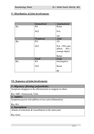
Downloaded 325 times


This document provides guidance on rheumatology history taking and clinical examination. It begins with 10 golden rules for rheumatology diagnosis and examination. It then describes rheumatology terminology and provides detailed guidance on taking a rheumatology history, including screening questions and analyzing symptoms such as pain, stiffness, swelling, deformity, movement and function, muscle weakness, sleep disturbance, and systemic or extra-articular manifestations. The history should explore personal factors, past medical history, present illness including specific symptoms, and review of other body systems. A thorough history and physical exam are essential for rheumatology diagnosis.


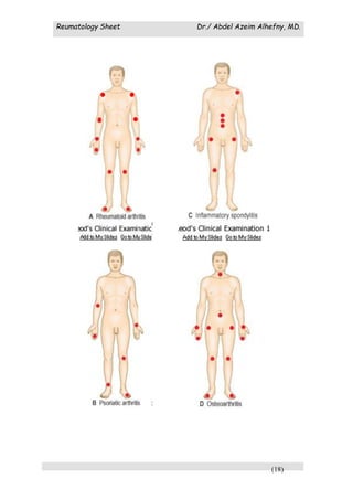
![[Int. med] approach to joint pain from SIMS Lahore](https://cdn.slidesharecdn.com/ss_thumbnails/064ftjyatacrqjgurlzo-signature-b01672da1ecf8b94befb115319b147a085de390b8cb403389bce6c156545fbb5-poli-150815171713-lva1-app6891-thumbnail.jpg?width=640&height=640&fit=bounds)


