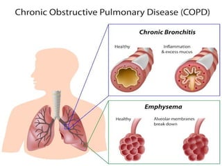
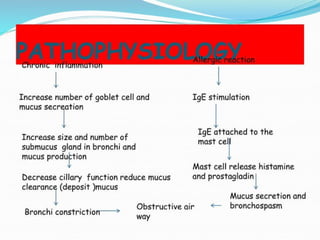
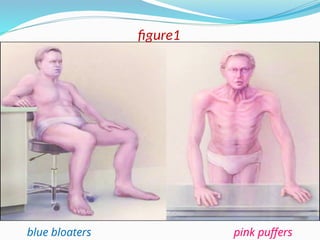
Prepared by : Rafed Abdullrzzaq AL-hendi
Faculty Of Nursing
4th level
Republic of Yemen
Jiblah university for
medical & health
sciences
first term
Chronic obstructive pulmonary disease
Medical and Surgical Nursing
 Definition:
Chronic obstructive pulmonary disease
is a preventable and treatable disease characterised by chronic
dyspnea with persistent expiratory airflow limitation that is
usually progressive, and irreversible disease.
Introduction
Classification
2- Emphysema :
A pathologic diagnosis: permanent enlargement of air spaces
distal to terminal bronchioles due to destruction of alveolar walls.
People with emphysema are also called “pink puffers”.
1- Chronic bronchitis
A clinical diagnosis: chronic cough productive of sputum for at
least 3 months per year for at least 2 consecutive years.
bronchitis is also termed as “blue bloaters”.
The two often coexist. Pure emphysema or pure chronic
bronchitis is rare.
(see at figure1)
(figure1)
5
blue bloaters pink puffers
figure1
o Tobacco smoke (indicated in almost 90% of COPD cases).
o α1-Antitrypsin deficiency.
o Environmental factors (e.g., second-hand smoke).
o Chronic asthma.
o Indoor air pollution.
o Occupational exposures, such as coal dust, silica.
o Lung growth: childhood infections or maternal smoking.
o Infections:HIV infection is associated with emphysema.
Risk Factors and Causes
o Chronic cough.
o Sputum production.
o Dyspnea (on exertion or at rest, depending on severity).
SIGN AND SYMPTOMS
 SYMPTOMS
 SIGN
o Tachypnea,
o tachycardia
o Cyanosis
o Use of accessory respiratory muscles.
o Prolonged forced expiratory time on physical examination
o During auscultation, end-expiratory wheezes on forced
expiration, decreased breath sounds, and/or inspiratory
crackles
o Hyperresonance on percussion
o Barrel chest
o Flushed face.
Cont..
(see at figure2).
figure2
 Pulmonary function testing (spirometry)
a. This is the definitive diagnostic test.
b. Obstruction is evident based on the following:
Decreased FEV1 and decreased FEV1/FVC ratio
If FEV1 is reduced to 70% of predicted value, mild disease is
suggested.
If FEV1 is reduced to 50% or less of predicted value, severe
disease is present.
Values in between indicate moderate disease
Diagnostic evaluation
 Chest X-ray :
 Low sensitivity for diagnosing COPD; only severe, advanced
emphysema will show the typical changes, which include:
Hyperinflation, flattened diaphragm
 Arterial blood gases (ABGs):
 Determines degree and severity of disease process, e.g.,
chronic pCO2 retention and decreased pO2.
(see at figure3).
Cont..
figure3
 Complete blood count (CBC) and differential:
Increased hemoglobin (advanced emphysema),.
 Screening for alpha1-antitrypsin deficiency.
 Sputum culture: Determines presence of infection
 Cytologic examination: for malignancy
 Electrocardiogram (ECG):,
 CT scan. may help in the differential diagnosis.
Cont..
 Acute exacerbations—most common causes are infection.
 Acute Respiratory Failure.
 Spontaneous Pneumothorax.
 Secondary polycythemia: compensatory response to chronic
hypoxemia
 Pulmonary HTN and cor pulmonale—may occur in patients
with severe, longstanding COPD who have chronic hypoxemia
Complications
 Smoking cessation:
• The most important intervention
• Disease progression slowed by cessation
• prolongs the survival rate
 Inhaled anticholinergic drugs (ipratropium bromide):
• Bronchodilatos
• Slower onset of action than the β-agonists, but last longer
 Inhaled β2-agonists (e.g., albuterol):
• bronchodilators
• Use long-acting agents (e.g., salmeterol) for patients
requiring frequent use.
MANAGEMENT
 Combination of β-agonist albuterol with ipratropium bromide
• More efficacious than either agent alone in bronchodilation
Cont..
 Corticosteroids: may be prescribed for patients to determine
whether pulmonary function improves and symptoms decrease.
 Other medications include alpha1-antitrypsin augmentation
therapy, antibiotic agents, mucolytic agents, antitussive agents,
 Oxygen therapy
• Some patients need continuous oxygen, whereas others only
require it during exertion or sleep.
 Theophylline (oral)—role is controversial
• Only modestly effective and has more side effects than other
bronchodilators.
Cont..
 Vaccination:
• Influenza vaccination annually for all patients.
• Vaccination against Streptococcus pneumoniae every 5 to 6
years should be offered to patients with COPD over 65 years
old, or under 65 who have severe disease .
 Pulmonary rehabilitation :
• Education, exercise, physiotherapy:
A major goal is to improve exercise tolerance.
• Pulmonary rehabilitation improves functional status and
quality of life .
1.Bronchodilators (β2-agonist) alone or in combination with
anticholinergics are first-line therapy
2.Systemic corticosteroids
Acute COPD exacerbation
 A persistent increase in dyspnea and Increased sputum
production and cough are common.
 Acute COPD exacerbation can lead to acute respiratory failure
requiring hospitalization, and possibly mechanical ventilation;
potentially fatal.
 Management:
3.Antibiotics (azithromycin or levofloxacin)
4.Supplemental oxygen is used to keep O2 saturation above 90%.
5.Intubation and mechanical ventilation may be required
Nursing intervention
 Regularly monitor vital signs and oxygen saturation
,ABG results , the severity of dyspnea and sputum color,odor
and character.
 Elevate head of bed, assist patient to assume position to
ease work of breathing
 Teach the client to use pursed-lip and diaphragmatic
breathing techniques
 Perform chest physiotherapy.
 note use of accessory muscles, pursed lip breathing, inability
to speak.
 Teach the client to wash his or her hands after contact with
potentially infectious material.
 Explain the patient about disease including cause, signs and
symptoms, medication, procedures, prevention
 Explain need for adequate nutritional intake
 Explain client about the importance of self care
 Notify the physician if any sign of infections occurs.
 Administer bronchodilators and supplemental oxygen therapy
if ordered
Cont..
Thank you

COPD سمنارجاهز جديد لطلبة كليةالتمريض.pptx

  • 1.
    Prepared by :Rafed Abdullrzzaq AL-hendi Faculty Of Nursing 4th level Republic of Yemen Jiblah university for medical & health sciences first term Chronic obstructive pulmonary disease Medical and Surgical Nursing
  • 2.
     Definition: Chronic obstructivepulmonary disease is a preventable and treatable disease characterised by chronic dyspnea with persistent expiratory airflow limitation that is usually progressive, and irreversible disease. Introduction
  • 3.
    Classification 2- Emphysema : Apathologic diagnosis: permanent enlargement of air spaces distal to terminal bronchioles due to destruction of alveolar walls. People with emphysema are also called “pink puffers”. 1- Chronic bronchitis A clinical diagnosis: chronic cough productive of sputum for at least 3 months per year for at least 2 consecutive years. bronchitis is also termed as “blue bloaters”. The two often coexist. Pure emphysema or pure chronic bronchitis is rare. (see at figure1) (figure1)
  • 5.
  • 6.
    blue bloaters pinkpuffers figure1
  • 7.
    o Tobacco smoke(indicated in almost 90% of COPD cases). o α1-Antitrypsin deficiency. o Environmental factors (e.g., second-hand smoke). o Chronic asthma. o Indoor air pollution. o Occupational exposures, such as coal dust, silica. o Lung growth: childhood infections or maternal smoking. o Infections:HIV infection is associated with emphysema. Risk Factors and Causes
  • 8.
    o Chronic cough. oSputum production. o Dyspnea (on exertion or at rest, depending on severity). SIGN AND SYMPTOMS  SYMPTOMS  SIGN o Tachypnea, o tachycardia o Cyanosis o Use of accessory respiratory muscles.
  • 9.
    o Prolonged forcedexpiratory time on physical examination o During auscultation, end-expiratory wheezes on forced expiration, decreased breath sounds, and/or inspiratory crackles o Hyperresonance on percussion o Barrel chest o Flushed face. Cont.. (see at figure2).
  • 10.
  • 11.
     Pulmonary functiontesting (spirometry) a. This is the definitive diagnostic test. b. Obstruction is evident based on the following: Decreased FEV1 and decreased FEV1/FVC ratio If FEV1 is reduced to 70% of predicted value, mild disease is suggested. If FEV1 is reduced to 50% or less of predicted value, severe disease is present. Values in between indicate moderate disease Diagnostic evaluation
  • 12.
     Chest X-ray:  Low sensitivity for diagnosing COPD; only severe, advanced emphysema will show the typical changes, which include: Hyperinflation, flattened diaphragm  Arterial blood gases (ABGs):  Determines degree and severity of disease process, e.g., chronic pCO2 retention and decreased pO2. (see at figure3). Cont..
  • 13.
  • 14.
     Complete bloodcount (CBC) and differential: Increased hemoglobin (advanced emphysema),.  Screening for alpha1-antitrypsin deficiency.  Sputum culture: Determines presence of infection  Cytologic examination: for malignancy  Electrocardiogram (ECG):,  CT scan. may help in the differential diagnosis. Cont..
  • 15.
     Acute exacerbations—mostcommon causes are infection.  Acute Respiratory Failure.  Spontaneous Pneumothorax.  Secondary polycythemia: compensatory response to chronic hypoxemia  Pulmonary HTN and cor pulmonale—may occur in patients with severe, longstanding COPD who have chronic hypoxemia Complications
  • 16.
     Smoking cessation: •The most important intervention • Disease progression slowed by cessation • prolongs the survival rate  Inhaled anticholinergic drugs (ipratropium bromide): • Bronchodilatos • Slower onset of action than the β-agonists, but last longer  Inhaled β2-agonists (e.g., albuterol): • bronchodilators • Use long-acting agents (e.g., salmeterol) for patients requiring frequent use. MANAGEMENT
  • 17.
     Combination ofβ-agonist albuterol with ipratropium bromide • More efficacious than either agent alone in bronchodilation Cont..  Corticosteroids: may be prescribed for patients to determine whether pulmonary function improves and symptoms decrease.  Other medications include alpha1-antitrypsin augmentation therapy, antibiotic agents, mucolytic agents, antitussive agents,  Oxygen therapy • Some patients need continuous oxygen, whereas others only require it during exertion or sleep.  Theophylline (oral)—role is controversial • Only modestly effective and has more side effects than other bronchodilators.
  • 18.
    Cont..  Vaccination: • Influenzavaccination annually for all patients. • Vaccination against Streptococcus pneumoniae every 5 to 6 years should be offered to patients with COPD over 65 years old, or under 65 who have severe disease .  Pulmonary rehabilitation : • Education, exercise, physiotherapy: A major goal is to improve exercise tolerance. • Pulmonary rehabilitation improves functional status and quality of life .
  • 19.
    1.Bronchodilators (β2-agonist) aloneor in combination with anticholinergics are first-line therapy 2.Systemic corticosteroids Acute COPD exacerbation  A persistent increase in dyspnea and Increased sputum production and cough are common.  Acute COPD exacerbation can lead to acute respiratory failure requiring hospitalization, and possibly mechanical ventilation; potentially fatal.  Management: 3.Antibiotics (azithromycin or levofloxacin) 4.Supplemental oxygen is used to keep O2 saturation above 90%. 5.Intubation and mechanical ventilation may be required
  • 20.
    Nursing intervention  Regularlymonitor vital signs and oxygen saturation ,ABG results , the severity of dyspnea and sputum color,odor and character.  Elevate head of bed, assist patient to assume position to ease work of breathing  Teach the client to use pursed-lip and diaphragmatic breathing techniques  Perform chest physiotherapy.  note use of accessory muscles, pursed lip breathing, inability to speak.
  • 21.
     Teach theclient to wash his or her hands after contact with potentially infectious material.  Explain the patient about disease including cause, signs and symptoms, medication, procedures, prevention  Explain need for adequate nutritional intake  Explain client about the importance of self care  Notify the physician if any sign of infections occurs.  Administer bronchodilators and supplemental oxygen therapy if ordered Cont..
  • 22.

Editor's Notes

  • #1 The word "cirrhosis" derives from Greek κίρῥος, meaning tawny (the orange-yellow colour of the diseased liver).