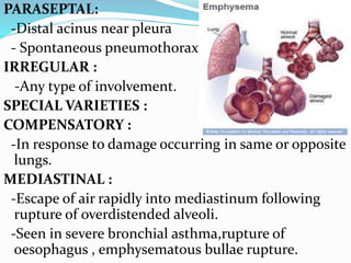
Dr. INDHUJA
I YEAR
M.D.,GEN.MEDICINE
OVERVIEW
DEFINITION
RISK FACTORS
PATHOPHYSIOLOGY
CLINICAL PRESENTATION
History
Physical findings
Lab findings
DIAGNOSIS
DIFFERENTIAL DIAGNOSIS
COMPLICATIONS
EXACERBATION OF COPD
TREATMENT
Pharmacological therapy
Non pharmacological therapy
DEFINITION
Disease state characterized by airflow
limitation that is not fully reversible.
RISK FACTORS
1. CIGARETTE SMOKING
 Pack years dose response relation
increases with advancing age.
 < 50 yrs: males > females
 >50 yrs: equal incidence.
Only 15% variability in FEV
 Additional environmental and genetic
factors.
Natural history:
 Severity of COPD depends on:
- Intensity
- Timing of exposure during
growth.
- Baseline lung function.
Affects both large airways (cough &
sputum), small airways & alveoli
(physiological).
2. PRIMARY RESPONSIVENESS:
 DUTCH hypothesis:
Asthma and COPD are variations of same
disease modulated by environmental and genetic
factors.
 BRITISH hypothesis:
Asthma and COPD are fundamentally different.
 Genetic predisposing factor: ADAM 33 & MMP12
(both asthma & COPD).
 Increased airway responsiveness = decreased
pulmonary function.
3. RESPIRATORY INFECTION :
 Needs to be proved.
4. OCCUPATIONAL EXPOSURE:
 Coaling, gold mining, cotton textile dust.
 Less important than the effect of smoking.
5.AMBIENT AIR POLLUTION:
 Biomass combustion
6. PASSIVE SMOKING:
 In utero smoking- reduced postnatal
pulmonary functions.
 Exposure of children to maternal smoking –
reduced lung growth.
7. GENETIC: Severe alpha 1 antitrypsin
deficiency.
 M allele - Normal levels.
 S allele - Reduced
 Z allele - Severely reduced.
 Null allele – Absent enzyme levels.
 Most common form : piZZ & piZZ/null forms
 Homozygous piZZ + smoking = early onset copd.
LAB: Immunologic levels of enzyme in serum
 Accompanying asthma & male gender worsens
prognosis.
Rx : Alpha 1 AT augmentation.
Heterozygous PiMZ : intermediate levels of
enzyme.
Others:
 Familial aggregation of airflow obstruction
within families of COPD pts.
 Minor allele SNP of MMP12 : reduced
expression of MMP12 - asthma & adult
smoker.
 COPD loci : near HHIP gene on chr. 21
chr. 15 ( near NAD receptor)
PATHOPHYSIOLOGY
AIRFLOW OBSTRUCTION:
 Most typical finding : persistent
reduction in forced expiratory flow rates.
 Increased residual volume, Residual
Volume/TLC, V/P mismatch.
Chronically reduced FEV1/FVC with no
major improvement on inhaled
bronchodilators.
Differentiate from asthma
Reduced max. expiratory flow rate
 Early - @ FRC. Advanced – entire breath
HYPERINFLATION:
 Air trapping- increased residual volume &
residual volume / TLC
 Late progressive hyperinflation : increased TLC
 Flattened diaphragm.
GAS EXCHANGE :
 Normal Pao2 – till FEV1 <50% of predicted
 Increased Paco2
 Chronic hypoxemia Pao2<55 FEV1 < 25%
 Pulmonary hypertension
 Cor pulmonale/RVH
EMPHYSEMA
PANACINAR :
 Non uniform ventilation and
VP mismatch
- Acini
- Alpha 1 antitrypsin deficiency
CENTRIACINAR :
- Smoking
-Respiratory bronchioles(focal)
-Upper lobes &
superior segments of lower lobes.
PARASEPTAL:
-Distal acinus near pleura
- Spontaneous pneumothorax.
IRREGULAR :
-Any type of involvement.
SPECIAL VARIETIES :
COMPENSATORY :
-In response to damage occurring in same or opposite
lungs.
MEDIASTINAL :
-Escape of air rapidly into mediastinum following
rupture of overdistended alveoli.
-Seen in severe bronchial asthma,rupture of
oesophagus , emphysematous bullae rupture.
CHRONIC BRONCHITIS:
 Cough & sputum occur on most days for at least 3
consecutive months for at least 2 successive years.
 PINK PUFFERS:
Thin, breathlessness , normal PaCO2 till late stages.
 BLUE BLOATERS:
Early hypercapnoea, oedema, secondary polycythemia.
PATHOGENESIS
Elastase – antielastase
hypothesis
Inflammation & extra
cellular matrix proteolysis
Cell death
Ineffective repair
CLINICAL
PRESENTATION
HISTORY
 Most common symptoms are:
-Cough : Nocturnal, productive, prolonged,
paroxysmal
-Sputum production : Early morning,
mucoid
-Exertional dyspnoea
 Symptoms are seen mainly
prior to acute exacerbation.
PHYSICAL EXAMINATION
Early stages- normal
Evidence of smoking - nicotine stained
nails, odour
Increased expiratory phase/ expiratory
wheeze
Signs of hyperinflation: barrel chest(1:1)
lung volumes, poor diaphragmatic
excursion, acc. Muscles of respiration used,
cyanosis, sits in tripod fashion
 Advanced : Systemic wasting, significant wt loss,
bitemporal wasting, diffuse loss of adipose tiisue,
Hoovers sign, high Reid index.
 Cor pulmonale
 Clubbing is not associated with COPD.
LABORATORY FINDINGS
Hallmark : airflow obstruction
Reduced FEV1(<80%) & reduced
FEV1/FVC(<70%)
Increased TLC, FRC, Residual volume.
Emphysema : Reduced DLCo2
ABG & oximetry : symptoms of
exacerabation
Increased hematocrit & signs of RVH –
chronic hypoxemia
GOLD CRITERIA FOR
COPD SEVERITY
BODE INDEX
VARIABLE 0 1 2 3
FEV1 >=65 50-64 36-49 <=35
DISTANCE WALKED IN
6 MIN (m) >=350 250-349 150-249 <=149
MMRC DYSPNOEA
SCALE
0-1 2 3 4
BODY MASS INDEX >21 <=21
RADIOLOGY
Chest
XRAY
-Bullae
-Paucity of
parenchymal
markings
-Hyperlucency
-Tubular heart
-Low flat diaphragm
CT : Quantification of emphysema
Sensitive in detecting bullae.
Definitive test.
Used in surgical therapy
ECG IN COPD
Chou’s ECG criteria for COPD
 P-pulmonale
 P wave axis ≥ +80°
 QRS amplitude less than 5 mm in all limb leads
 QRS axis > +90°
 QRS amplitude less than 5 mm in V5, V6
 S1-S2-S3 pattern with R/S <1 in lead I, II, III
 Atrial arrhythmias (especially Multifocal Atrial
Tachycardia or MAT)
Schamroth’s Sign Criteria for COPD:
Isoelectric P wave in lead I
Very small QRS complex of less than 1.5 mm
total deflection
T wave of less than 0.5 mm in lead I
OTHERS
Alpha 1 antitrypsin measurement –
recent advancement
 When reduced , determine type of
protease inhibitor.
Isoelectric focusing of serum is
done.
Molecular genotypes of DNA
DIFFERENTIAL DIAGNOSIS
ASTHMA :
Early age of onset, presence of atopy, lack
of smoking history, variability of symptoms
over time, highly reversible airflow
obstruction, worm like sputum with casts.
BRONCHIECTASIS :
Fetid sputum, differentiate by CT.
BRONCHIOLITIS OBLITERANS
EXACERBATIONS OF COPD
 Episodes of severe dyspnoea and cough with
change in amount and character of sputum.
 Increased ratio of diameter of pulmonary artery to
aorta on chest ct - increased r/o COPD.
 Cause : infections.
 Bacterial – S.pneumoniae, H.influenza,
M.catarrhalis,
 Viral
 Assess severity of preexisting disease & recent
exacerbation.
COMPLICATIONS
Pneumothorax
Respiratory failure
Cor pulmonale
Polycythemia
TREATMENT
STABLE PHASE
Smoking cessation
Oxygen therapy
Lung volume reduction
surgery
PHARMACOTHERAPY
Smoking cessation :
 Bupropion
 Nicotine replacement Rx
 Varenicline – nicotinic
acid receptor agonist-
antagonist
Recomm : For all adult
non pregnant smokers
considering quitting ,
in the absence of any
contraindication,
pharmacotherapy is advised.
2. BRONCHODILATORS :
 Inhaled route preferred due to reduced
side effects.
3 . ANTICHOLINERGICS :
 Ipratropium bromide – acute
improvement of symptoms and FEV1
 Tiotropium reduces exacerbation
 Both do not affect rate of decline of FEV1
 Reduced mortality rate in tiotropium
treatment but statistically insignificant
4. BETA AGONISTS :
 Symptomatic benefit
 S/E : tremor & tachycardia
 LABA preferred over SA drugs
 Beta agonist + inhaled
anticholinergic
Incremental benefit
 LABA without
concomitant inhaled steroids
Increased r/o death.
5. INHALED GLUCOCORTICOID :
 Regular use – doubtful improvement in rate of
decline of lung function
 Increased r/o oropharyngeal candidiasis
 Reduce exacerbation frequency by 25 %
6 . ORAL GLUCOCORTICOID :
 Chronic use c/i due to adverse risk/benefit ratio.
SIDE EFFECTS
 Weight gain
 Osteoporosis,
 Glucose intolerance
 Infection
7. THEOPHYLLINE :
 Modest improvement in expiratory flow rate &
VC
 Slight improvement in O2 & CO2 levels
 most common side effect- tachycardia, tremor.
 Therapeutic drug monitoring to reduce toxicity
 Selective PDE4 inhibitor Roflumilast – reduces
exacerbation frequency.
8. ANTIBIOTICS :
 RCT with azithromycin daily in pts with
exacerbation in past 6 months has reduced its
frequency
9.SUPPLEMENTAL OXYGEN :
 Reduced mortality in pts with resting
hypoxemia & signs of pulmonary HT/ RV
failure
 Mortality benefit = no. of hours/ day O2
used
 Used in exertional/ nocturnal hypoxemia.
10. OTHER AGENTS :
 NAC – mucolytic & anti oxidant
 IV alpha1 AT augmentation therapy if
serum levels < 11 microns PiZ
 HBV vaccination prior to augmentation
therapy
NON PHARMACOLOGIC
THERAPY
1 .GENERAL MEDICAL CARE :
Annual influenza vaccine
Polyvalent pneumococcal vaccine
Bordetella pertussis vaccine
2 . PULMONARY REHABILITATION :
 Education & cardiovascular
conditioning
 Reduced rate of hospitalization over 6-
12 months
3. LUNG VOLUME REDUCTION SURGERY
 C/I in significant pleural disease
-Pulmonary artery systolic pressure > 45 mmhg
-Ccf, FEV1 20 % of predicted
-Diffusely distributed emphysema in CT
 Reduced mortality rates & symptomatic benefit.
 Prognosis – anatomic distribution of
emphysema & post rehab exercise capacity
 Benefits in upper lobe predominant emphysema
& low post rehab exercise capacity
4 . LUNG
TRANSPLANTAION :
 2nd leading indication – COPD
Severe disability despite maximal
medical treatment
Free of comorbid conditions
TREATMENT OF EXACERBATION
1.Bronchodilators-
Inhaled SABA+anticholinergics
methylxanthines.
2.Antibiotics – According to susceptibility
Moderate to severe infections.
3.Glucocorticoids – 30 -40 mg of oral prednisolone or
its equivalent for 10- 14 days reduces relapse upto 6
months
S/E : hyperglycemia in DM patients
4. Supplemental O2 till spO2 levels >90%
5 . Mechanical ventilatory support :
 NIPPV in PaCO2>45 mmhg (respiratory failure )
reduces mortality rates.
 C/I : CVS instability, impaired mental status,
craniofacial deformity, extreme obesity, burns &
copious secretions.
 Invasive ventilation with ET tube – severe resp.
distress
 Sufficient expiratory time in pts with severe airflow
obstruction & presence of auto PEEP.
 Mortality rates : 17 – 30% & 60% in >65 yrs
PROGNOSIS
 Main prognostic factor : Degree of airway
obstruction
 Directly proportional to post bronchodilator
FEV.
 Inversely related to the age of the patient.
 BODE INDEX : Better predictor of mortality.
 Other factors : Degree of weight loss
pulmonary hypertension.
REFERENCES
HARRISON’S PRINCIPLES OF
INTERNAL MEDICINE 18th edition.
DAVIDSON’S PRINCIPLES &
PRACTICE OF MEDICINE 21st edition
GOLDMAN & CECIL MEDICINE 25th
edition.
COPD ppt.pptx
COPD ppt.pptx

COPD ppt.pptx

  • 1.
  • 2.
    OVERVIEW DEFINITION RISK FACTORS PATHOPHYSIOLOGY CLINICAL PRESENTATION History Physicalfindings Lab findings DIAGNOSIS DIFFERENTIAL DIAGNOSIS COMPLICATIONS EXACERBATION OF COPD TREATMENT Pharmacological therapy Non pharmacological therapy
  • 3.
    DEFINITION Disease state characterizedby airflow limitation that is not fully reversible.
  • 4.
  • 5.
    1. CIGARETTE SMOKING Pack years dose response relation increases with advancing age.  < 50 yrs: males > females  >50 yrs: equal incidence. Only 15% variability in FEV  Additional environmental and genetic factors.
  • 6.
    Natural history:  Severityof COPD depends on: - Intensity - Timing of exposure during growth. - Baseline lung function. Affects both large airways (cough & sputum), small airways & alveoli (physiological).
  • 8.
    2. PRIMARY RESPONSIVENESS: DUTCH hypothesis: Asthma and COPD are variations of same disease modulated by environmental and genetic factors.  BRITISH hypothesis: Asthma and COPD are fundamentally different.  Genetic predisposing factor: ADAM 33 & MMP12 (both asthma & COPD).  Increased airway responsiveness = decreased pulmonary function.
  • 9.
    3. RESPIRATORY INFECTION:  Needs to be proved. 4. OCCUPATIONAL EXPOSURE:  Coaling, gold mining, cotton textile dust.  Less important than the effect of smoking. 5.AMBIENT AIR POLLUTION:  Biomass combustion 6. PASSIVE SMOKING:  In utero smoking- reduced postnatal pulmonary functions.  Exposure of children to maternal smoking – reduced lung growth.
  • 10.
    7. GENETIC: Severealpha 1 antitrypsin deficiency.  M allele - Normal levels.  S allele - Reduced  Z allele - Severely reduced.  Null allele – Absent enzyme levels.  Most common form : piZZ & piZZ/null forms  Homozygous piZZ + smoking = early onset copd. LAB: Immunologic levels of enzyme in serum  Accompanying asthma & male gender worsens prognosis.
  • 11.
    Rx : Alpha1 AT augmentation. Heterozygous PiMZ : intermediate levels of enzyme. Others:  Familial aggregation of airflow obstruction within families of COPD pts.  Minor allele SNP of MMP12 : reduced expression of MMP12 - asthma & adult smoker.  COPD loci : near HHIP gene on chr. 21 chr. 15 ( near NAD receptor)
  • 12.
  • 13.
    AIRFLOW OBSTRUCTION:  Mosttypical finding : persistent reduction in forced expiratory flow rates.  Increased residual volume, Residual Volume/TLC, V/P mismatch. Chronically reduced FEV1/FVC with no major improvement on inhaled bronchodilators. Differentiate from asthma Reduced max. expiratory flow rate  Early - @ FRC. Advanced – entire breath
  • 14.
    HYPERINFLATION:  Air trapping-increased residual volume & residual volume / TLC  Late progressive hyperinflation : increased TLC  Flattened diaphragm. GAS EXCHANGE :  Normal Pao2 – till FEV1 <50% of predicted  Increased Paco2  Chronic hypoxemia Pao2<55 FEV1 < 25%  Pulmonary hypertension  Cor pulmonale/RVH
  • 15.
    EMPHYSEMA PANACINAR :  Nonuniform ventilation and VP mismatch - Acini - Alpha 1 antitrypsin deficiency CENTRIACINAR : - Smoking -Respiratory bronchioles(focal) -Upper lobes & superior segments of lower lobes.
  • 16.
    PARASEPTAL: -Distal acinus nearpleura - Spontaneous pneumothorax. IRREGULAR : -Any type of involvement. SPECIAL VARIETIES : COMPENSATORY : -In response to damage occurring in same or opposite lungs. MEDIASTINAL : -Escape of air rapidly into mediastinum following rupture of overdistended alveoli. -Seen in severe bronchial asthma,rupture of oesophagus , emphysematous bullae rupture.
  • 17.
    CHRONIC BRONCHITIS:  Cough& sputum occur on most days for at least 3 consecutive months for at least 2 successive years.  PINK PUFFERS: Thin, breathlessness , normal PaCO2 till late stages.  BLUE BLOATERS: Early hypercapnoea, oedema, secondary polycythemia.
  • 18.
    PATHOGENESIS Elastase – antielastase hypothesis Inflammation& extra cellular matrix proteolysis Cell death Ineffective repair
  • 19.
  • 20.
    HISTORY  Most commonsymptoms are: -Cough : Nocturnal, productive, prolonged, paroxysmal -Sputum production : Early morning, mucoid -Exertional dyspnoea  Symptoms are seen mainly prior to acute exacerbation.
  • 21.
    PHYSICAL EXAMINATION Early stages-normal Evidence of smoking - nicotine stained nails, odour Increased expiratory phase/ expiratory wheeze Signs of hyperinflation: barrel chest(1:1) lung volumes, poor diaphragmatic excursion, acc. Muscles of respiration used, cyanosis, sits in tripod fashion
  • 22.
     Advanced :Systemic wasting, significant wt loss, bitemporal wasting, diffuse loss of adipose tiisue, Hoovers sign, high Reid index.  Cor pulmonale  Clubbing is not associated with COPD.
  • 23.
  • 24.
    Hallmark : airflowobstruction Reduced FEV1(<80%) & reduced FEV1/FVC(<70%) Increased TLC, FRC, Residual volume. Emphysema : Reduced DLCo2 ABG & oximetry : symptoms of exacerabation Increased hematocrit & signs of RVH – chronic hypoxemia
  • 25.
  • 26.
    BODE INDEX VARIABLE 01 2 3 FEV1 >=65 50-64 36-49 <=35 DISTANCE WALKED IN 6 MIN (m) >=350 250-349 150-249 <=149 MMRC DYSPNOEA SCALE 0-1 2 3 4 BODY MASS INDEX >21 <=21
  • 27.
  • 28.
  • 29.
    CT : Quantificationof emphysema Sensitive in detecting bullae. Definitive test. Used in surgical therapy
  • 30.
    ECG IN COPD Chou’sECG criteria for COPD  P-pulmonale  P wave axis ≥ +80°  QRS amplitude less than 5 mm in all limb leads  QRS axis > +90°  QRS amplitude less than 5 mm in V5, V6  S1-S2-S3 pattern with R/S <1 in lead I, II, III  Atrial arrhythmias (especially Multifocal Atrial Tachycardia or MAT)
  • 31.
    Schamroth’s Sign Criteriafor COPD: Isoelectric P wave in lead I Very small QRS complex of less than 1.5 mm total deflection T wave of less than 0.5 mm in lead I
  • 32.
    OTHERS Alpha 1 antitrypsinmeasurement – recent advancement  When reduced , determine type of protease inhibitor. Isoelectric focusing of serum is done. Molecular genotypes of DNA
  • 33.
    DIFFERENTIAL DIAGNOSIS ASTHMA : Earlyage of onset, presence of atopy, lack of smoking history, variability of symptoms over time, highly reversible airflow obstruction, worm like sputum with casts. BRONCHIECTASIS : Fetid sputum, differentiate by CT. BRONCHIOLITIS OBLITERANS
  • 35.
  • 36.
     Episodes ofsevere dyspnoea and cough with change in amount and character of sputum.  Increased ratio of diameter of pulmonary artery to aorta on chest ct - increased r/o COPD.  Cause : infections.  Bacterial – S.pneumoniae, H.influenza, M.catarrhalis,  Viral  Assess severity of preexisting disease & recent exacerbation.
  • 37.
  • 38.
  • 39.
    STABLE PHASE Smoking cessation Oxygentherapy Lung volume reduction surgery
  • 41.
  • 42.
    Smoking cessation : Bupropion  Nicotine replacement Rx  Varenicline – nicotinic acid receptor agonist- antagonist Recomm : For all adult non pregnant smokers considering quitting , in the absence of any contraindication, pharmacotherapy is advised.
  • 43.
    2. BRONCHODILATORS : Inhaled route preferred due to reduced side effects. 3 . ANTICHOLINERGICS :  Ipratropium bromide – acute improvement of symptoms and FEV1  Tiotropium reduces exacerbation  Both do not affect rate of decline of FEV1  Reduced mortality rate in tiotropium treatment but statistically insignificant
  • 44.
    4. BETA AGONISTS:  Symptomatic benefit  S/E : tremor & tachycardia  LABA preferred over SA drugs  Beta agonist + inhaled anticholinergic Incremental benefit  LABA without concomitant inhaled steroids Increased r/o death.
  • 45.
    5. INHALED GLUCOCORTICOID:  Regular use – doubtful improvement in rate of decline of lung function  Increased r/o oropharyngeal candidiasis  Reduce exacerbation frequency by 25 % 6 . ORAL GLUCOCORTICOID :  Chronic use c/i due to adverse risk/benefit ratio. SIDE EFFECTS  Weight gain  Osteoporosis,  Glucose intolerance  Infection
  • 46.
    7. THEOPHYLLINE : Modest improvement in expiratory flow rate & VC  Slight improvement in O2 & CO2 levels  most common side effect- tachycardia, tremor.  Therapeutic drug monitoring to reduce toxicity  Selective PDE4 inhibitor Roflumilast – reduces exacerbation frequency. 8. ANTIBIOTICS :  RCT with azithromycin daily in pts with exacerbation in past 6 months has reduced its frequency
  • 47.
    9.SUPPLEMENTAL OXYGEN : Reduced mortality in pts with resting hypoxemia & signs of pulmonary HT/ RV failure  Mortality benefit = no. of hours/ day O2 used  Used in exertional/ nocturnal hypoxemia. 10. OTHER AGENTS :  NAC – mucolytic & anti oxidant  IV alpha1 AT augmentation therapy if serum levels < 11 microns PiZ  HBV vaccination prior to augmentation therapy
  • 48.
  • 49.
    1 .GENERAL MEDICALCARE : Annual influenza vaccine Polyvalent pneumococcal vaccine Bordetella pertussis vaccine 2 . PULMONARY REHABILITATION :  Education & cardiovascular conditioning  Reduced rate of hospitalization over 6- 12 months
  • 50.
    3. LUNG VOLUMEREDUCTION SURGERY  C/I in significant pleural disease -Pulmonary artery systolic pressure > 45 mmhg -Ccf, FEV1 20 % of predicted -Diffusely distributed emphysema in CT  Reduced mortality rates & symptomatic benefit.  Prognosis – anatomic distribution of emphysema & post rehab exercise capacity  Benefits in upper lobe predominant emphysema & low post rehab exercise capacity
  • 52.
    4 . LUNG TRANSPLANTAION:  2nd leading indication – COPD Severe disability despite maximal medical treatment Free of comorbid conditions
  • 53.
    TREATMENT OF EXACERBATION 1.Bronchodilators- InhaledSABA+anticholinergics methylxanthines. 2.Antibiotics – According to susceptibility Moderate to severe infections. 3.Glucocorticoids – 30 -40 mg of oral prednisolone or its equivalent for 10- 14 days reduces relapse upto 6 months S/E : hyperglycemia in DM patients 4. Supplemental O2 till spO2 levels >90%
  • 54.
    5 . Mechanicalventilatory support :  NIPPV in PaCO2>45 mmhg (respiratory failure ) reduces mortality rates.  C/I : CVS instability, impaired mental status, craniofacial deformity, extreme obesity, burns & copious secretions.  Invasive ventilation with ET tube – severe resp. distress  Sufficient expiratory time in pts with severe airflow obstruction & presence of auto PEEP.  Mortality rates : 17 – 30% & 60% in >65 yrs
  • 55.
    PROGNOSIS  Main prognosticfactor : Degree of airway obstruction  Directly proportional to post bronchodilator FEV.  Inversely related to the age of the patient.  BODE INDEX : Better predictor of mortality.  Other factors : Degree of weight loss pulmonary hypertension.
  • 56.
    REFERENCES HARRISON’S PRINCIPLES OF INTERNALMEDICINE 18th edition. DAVIDSON’S PRINCIPLES & PRACTICE OF MEDICINE 21st edition GOLDMAN & CECIL MEDICINE 25th edition.