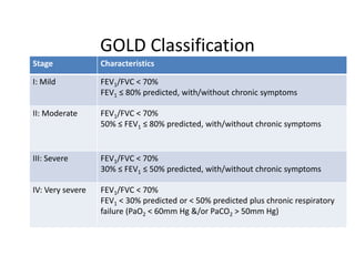
COPD pathophysiology &
anesthetic management
Moderator: Prof. Dr Bisharad man
Shrestha
3rd july,2014
COPD
Chronic obstructive pulmonary disease
• Chronic progressive Inflammatory airway
disease with airflow limitation that is NOT
FULLY reversible
COPD
Chronic Bronchitis: (Clinical Definition)
• Chronic productive cough with sputum on
most days of 3 consecutive months in 2
successive years
Emphysema: (Pathological Definition)
• Permanent enlargement of the airspaces distal
to the terminal bronchioles by destruction of
their walls and without obvious fibrosis
COPD: Risk factors
Host factos:
•Genetic factors: e.g. α1 Antitrypsin Deficiency
•Sex : Prevalence more in males.
?Females more susceptible
•Airway hyperactivity,
Immunoglobulin E and asthma
Exposures:
•Smoking: Most Important Risk Factor
•Socioeconomic status
•Occupation
•Environmental pollution
•Perinatal events and childhood illness
•Recurrent bronchopulmonary infections
•Diet
Pathogenesis:
Tobacco smoke & other
noxious gases
Inflammatory
response in airways
Tissue Destruction
Impaired defense against tissue destruction
Impaired repair mechanisms
Proteinase & Antiproteinase
imbalance
Oxidative
Stress
Alpha 1
antitrypsin def.
Pathology:
• Large airways
– hypertrophy and hyperplasia of mucus glands and
goblet cells
– Squamous metaplasia: impaired mucociliary
clearance,loss of cilia
– Smooth muscle hypertrophy
– Neutrophil influx
• Small airways:
– Goblet cell metaplasia
– Replacement of surfactant secreting Clara cells
with mucus secreting and infiltrating
mononuclear inflammatory cells
– Fibrosis
– Airway narrowing
• Parenchyma:
– Destruction of gas exchanging airspaces
Outcomes
• Airflow obstruction:
– Decrease in FEF25-75% FEV1 and FEV1/FVC ratio
• Hyperinflation :
– Air trapping due to premature closure of airways with
obstruction
– Increase in RV and RV/TLC ratio
• Impaired Gas exchange: non uniform ventilation,
V/Q mismatch
• Pulmonary hypertension
– Hypoxic Vasoconstriction,Endothelial dysfunction
– Remodelling of arteries & capillary destruction
Comparative features of COPD
Mechanism of airway
obstruction
Dyspnea
FEV1
PaO2
PaCO2
Diffusing capacity
Hematocrit
Cor pulmonale
Prognosis
Mucus & inflammation
Moderate
Decreased
Marked decrease
Increased
Normal
Increased
Marked
Poor
loss of elastic recoil
Severe
Decreased
Modest decrease
normal to decreased
Decreased
Normal
Mild
Better
feature Ch. Bronchitis Emphysema
Flow-vol loop
GOLD Classification
Stage Characteristics
I: Mild FEV1/FVC < 70%
FEV1 ≤ 80% predicted, with/without chronic symptoms
II: Moderate FEV1/FVC < 70%
50% ≤ FEV1 ≤ 80% predicted, with/without chronic symptoms
III: Severe FEV1/FVC < 70%
30% ≤ FEV1 ≤ 50% predicted, with/without chronic symptoms
IV: Very severe FEV1/FVC < 70%
FEV1 < 30% predicted or < 50% predicted plus chronic respiratory
failure (PaO2 < 60mm Hg &/or PaCO2 > 50mm Hg)
Treatment
• Drugs Modifying natural history of Disease:
– Smoking cessation
– Long term oxygen therapy
• Symptomatic:
– Bronchodilators: Anticholinergics,Beta
Agonists,Methylxanthines
– Antibiotics
– Others e.g. mucolytics
• Pulmonary Rehabilitation
• Nutrition
COPD: Exacerbations
Definition:
• Event characterised by a change in the patient’s
baseline dyspnoea, cough and/or sputum beyond
day-to-day variability sufficient to warrant a change
in management
Precipitating Causes:
• Infections: Bacterial, Viral
• Air pollution exposure
• Non compliance with LTOT(long term O2 therapy)
Anesthetic management
Pre-operative assessment:
History:
- co-morbidities
– Smoking (pack years)
– Cough: Type, Progression, Recent RTI
– Sputum: Quantity, color
– Dyspnea
– Exercise tolerance
– Occupation, Allergies
– Symptoms of cardiac or respiratory failure
– Drugs
Pre-operative assessment: Examination
Physical Examination: Better at assessing chance of post op
complications
Airway obstruction
– hyperinflation of chest, Barrel chest
– Decreased breath sounds
– Expiratory rhonchi
– Prolonged expiration: Watch & Stethoscope test, >4 sec
↑WOB
– ↑ RR, ↑HR
– Accessory muscles used
– Tracheal tug
– Intercostal indrawing
– Tripod sitting posture
• Body Habitus
Obesity/ Malnourished
S. albumin (?<3.5gm/dl)
• Active infection
– Sputum- change in quantity,
nature
– Fever
– Crepitations
Respiratory failure
Hypercapnia
Hypoxia
Cyanosis
Cor Pulmonale and Right heart
failure
Dependent edema
tender hepatomegaly
Pulmonary hypertension
Loud P2
Right Parasternal heave
Tricuspid regurgitation
Pre-operative assessment: Examination
Preoperative Assessment: Investigations
• Complete Blood count
• Serum Electrolytes
• Blood Sugar
• Urinalysis
• ECG
• Arterial Blood Gases
• Diagnostic Radiology
– Chest X Ray
– Spiral CT
• Preoperative Pulmonary Function Tests
– Tool for optimisation of pre-op lung function
– Not to assess risk of post op pulmonary complications
Investigations: Chest X-Ray
• Overinflation
• Depression or flattening of
diaphragm
• Increase in length of lung
• ↑ size of retrosternal airspace
• ↑ lung markings- dirty lung
• Bullae +/-
• Vertical Cardiac silhouette
• ↑ transverse diameter of chest,
ribs horizontal, square chest
• Enlarged pulmonary artery with
rapid tapering in MZ
Pulmonary Function Tests:
Measuremen
t
Normal Obstructive Restrictive
FVC (L) 80% of TLC (4800)   
FEV1 (L) 80% of FVC  
FEV1/FVC(%) 75- 85% N to  N to 
FEV25%-75%(L/sec) 4-5 L/ sec  N to 
PEF(L/sec) 450- 700 L/min  N to 
Slope of FV
curve
 
MVV(L/min) 160-180 L/min  N to 
TLC 6000 ml N to  
RV 1500 mL  
RV/TLC(%) 0.25  N
Preoperative Assessment: Investigations
ECG
• Signs of RVH:
– RAD
– p Pulmonale in Lead II
– Predominant R wave in V1-3
– RS pattern in precordial leads
Arterial Blood Gases:
• In moderate-severe disease
• Nocturnal sample in cor Pulmonale
– Increased PaCO2 is prognostic marker
– Strong predictor of potential intra op respiratory failure & post
op Ventilatory failure
– Also, increased d/t post op pain, shivering, fever,respiratory
depressants
Pre-operative preparation
• Cessation of smoking (6 weeks preferably)
• Dilation of airways
• Loosening & Removal of secretions
• Eradication of infection
• Recognition of Cor Pulmonale and treatment
• Improve strength of skeletal muscles – nutrition,
exercise
• Correct electrolyte imbalance
• Familiarization with respiratory therapy, education,
motivation & facilitation of patient care
Risk reduction strategies
• Pre-operative optimisation:
– Smoking cessation:
• Decreases carboxy-Hb levels (6.5%-1.1%): -ve inotropic
effect
– Optimisation of drug therapy
– Treatment of infection
– Respiratory physiotherapy
• Intraoperative :
– Selection of anesthetic technique
• Use of regional anesthesia, if feasible
– Precautions: Large doses of sedatives , High blocks
• GA:
– Inhalational agents: sevoflurane, desflurane, nitrous oxide
– Opioids
– Humidification of inspired gases
– Optimal fluid management
– Large tidal volumes (8-10) with slow RR (6-10/min)
Risk reduction strategies:
– Use of minimal invasive techniques (surgery)
when possible
– Avoiding use of long acting NMBDs
– Avoiding surgical procedures likely to require > 3
hours
Risk reduction strategies:
• Post operative:
– Maximization of analgesia control:
• Epidural analgesia
• Intercostal nerve blocks
• Patient controlled analgesia
– Lung volume expansion maneuvers
• Voluntary deep breathing
• Incentive spirometry
• CPAP
Thank you.

Copd&anesthesia

  • 1.
    COPD pathophysiology & anestheticmanagement Moderator: Prof. Dr Bisharad man Shrestha 3rd july,2014
  • 2.
    COPD Chronic obstructive pulmonarydisease • Chronic progressive Inflammatory airway disease with airflow limitation that is NOT FULLY reversible
  • 3.
    COPD Chronic Bronchitis: (ClinicalDefinition) • Chronic productive cough with sputum on most days of 3 consecutive months in 2 successive years Emphysema: (Pathological Definition) • Permanent enlargement of the airspaces distal to the terminal bronchioles by destruction of their walls and without obvious fibrosis
  • 4.
    COPD: Risk factors Hostfactos: •Genetic factors: e.g. α1 Antitrypsin Deficiency •Sex : Prevalence more in males. ?Females more susceptible •Airway hyperactivity, Immunoglobulin E and asthma Exposures: •Smoking: Most Important Risk Factor •Socioeconomic status •Occupation •Environmental pollution •Perinatal events and childhood illness •Recurrent bronchopulmonary infections •Diet
  • 5.
    Pathogenesis: Tobacco smoke &other noxious gases Inflammatory response in airways Tissue Destruction Impaired defense against tissue destruction Impaired repair mechanisms Proteinase & Antiproteinase imbalance Oxidative Stress Alpha 1 antitrypsin def.
  • 6.
    Pathology: • Large airways –hypertrophy and hyperplasia of mucus glands and goblet cells – Squamous metaplasia: impaired mucociliary clearance,loss of cilia – Smooth muscle hypertrophy – Neutrophil influx
  • 7.
    • Small airways: –Goblet cell metaplasia – Replacement of surfactant secreting Clara cells with mucus secreting and infiltrating mononuclear inflammatory cells – Fibrosis – Airway narrowing • Parenchyma: – Destruction of gas exchanging airspaces
  • 8.
    Outcomes • Airflow obstruction: –Decrease in FEF25-75% FEV1 and FEV1/FVC ratio • Hyperinflation : – Air trapping due to premature closure of airways with obstruction – Increase in RV and RV/TLC ratio • Impaired Gas exchange: non uniform ventilation, V/Q mismatch • Pulmonary hypertension – Hypoxic Vasoconstriction,Endothelial dysfunction – Remodelling of arteries & capillary destruction
  • 9.
    Comparative features ofCOPD Mechanism of airway obstruction Dyspnea FEV1 PaO2 PaCO2 Diffusing capacity Hematocrit Cor pulmonale Prognosis Mucus & inflammation Moderate Decreased Marked decrease Increased Normal Increased Marked Poor loss of elastic recoil Severe Decreased Modest decrease normal to decreased Decreased Normal Mild Better feature Ch. Bronchitis Emphysema Flow-vol loop
  • 10.
    GOLD Classification Stage Characteristics I:Mild FEV1/FVC < 70% FEV1 ≤ 80% predicted, with/without chronic symptoms II: Moderate FEV1/FVC < 70% 50% ≤ FEV1 ≤ 80% predicted, with/without chronic symptoms III: Severe FEV1/FVC < 70% 30% ≤ FEV1 ≤ 50% predicted, with/without chronic symptoms IV: Very severe FEV1/FVC < 70% FEV1 < 30% predicted or < 50% predicted plus chronic respiratory failure (PaO2 < 60mm Hg &/or PaCO2 > 50mm Hg)
  • 11.
    Treatment • Drugs Modifyingnatural history of Disease: – Smoking cessation – Long term oxygen therapy • Symptomatic: – Bronchodilators: Anticholinergics,Beta Agonists,Methylxanthines – Antibiotics – Others e.g. mucolytics • Pulmonary Rehabilitation • Nutrition
  • 12.
    COPD: Exacerbations Definition: • Eventcharacterised by a change in the patient’s baseline dyspnoea, cough and/or sputum beyond day-to-day variability sufficient to warrant a change in management Precipitating Causes: • Infections: Bacterial, Viral • Air pollution exposure • Non compliance with LTOT(long term O2 therapy)
  • 13.
  • 14.
    Pre-operative assessment: History: - co-morbidities –Smoking (pack years) – Cough: Type, Progression, Recent RTI – Sputum: Quantity, color – Dyspnea – Exercise tolerance – Occupation, Allergies – Symptoms of cardiac or respiratory failure – Drugs
  • 15.
    Pre-operative assessment: Examination PhysicalExamination: Better at assessing chance of post op complications Airway obstruction – hyperinflation of chest, Barrel chest – Decreased breath sounds – Expiratory rhonchi – Prolonged expiration: Watch & Stethoscope test, >4 sec ↑WOB – ↑ RR, ↑HR – Accessory muscles used – Tracheal tug – Intercostal indrawing – Tripod sitting posture
  • 16.
    • Body Habitus Obesity/Malnourished S. albumin (?<3.5gm/dl) • Active infection – Sputum- change in quantity, nature – Fever – Crepitations Respiratory failure Hypercapnia Hypoxia Cyanosis Cor Pulmonale and Right heart failure Dependent edema tender hepatomegaly Pulmonary hypertension Loud P2 Right Parasternal heave Tricuspid regurgitation Pre-operative assessment: Examination
  • 17.
    Preoperative Assessment: Investigations •Complete Blood count • Serum Electrolytes • Blood Sugar • Urinalysis • ECG • Arterial Blood Gases • Diagnostic Radiology – Chest X Ray – Spiral CT • Preoperative Pulmonary Function Tests – Tool for optimisation of pre-op lung function – Not to assess risk of post op pulmonary complications
  • 18.
    Investigations: Chest X-Ray •Overinflation • Depression or flattening of diaphragm • Increase in length of lung • ↑ size of retrosternal airspace • ↑ lung markings- dirty lung • Bullae +/- • Vertical Cardiac silhouette • ↑ transverse diameter of chest, ribs horizontal, square chest • Enlarged pulmonary artery with rapid tapering in MZ
  • 19.
    Pulmonary Function Tests: Measuremen t NormalObstructive Restrictive FVC (L) 80% of TLC (4800)    FEV1 (L) 80% of FVC   FEV1/FVC(%) 75- 85% N to  N to  FEV25%-75%(L/sec) 4-5 L/ sec  N to  PEF(L/sec) 450- 700 L/min  N to  Slope of FV curve   MVV(L/min) 160-180 L/min  N to  TLC 6000 ml N to   RV 1500 mL   RV/TLC(%) 0.25  N
  • 20.
    Preoperative Assessment: Investigations ECG •Signs of RVH: – RAD – p Pulmonale in Lead II – Predominant R wave in V1-3 – RS pattern in precordial leads Arterial Blood Gases: • In moderate-severe disease • Nocturnal sample in cor Pulmonale – Increased PaCO2 is prognostic marker – Strong predictor of potential intra op respiratory failure & post op Ventilatory failure – Also, increased d/t post op pain, shivering, fever,respiratory depressants
  • 21.
    Pre-operative preparation • Cessationof smoking (6 weeks preferably) • Dilation of airways • Loosening & Removal of secretions • Eradication of infection • Recognition of Cor Pulmonale and treatment • Improve strength of skeletal muscles – nutrition, exercise • Correct electrolyte imbalance • Familiarization with respiratory therapy, education, motivation & facilitation of patient care
  • 22.
    Risk reduction strategies •Pre-operative optimisation: – Smoking cessation: • Decreases carboxy-Hb levels (6.5%-1.1%): -ve inotropic effect – Optimisation of drug therapy – Treatment of infection – Respiratory physiotherapy
  • 23.
    • Intraoperative : –Selection of anesthetic technique • Use of regional anesthesia, if feasible – Precautions: Large doses of sedatives , High blocks • GA: – Inhalational agents: sevoflurane, desflurane, nitrous oxide – Opioids – Humidification of inspired gases – Optimal fluid management – Large tidal volumes (8-10) with slow RR (6-10/min) Risk reduction strategies:
  • 24.
    – Use ofminimal invasive techniques (surgery) when possible – Avoiding use of long acting NMBDs – Avoiding surgical procedures likely to require > 3 hours
  • 25.
    Risk reduction strategies: •Post operative: – Maximization of analgesia control: • Epidural analgesia • Intercostal nerve blocks • Patient controlled analgesia – Lung volume expansion maneuvers • Voluntary deep breathing • Incentive spirometry • CPAP
  • 26.

Editor's Notes

  • #8 DUTCH hypothesis:Asthma, chronic bronchitis and emphysema are variations of same basic disease which is modulated by environmental and genetic factors to produce pathologically distinct entities British hypothesis: Asthma and COPD are different diseases: asthma: allergic COPD: smoking related inflammation and damage
  • #10 Anesthesia & co-existing diseases, stoelting sixth edn
  • #11 Chronic symptoms (cough,sputum production) , GLOBAL INITIATIVE FOR OBSTRUCTIVE LUNG DISEASES
  • #12 Diuretics if in failure
  • #13 Infectious process [7, 8]: viral (Rhinovirus spp., influenza); bacteria (Haemophilus influenzae, Streptococcus pneumoniae, Moraxella catarrhalis, Enterobacteriaceae spp., Pseudomonas spp.).
  • #15 >60 pack yrs correlated with double risk of post-op pulm complications
  • #16 In tripod position, one sits or stands leaning forward and supporting the upper body with hands on the knees or on another surface.[2] Among medical professionals, a patient adopting the tripod position is considered an indication that the patient may be in respiratory distress
  • #17 S. albumin (?<3.5gm/dl) is a strong predictor of post-operative pulmonary complication
  • #22 6 wks smoking cessation required for better ciliary function and immune responses
  • #23 CO; t ½: 4-6 hrs, P50 increases from 22.9 to 26.4 mm Hg with smoking cessation for 12 hrs. but short term cessation does not decrease risk of post op pulmonary complications
  • #24 Inhaled anesthetics may attenuate HPV. Slow RR with high Vt: minimise likelihood of turbulent airflow and maintain V/Q ratio. Also, it allows sufficient time for venous return to the heart and are less likely to be associated with hyperventilation. There is sufficient time for exhalation so that air trapping can be minimised.
  • #26 PAIN CONTROL: ambulation increases FRC and improves arterial oxygenation presumably by improving ventilation to perfusion matching. Vital capacity is decreased by 40% which returns to near normal level after 14 days. FRC decreases only 16 hrs after surgery which may be due to altered respiratory ms co-ordination. Alleviating pain does not improve Vital capacity or FRC, suggesting that trauma from surgical procedure contributes to decreased lung volumes.