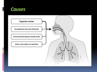
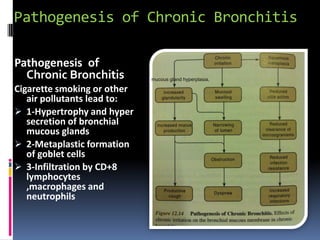
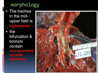
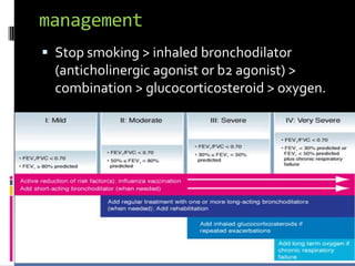
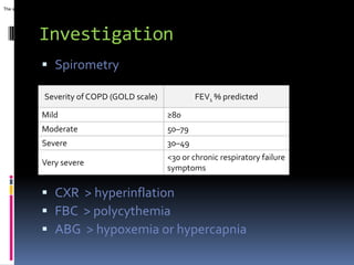
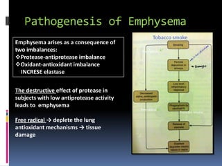
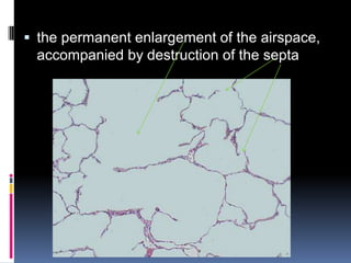
Free syria
RAHEEF ALATASSI
objectives
   chronic bronchitis :
       definition
       causes
       path physiology
       morphology
       mechanism of obstruction
       management
       complication
       S/S
       Investigation
   Emphysema:
       definition
       Causes
       path physiology
       morphology
       types
       management
       complication
       S/S
       Investigation
   Pulmonary hypertension:
       Definition
CHRONIC BRONCHITIS
Definition
    It is defined as a persistent productive
        cough for at least 3 consecutive months in
        at least 2 consecutive years
forms                            features

Simple chronic bronchitis        the productive cough raises
                                 mucoid sputum, but airflow is not
                                 obstructed
Chronic asthmatic bronchitis      hyper-responsive airways with
                                 intermittent bronchospasm and
                                 wheezing

Chronic obstructive bronchitis   develops chronic outflow
                                 obstruction
                                 Associated with emphysema
Causes
Pathogenesis of Chronic Bronchitis

Pathogenesis of
  Chronic Bronchitis         mucous gland hyperplasia,

Cigarette smoking or other
   air pollutants lead to:
 1-Hypertrophy and hyper
   secretion of bronchial
   mucous glands
 2-Metaplastic formation
   of goblet cells
 3-Infiltration by CD+8
   lymphocytes
   ,macrophages and
   neutrophils
morphology
 The trachea
  in the mid-
  upper field is
  hyperemic
 the
  bifurcation &
  bronchi
  contain
  mucopurulent
  exudate
  secretion.
 enlargement of the mucus-secreting
 glands
management
 Stop smoking > inhaled bronchodilator
  (anticholinergic agonist or b2 agonist) >
  combination > glucocorticosteroid > oxygen.
Complications:
 Pulmonary hypertension and cardiac failure
 Recurrent infections
 Respiratory failure

S/S :
 cough , sputum ,frequent infections ,
  intermittent dyspnea, wheeze.
 Some patients develop significant COPD
  with outflow obstruction: hypercapnia,
  hypoxemia, cyanosis
The severity of COPD also depends on the severity of dyspnea and exercise limitation. These and


                 Investigation
                   Spirometry

                    Severity of COPD (GOLD scale)                                             FEV1 % predicted

                   Mild                                                          ≥80
                   Moderate                                                      50–79
                   Severe                                                        30–49
                                                                                 <30 or chronic respiratory failure
                   Very severe
                                                                                 symptoms

                   CXR > hyperinflation
                   FBC > polycythemia
                   ABG > hypoxemia or hypercapnia
Spirometry
EMPHYSEMA
definition
• It is permanent enlargement of air spaces
  distal to terminal bronchioles, accompanied
  by destruction of their walls.
• not associate with fibrosis

Causes:
Smoking
Deficinecy of a1 antitrypsin
path physiology
Pathogenesis of Emphysema
                                                   Tobacco smoke
Emphysema arises as a consequence of
two imbalances:                           free radical
Protease-antiprotease imbalance
Oxidant-antioxidant imbalance
  INCRESE elastase


The destructive effect of protease in
subjects with low antiprotease activity
leads to emphysema
                                          free radical
                                                             Protease release

Free radical → deplete the lung
antioxidant mechanisms → tissue
damage
morphology

 The external surfaces of the upper lobes
  of both the right and left lungs have large
  bullae.
 Distal type.
 bullae
Cyst
more than
1 cm.
 the permanent enlargement of the airspace,
 accompanied by destruction of the septa
types
type            features
                It involve the central or proximal parts of the acini
Centriacinar    The lesions are more common and severe in the upper
                lobes
                This type is seen as a result of cigarette smoking

                It occurs more commonly in the lower lung zones
Panacinar       The type of emphysema occurs in α1-antitrypsin
                deficiency

                   The distal part of the acinus is involved
Distal Acinar      It is seen adjacent to the pleura, along the lobular
                    margins,scarring.
                   Cystlike structures can be formed (bullae)

                The acinus is irregularly involved
Irregular       It is associated with scarring
                Clinically asymptomatic
                The most common form of emphysema
Management & Investigation


    The same treatment &
  Investigation as the chronic
           bronchitis
Pulmonary Hypertension in COPD
        Chronic hypoxia


   Pulmonary vasoconstriction
                                    Muscularization

   Pulmonary hypertension           Intimal
                                    hyperplasia
                                    Fibrosis
       Cor pulmonale                Obliteration

                            Edema
             Death
complication

I.   Pulmonary hypertension;
      hypoxia-induced pulmonary vascular spasm
      loss of capillary surface area due to alveolar wall
       destruction
II. Right side heart failure
III. Respiratory failure
S/S
   Patients present with dyspnea, and
     abnormal pulmonary function tests

                            Patients with no bronchitis
Patients with underlying
chronic bronchitis          They have more severe
                            dyspnea and
They have less dyspnea     hyperventilation
and retain more CO2
 They are always hypoxic    They have normal gas
and cyanotic (blue            exchange and adequate
bloaters)                     oxygenation of
                              hemoglobin (pink puffers)
Pulmonary hypertension:

 The pulmonary circulation is normally one of
  low resistance, with pulmonary blood
  pressures being only about one-eighth of
  systemic pressure. Pulmonary hypertension
  (when mean pulmonary pressures reach one-
  fourth or more of systemic levels) is most
  often secondary to a decrease in the cross-
  sectional area of the pulmonary vascular bed,
  or to increased pulmonary vascular blood
  flow
morphology

 A, Gross photograph of
  atheroma formation, a
  finding usually limited to
  large vessels.
 B, Marked medial
  hypertrophy.
 C, Plexogenic lesion
  characteristic of advanced
  pulmonary hypertension
  seen in small arteries.

COPD lecture

  • 1.
  • 2.
    objectives  chronic bronchitis :  definition  causes  path physiology  morphology  mechanism of obstruction  management  complication  S/S  Investigation  Emphysema:  definition  Causes  path physiology  morphology  types  management  complication  S/S  Investigation  Pulmonary hypertension:  Definition
  • 3.
  • 4.
    Definition  It is defined as a persistent productive cough for at least 3 consecutive months in at least 2 consecutive years forms features Simple chronic bronchitis the productive cough raises mucoid sputum, but airflow is not obstructed Chronic asthmatic bronchitis hyper-responsive airways with intermittent bronchospasm and wheezing Chronic obstructive bronchitis develops chronic outflow obstruction Associated with emphysema
  • 5.
  • 6.
    Pathogenesis of ChronicBronchitis Pathogenesis of Chronic Bronchitis mucous gland hyperplasia, Cigarette smoking or other air pollutants lead to:  1-Hypertrophy and hyper secretion of bronchial mucous glands  2-Metaplastic formation of goblet cells  3-Infiltration by CD+8 lymphocytes ,macrophages and neutrophils
  • 7.
    morphology  The trachea in the mid- upper field is hyperemic  the bifurcation & bronchi contain mucopurulent exudate secretion.
  • 8.
     enlargement ofthe mucus-secreting glands
  • 9.
    management  Stop smoking> inhaled bronchodilator (anticholinergic agonist or b2 agonist) > combination > glucocorticosteroid > oxygen.
  • 10.
    Complications:  Pulmonary hypertensionand cardiac failure  Recurrent infections  Respiratory failure S/S :  cough , sputum ,frequent infections , intermittent dyspnea, wheeze.  Some patients develop significant COPD with outflow obstruction: hypercapnia, hypoxemia, cyanosis
  • 11.
    The severity ofCOPD also depends on the severity of dyspnea and exercise limitation. These and Investigation  Spirometry Severity of COPD (GOLD scale) FEV1 % predicted Mild ≥80 Moderate 50–79 Severe 30–49 <30 or chronic respiratory failure Very severe symptoms  CXR > hyperinflation  FBC > polycythemia  ABG > hypoxemia or hypercapnia
  • 12.
  • 13.
  • 14.
    definition • It ispermanent enlargement of air spaces distal to terminal bronchioles, accompanied by destruction of their walls. • not associate with fibrosis Causes: Smoking Deficinecy of a1 antitrypsin
  • 15.
  • 16.
    Pathogenesis of Emphysema Tobacco smoke Emphysema arises as a consequence of two imbalances: free radical Protease-antiprotease imbalance Oxidant-antioxidant imbalance INCRESE elastase The destructive effect of protease in subjects with low antiprotease activity leads to emphysema free radical Protease release Free radical → deplete the lung antioxidant mechanisms → tissue damage
  • 17.
    morphology  The externalsurfaces of the upper lobes of both the right and left lungs have large bullae.  Distal type.  bullae Cyst more than 1 cm.
  • 18.
     the permanentenlargement of the airspace, accompanied by destruction of the septa
  • 19.
    types type features It involve the central or proximal parts of the acini Centriacinar The lesions are more common and severe in the upper lobes This type is seen as a result of cigarette smoking It occurs more commonly in the lower lung zones Panacinar The type of emphysema occurs in α1-antitrypsin deficiency  The distal part of the acinus is involved Distal Acinar  It is seen adjacent to the pleura, along the lobular margins,scarring.  Cystlike structures can be formed (bullae) The acinus is irregularly involved Irregular It is associated with scarring Clinically asymptomatic The most common form of emphysema
  • 21.
    Management & Investigation The same treatment & Investigation as the chronic bronchitis
  • 22.
    Pulmonary Hypertension inCOPD Chronic hypoxia Pulmonary vasoconstriction Muscularization Pulmonary hypertension Intimal hyperplasia Fibrosis Cor pulmonale Obliteration Edema Death
  • 23.
    complication I. Pulmonary hypertension;  hypoxia-induced pulmonary vascular spasm  loss of capillary surface area due to alveolar wall destruction II. Right side heart failure III. Respiratory failure
  • 24.
    S/S Patients present with dyspnea, and abnormal pulmonary function tests Patients with no bronchitis Patients with underlying chronic bronchitis They have more severe dyspnea and They have less dyspnea hyperventilation and retain more CO2  They are always hypoxic  They have normal gas and cyanotic (blue exchange and adequate bloaters) oxygenation of hemoglobin (pink puffers)
  • 25.
    Pulmonary hypertension:  Thepulmonary circulation is normally one of low resistance, with pulmonary blood pressures being only about one-eighth of systemic pressure. Pulmonary hypertension (when mean pulmonary pressures reach one- fourth or more of systemic levels) is most often secondary to a decrease in the cross- sectional area of the pulmonary vascular bed, or to increased pulmonary vascular blood flow
  • 26.
    morphology  A, Grossphotograph of atheroma formation, a finding usually limited to large vessels.  B, Marked medial hypertrophy.  C, Plexogenic lesion characteristic of advanced pulmonary hypertension seen in small arteries.

Editor's Notes

  • #5 Clinical defethion1- just productive caught 2-Chronic asthmatic bronchitis  associated hyperresponsive (hyperreactive) airways , bronchospasm3- - obstructive lesion
  • #12 A diagnosis of COPD should be considered in any patient who has cough, sputum production, or dyspnea and/or a history of exposure to risk factors. The diagnosis is confirmed by spirometry. To help identify individuals earlier in the course of disease, spirometry should be performed for patients who have chronic cough and sputum production even if they do not have dyspnea. Spirometry is the best way to diagnose COPD and to monitor its progression and health care workers to care for COPD patients should have assess to spirometry.
  • #15 air spaces distal to terminal bronchioles is  (acini) respiratory bronchiole , alveolar duct , alveoli
  • #24 Hypoxia in lung is special will induce pulmonary vasoconstriction that will elevate pulmonary hypertension, pressure overload right side heart failure