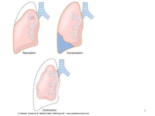
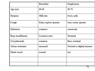
Atelectasis (Collapse)
• Loss of lung volume caused by inadequate
expansion of air spaces
• Ventilation perfusion imbalance and hypoxia.
• classified into three forms
Resorption atelectasis
• Occurs when an obstruction prevents air from reaching
distal airways.
• Any air present gradually becomes absorbed, and alveolar
collapse follows.
• The most common cause of resorption collapse is
obstruction of a bronchus.
• Resorption atelectasis most frequently occurs
postoperatively due to intrabronchial mucous or
mucopurulent plugs
• May also result from foreign body aspiration (particularly
in children), bronchial asthma, bronchiectasis, chronic
bronchitis, or intrabronchial tumor.
Compression atelectasis
• Is usually associated with accumulation of fluid, blood,
or air within the pleural cavity.
• A frequent cause is pleural effusions occurring in the
setting of congestive heart failure.
• Leakage of air into the pleural cavity (pneumothorax)
also leads to compression atelectasis.
• Basal atelectasis resulting from a failure to breath
deeply commonly occurs in bedridden patients, in
patients with ascites, and during and after surgery.
• Contraction atelectasis (or cicatrization
atelectasis)
• Occurs when local or diffuse fibrosis affecting
the lung or the pleura hamper lung expansion
5
6
OBSTRUCTIVE Vs RESTRICTIVE
PULMONARY DISEASES
Classification is based on pulmonary function tests
• Obstructive disease (airway disease)
• Characterized by an increase in resistance caused by
partial or complete obstruction at any level
• Decreased expiratory flow rate, usually measured by
forced expiratory volume at 1 second (FEV1)
• Thus, the ratio of FEV to FVC is characteristically
decreased
• The major diffuse obstructive disorders
- Emphysema
- Chronic bronchitis
- Bronchiectasis
- Asthma
6
7
Restrictive disease
• Characterized by reduced expansion of lung
parenchyma, with decreased total lung capacity.
• FVC is reduced and the expiratory flow rate is normal
• Restrictive diseases occur in
 Chest disorders – kyphoscoliosis, polio, obesity, pleural
disease
 Acute or chronic interstitial & infiltrative diseases
ARDS, pneumoconioses, interstitial fibrosis of unknown
etiology, infiltrative conditions (e.g., sarcoidosis).
7
8
Chronic obstructive pulmonary disease (COPD)
Emphysema
• Emphysema is characterized by permanent
enlargement of the air spaces distal to the
terminal bronchioles, accompanied by destruction
of their walls without significant fibrosis
• Four main types
1.Centriacinar
• Destruction of central portion with sparing of distal
airways
• Upper lobes > lower
• Cause: smoking
2.Panacinar
• Uniform injury
• Lower lobes > upper
• Cause: alpha-1-antitrypsin deficiency
8
9
3.Distal Acinar (paraseptal) Emphysema
• The proximal portion of the acinus is normal, but
the distal part is predominantly involved.
• More striking adjacent to the pleura
• Occurs adjacent to areas of fibrosis, scarring or
atelectasis
• More sever in the upper half of the lungs
• Sometimes form cyst like structures(bullae)
• Underlies many of the spontaneous pneumothorax
9
10
4.Airspace enlargement with fibrosis (
Irregular)
• The acinus is irregularly involved
• Associated with scarring
• Asymptomatic & clinically insignificant
10
11
12
Pathogenesis
• The pathogenesis of centriacinar and
panacinar is not completely understood.
• Current opinion favors emphysema arising as
a consequence of two critical imbalances:
• Protease-antiprotease imbalance
• Oxidant-antioxidant imbalance
• COPD is characterized by mild chronic
inflammation through out the airways,
parenchyma, & pulmonary vasculature.
12
13
Protease – antiprotease theory
• Alveolar wall destruction occurs from an
imbalance between proteases (mainly
elasteases) & antiproteases in the lungs
• Any stimulus that increases either the
number of leukocytes in the lung or the
release of their elastase – containing granules
increases elastolytic activity
13
14
Oxidant-antioxidant imbalance
• Stimulated neutrophils also release oxygen
free radicals which inhibit α1- AT activity
• With low levels of serum α1- AT the process
of elastic tissue destruction is unchecked,
with consequent emphysema
14
15
Ctd…
• In smokers – both increased elastase availability &
decreased anti elastase activity occur
• In smokers - both neutrophils & macrophages
accumulate in the alveoli
• It may involve the direct chemo attractant effect of
nicotine as well as reactive oxygen species contained in
smoke
• Accumulated neutrophils are activated & release there
enzymes ( elastase, proteinase 3, cathepsin G )
• Smoking also plays a role in perpetuating the oxidant –
anti oxidant imbalance
• Oxidative injury also results in functional α1- AT
deficiency
15
16
17
Clinical course
• Pulmonary function tests reveal reduced FEV1 with normal
or near-normal FVC.
• Hence, the FEV1 to FVC ratio is reduced.
• Do not appear until one third of pulmonary parenchyma is
damaged
• Dyspnea is first symptom & insidious
• Cough or wheezing
• Weight loss
• Exiparatory air flow limitation by spirometry
• Development of cor-pulmonale & CHF secondary to
pulmonary HTN
17
18
Morphology
• Panacinar emphysema produces voluminous
lungs
• Large apical blebs or bullae are characterstic
of irregular emphysema & distal acinar
emphysema
• Respiratory bronchioles & vasculature of the
lung are deformed
18
19
21
Cause of death in COPD
• Respiratory acidosis & coma
• Rt sided heart failure
• Massive collapse of lungs secondary to pneumothorax
Conditions Related to Emphysema
• Compensatory hyperinflation
• Obstructive hyperinflation
• Bullous emphysema
• Interstitial emphysema
21
22
Chronic bronchitis
• Common in habitual smokers & inhabitants of smog ridden
cities
• When persists for years
1. progress to COPD
2. lead to heart failure
3. fertile ground for cancer
• Defined as persistent cough with sputum production for at
least 3 months in at least 2 consecutive yrs, in the absence
of any other identifiable cause.
22
23
Pathogenesis
• Chronic irritation by inhaled substances-
smoking in 90%
• Bacterial & viral infections are important in
triggering acute exacerbation
• Most frequent in middle aged male
• More common in heavy smokers ( 4-10X )
23
24
Ctd…
• Hypersecretion of mucus in the large
airways associated with hypertrophy of the
submucosal glands
• Proteases from neutrophils & matrix
metalloproteinase stimulate this
hypersecretion
• Role of infections appears to be secondary
24
25
Morphology
• Hyperemia, swelling, edema of mucus
membranes accompanied by excessive
secretion
• Chronic inflammation of the airways, &
enlargement of mucus secreting glands
• Reid index ratio i.e the thickness of the
mucus gland layer to the bronchial wall
25
26
Ctd…
• Reid index (normally 0.4) is increased
• Bronchial epithelium may exhibit squamous
metaplasia & dysplasia
• Marked narrowing of bronchioles
• In severe cases, bronchiolitis obliterans
26
27
28
Clinical features
• Persistent productive cough
• Dyspnea
• COPD(hypoxemia,hypercapnia,cyanosis)
• Col pulmonale with cardiac failure
28
29
yr
Bronchitis Emphysema
Age (yrs) 40-45 50-75
Dyspnea Mild, late Sever, early
Cough Early, copious sputum Late, scanty sputum
Infections common occasional
Resp. insufficiency Common treat Terminal
Col pulmonale common Rare, terminal
Airway resistance increased Normal or slightly increase
Elastic recoil normal low
29
30
Asthma
• It is a chronic inflammatory disorder of the airways
that causes recurrent episode of wheezing,
breathlessness, chest tightness & cough particularly at
night &/or in the early morning
• The hallmarks of asthma are intermittent, reversible
airway obstruction; chronic bronchial inflammation
with eosinophils; bronchial smooth muscle cell
hypertrophy and hyperreactivity; and increased mucus
secretion.
• Inflammation causes an increase in airway
responsiveness to a variety of stimuli
• Eosinophils, mast cells, macrophages, T lymphocytes,
neutrophils & epithelial cells play a role
30
31
Categorization
1. depending on the frequency & severity of
symptoms
- Mild
- Moderate
- Severe
2. inciting agent
- Intrinsic (non atopic)
- Extrinsic (atopic)
31
32
Atopic Asthma
• Usually begins in childhood
• Triggered by env’tal pollutants
• Positive family history is common
• Asthmatic attacks often preceded by allergic
rhinitis, urticaria or eczema
• Skin test is positive to allergen injection
32
• The classic atopic form is associated with excessive type 2 helper T (TH2) cell
activation.
• Cytokines produced by TH2 cells account for most of the features of atopic
asthma— IL-4 and IL-13 stimulate IgE production, IL-5 activates eosinophils, and IL-
13 also stimulates mucus production.
• IgE coats submucosal mast cells, which on exposure to allergen release their
granule contents and secrete cytokines and other mediators.
• Mast cell–derived mediators produce two waves of reaction: an early (immediate)
phase and a late phase
• The early-phase reaction is dominated by bronchoconstriction, increased mucus
production, and vasodilation.
• Bronchoconstriction is triggered by mediators released from mast cells, including
histamine, prostaglandin D2, and leukotrienes LTC4, D4, and E4, and also by reflex
neural pathways.
• The late-phase reaction is inflammatory in nature.
• Inflammatory mediators stimulate epithelial cells to produce chemokines
(including eotaxin, a potent chemoattractant and activator of eosinophils) that
promote the recruitment of TH2 cells, eosinophils, and other leukocytes, thus
amplifying an inflammatory reaction that is initiated by resident immune cells.
34
Ctd…
• Initial sensitization to inhaled antigens which
stimulate induction of TH2 type cells that release
cytokines such as IL 4 & 5
• These cytokines promote IgE production by B cells,
growth of mast cells, growth & activation of
eosinophils
• Subsequent IgE mediated reaction to inhaled
allergens elicits an acute response & late phase
reactions
• Sub epithelial vagal (parasymphatetic) stimulation
provokes broncho constriction
34
35
Ctd…
• Sub epithelial vagal (parasymphatetic)
stimulation provokes broncho constriction
• Acute or immediate response consists of
broncho constriction, edema, mucus
secretion & hypotension
• Expose of pre sensitized IgE coated mast cells
to antigen stimulates release of chemical
mediators
35
36
Ctd…
• Mast cells also release cytokines that cause the
influx of other leukocytes & set the stage of the
late phase reaction
(4-8hrs later)
• Mediators can also be produced by other cells
• Inflammatory cells already present in asthmatics
• Vascular endothelium
• Airway epithelial cells (eotaxin)
• MBP (major basic protein) causes epithelial cell
damage & airway constriction
36
37
39
Non-Atopic Asthma
• Most frequently triggered by viral respiratory infection
• Positive family history is uncommon
• Serum IgE level is normal, Skin test is negative
• No associated allergens
• Virus induced inflammation of the respiratory mucosa
lowers the threshold of the sub epithelial vagal receptors
to irritants
• Inhaled air pollutant may also contribute
39
40
Other forms
Drug induced Asthma
• Aspirin – tipping the balance toward bronco
constrictor leukotrines
Occupational asthma
• Fumes ,dusts
40
41
Morphology
• Occlusion of bronchi and bronchioles by
mucus plug
• Mucus plug contain whorls of shed
epithelium - curschmann spirals
• Eosinophils and charcot leyden crystals
(Crystalloid made up of eosinophil
membrane protein)
41
42
Airway remodeling
Is characteristic finding of asthma
Thickening of the basement membrane of
the bronchial epithelium
Edema and inflammatory infiltrate in the
bronchial walls, with prominence of
eosinophils and mast cells
Increase in size of the submucosal glands
 Hypertrophy of the bronchial muscle wall
42
43
44
Clinical course
• An attack of asthma is characterized by severe dyspnea and
wheezing due to bronchoconstriction and mucus plugging, which
leads to trapping of air in distal airspaces and progressive
hyperinflation of the lungs.
• In the usual case, attacks last from 1 to several hours and subside
either spontaneously or with therapy.
• Intervals between attacks are characteristically free from overt
respiratory difficulties, but persistent, subtle deficits can be
detected by pulmonary function tests.
• Occasionally a severe paroxysm occurs that does not respond to
therapy and persists for days and even weeks (status
asthmaticus).
• The associated hypercapnia, acidosis, and severe hypoxia may be
fatal, although in most cases the condition is more disabling than
lethal.
44
• Standard therapies include anti-inflammatory
drugs, particularly glucocorticoids and
bronchodilators such as beta-adrenergic drugs
and leukotriene inhibitors
46
Bronchiectasis
• A disease characterized by permanent dilation of
bronchi and bronchioles caused by destruction
of the muscle and elastic tissue resulting from or
associated with chronic necrotizing infection
manifested by cough, fever, &copious amounts
of foul smelling position dependent,purulent
sputum
• It is not a primary disorder, as it always occurs
secondary to persistent infection or obstruction
caused by a variety of conditions.
46
47
Bronchiectasis
47
48
ETIOLOGY and PATHOGENESIS
• Obstruction & infection are major influences
• Pooling of secretions distal to obstruction ,& there
is inflammation of the airway
• Severe infections of the airway lead to
inflammation, often with necrosis,fibrosis,and
eventually dilation of the airways
• In primary ciliary dyskinesia ,poorly functioning
cillia contribute to the retention of secretions &
recurrent infection that in turn gives brochiectasis
48
49
ctd
Bronchiectasis develops in association with
• Congenital or hereditary conditions including cystic
fibrosis, primary ciliary dyskinesia , kartagner
syndrome, immunodeficiency, intralobular
sequestration of lung
• Post infectious conditions- bacterial, viral, & fungal
infections
• Bronchial obstruction – tumors, foreign bodies
49
50
Morphology
• Usually affects the lower lobes
• More severe in the more distal bronchi & bronchioles
• The airways are dilated
• Cut surface of the lung reveals dilated bronchi appearing as
cysts filled mucopurulent secrations
• Active case- intense acute & chronic inflammatory
exudation associated with desquamation of lining
epithelium and areas of ulceration
• Chronic cases-bronchial and peribronchial wall fibrosis
50
51
Ctd…
• The airways are dilated sometimes four
times than the normal size
• Long tube like enlargement is called
cylinderical bronchiectasis
• Saccular distension is the other variant of
bronchiectasis
51
52
53
Clinical course
• Sever, persistent cough
• Expectoration of foul smelling, sometimes bloody sputum
• Dyspnea, orthopnea, Fever
• Mixed flora can be cultured
Complications
• Cor pulmonale
• Metastatic brain abscess
• amyloidosis
53
Disorders Associated With Airflow Obstruction: The
Spectrum of Chronic Obstructive Pulmonary Disease
55
Acute restrictive disease
ARDS
• Synonyms – shock lung, diffuse alveolar damage, acute
alveolar injury ,acute lung injury
• Respiratory failure occurring within 1 week of a known
clinical insult
• Caused by diffuse alveolar capillary damage
• Rapid onset of sever life threatening respiratory
insufficiency, cyanosis, severe arterial hypoxemia refractory
to oxygen therapy & may progress to multisystem organ
failure
55
• Severe ARDS is characterized by rapid onset of life threatening
respiratory insufficiency, cyanosis, and severe arterial hypoxemia
that is refractory to oxygen therapy.
• The histologic manifestation of ARDS in the lungs is known as
diffuse alveolar damage (DAD).
• ARDS may occur in a multitude of clinical settings and is associated
with primary pulmonary diseases and severe systemic inflammatory
disorders such as sepsis.
• The most frequent triggers of ARDS are pneumonia (35%–45%) and
sepsis (30%–35%), followed by aspiration, trauma (including brain
injury, abdominal surgery, and multiple fractures), pancreatitis, and
transfusion reactions.
• ARDS should not be confused with respiratory distress syndrome of
the newborn; which is caused by a deficiency of surfactant caused
by prematurity.
Morphology
• In the acute phase of ARDS, the lungs are dark
red, firm, airless, and heavy.
• Microscopic examination reveals capillary
congestion, necrosis of alveolar epithelial cells,
interstitial and intraalveolar edema and
hemorrhage, and (particularly with sepsis)
collections of neutrophils in capillaries.
• The most characteristic finding is the presence of
hyaline membranes, particularlylining the
distended alveolar ducts
• Such membranes consist of fibrin-rich edema fluid
admixed with remnants of necrotic epithelial cells.
• Overall, the picture is remarkably similar to that seen in
respiratory distress syndrome of the newborn
• In the organizing stage, type II pneumocytes proliferate
vigorously in an attempt to regenerate the alveolar
lining.
• Resolution is unusual; more commonly, the fibrin-rich
exudates organize into intra alveolar fibrosis.
• Marked thickening of the alveolar septa ensues due to
proliferation of interstitial cells and deposition of
collagen.
59
Ctd…
• Numerous & diverse conditions are
associated with ARDS but the commonest
four are
Sepsis
Diffuse pulmonary infections
Gastric aspiration
Mechanical injury including head injury
59
60
Pathogenesis
• Central causation is diffuse damage to the alveolar
capillary walls
• Alveolar capillary damage causes increased vascular
permeability
• Acute lung injury occurs as a result of cellular
events initiated by inflammatory stimuli
• Following an acute insult, pulmonary macrophages
synthesize IL-8
60
61
Ctd…
• Release of mediators lead to activations of
neutrophils
• Activated neutrophils releases of oxidases,
proteases, PAF, leukotrienes that cause tissue
damage
• Exudate & diffuse tissue destruction results in
organization, scarring, producing chronic lung
diseases
• Transudate of cardiogenic pulmonary edema
usually resolves
61
• The destructive forces unleashed by
neutrophils can be counteracted by an array
of endogenous anti-proteases and anti-
oxidants that are upregulated by
proinflammatory cytokines.
• It is the balance between the destructive and
protective factors that determines the degree
of tissue injury and clinical severity of the
ARDS.
63
Morphology
• Lungs are heavy, firm, red & boggy
• Congestion, interstitial & intra-alveolar edema,
inflammation & fibrin deposition
• The alveolar walls become lined with waxy hyaline
membranes
• Resolution is unusual, more commonly intra-
alveolar fibrosis
• Thickened alveolar septa caused by proliferation of
interstitial cells
63
66
Clinical course
• Profound dyspnea & tachypnea herald ARDS
• Cyanosis, hypoxemia, respiratory failure
• Hypoxemia becomes unresponsive to oxygen
therapy
• Respiratory acidosis
• Mortality rate is about 60%
• X- ray may be normal initially, later there will be
diffuse bilateral infiltrates
66
C/F
• In 85% of cases, it develops within 72 hours of the
initial insult.
• The overall hospital mortality rate is 38.5% (27%, 32%,
and 45% for mild, moderate, and severe ARDS,
respectively).
• Predictors of poor prognosis include advanced age,
bacteremia (sepsis), and the development of
multiorgan failure.
• Most patients who survive the acute insult recover
normal respiratory function within 6 to 12 months, but
the rest develop diffuse interstitial fibrosis leading to
chronic respiratory insufficiency.
CHRONIC INTERSTITIAL(RESTRICTIVE, INFILTRATIVE)
LUNG DISEASES
• Chronic interstitial diseases are
heterogeneous group of disorders
characterized by bilateral, often patchy,
pulmonary fibrosis mainly affecting the walls
of the alveoli
• Many of the entities in this group are of
unknown cause and pathogenesis; some have
an intraalveolar and an interstitial component
68
69
Ctd…
• The hallmark of these disorders is reduced compliance
(i.e., more pressure is required to expand the lungs
because they are stiff), which in turn necessitates
increased effort of breathing (dyspnea).
• Patients have dyspnea, tachypnea & respiratory
crackles, cyanosis with out wheezing or other evidence
of airway obstruction
• CXR shows diffuse infiltration by small nodules,
irregular lines or ground glass shadows
• With progression, patients may develop respiratory
failure, pulmonary hypertension, and cor pulmonale
69
70
71
Major categories of chronic interstitial lung
disease
71
72
Pneumoconiosis
• Used to describe the non neoplastic lung rxn
to inhalation of mineral dusts, organic
particulates, chemical fumes & vapors
72
73
Pathogenesis
• Regardless of the cause the earliest
manifestation of interstitial disease is
alveolitis i.e accumulation of inflammatory &
immune effector cells with in the alveolar
walls & spaces.
• Leukocyte accumulation distorts alveolar
structure & release of mediators resulting
fibrosis
73
74
General pathogenesis
• The development of pneumoconiosis depends on
 dust retained in the lungs & airways
 The size, shape of the particles
 particle solubility & physiochemical reactivity
 Additional effects of other irritants eg. Smoking
74
75
1. Coal workers’ pneumoconiosis
• There are three spectrum of this disease
I. Asymptomatic anthracosis
II.Simple coal workers’ pneumoconiosis
III.Complicated coal workers’
75
76
Morphology
• Anthracosis – innocuous, coal induced pulmonary lesion,
commonly seen in all urban dwellers & tobacco smokers &
the inhaled carbon accumulate in connective tissue.
• Simple CWP – coal macules containing carbon laden
macrophages 1-2mm in diameter, coal nodules, upper lobe
heavily involved
• Complicated CWP – generally requires many years to
develop , intense black scars, dense collagen & pigment with
areas of necrosis
76
78
Clinical course
• CWP benign disease with little decrement in lung
function.
• Its exposure is during coal mining
• < 10% develop PMF leading to pulmonary
dysfunction, pulmonary HTN & cor pulmonale
• Increased risk of chronic bronchitis & emphysema
• CWP doesn’t predispose to cancer independent of
smoking
78
79
2. Silicosis
• Caused by inhalation of crystalline silicon dioxide
(silica)
• Exposure sand blasting, hard rock mining, stone cutting
• There are two forms of silica-crystalline form which is
the most fibrogenic & amorphous form
• crystalline forms (including quartz, cristobalite, and
tridymite) are by far the most toxic and fibrogenic.
• Of these, quartz is most commonly implicated in
silicosis.
• After inhalation the particles will interact with
epithelial cells & macrophages
79
80
Ctd…
• Some of the cytokines include IL-1, TNF,
fibronectin, lipid mediators, oxygen derived
free radical & fibrogenic cytokines
80
81
Morphology
• Hard collagenous scar
• Some nodules undergo central softening &
cavitations
• CXR egg shell calcification
• Examination by polarized microscopy
birefrigent silica
81
83
Clinical feature
• Most prevalent chronic occupational disease in
the world
• Occurs after decades of exposure & cause nodular
fibrosing pneumoconiosis
• It increases susceptibility to TB
• Most patients do not develop dyspnea until late
in the course
• Its relation with cancer is controversial
83
84
3. Asbestosis related disease
• Its exposure is linked to
Pleural effusion or fibrosis
Asbestosis
Lung cancer (5x)
Mesothelioma
Laryngeal or colonic cancer
• It causes diffuse interstitial disease unlike silicosis which
causes nodular fibrosing disease
• Asbestosis can act as tumor initiator or promoter
84
85
Morphology
• It is difficult to differentiate from other diffuse interstitial
disease unless there is asbestos bodies
• Asbestosis is marked by diffuse pulmonary interstitial fibrosis,
characterized by the presence of asbestos bodies, which are
seen as golden brown, fusiform or beaded rods with a
translucent center.
• They consist of asbestos fibers coated with an iron-containing
proteinaceous material
• Fibrosis begins around the bronchioles & alveolar ducts
• Unlike other forms of pneumoconiosis it affects the lower lobes
• Pleural plaques are the most common manifestations of
asbestosis
• May be complicated by pulmonary HTn & cor pulmonale
85
87
Idiopathic pulmonary fibrosis
• Also called cryptogenic fibrosing alveolitis
• Patchy, progressive bilateral interstitial fibrosis
• It refers to a clinicopathologic syndrome with a
characteristic radiologic, pathologic & clinical
features
• The radiologic and histologic pattern of fibrosis is
referred to as usual interstitial pneumonia (UIP),
which is required for the diagnosis of IPF
• Histological features of usually interstitial
pneumonia is required for its diagnosis
87
Pathogenesis
• Unknown etiology
• It is caused by repeated cycles of acute lung injury
(alveolitis) by some undefined agents
• Wound healing at these sites give rise to exuberant
fibrosis
• The inflammatory response is mediated by TH2 cell
types
• Genetic & environmental factors modify the
phenomenon
88
Morphology
• Pleural surface grossly cobblestoned owing to
retraction of scars along the interlobular septa
• Microscopically the hallmark UIP is patchy
interstitial fibrosis & the earliest
manifestation is fibroblastic foci
89
Ctd…
• Honey comb fibrosis dense, causes collapse of
alveolar walls & formation of cystic spaces
lined by hyperplastic type II pneumocytes or
bronchial epithelium.
• Infiltration of lymphocytes
• Foci of squamous metaplasia & smooth
muscle hyperplasia
• Secondary pulmonary HTn due to intimal
fibrosis & medial thickening of the pulmonary
arteries
90
Clinical course
• Age 40-70 yrs ( >60yrs )
• Dyspnea
• Dry cough
• Hypoxemia
• Cyanosis
• Clubbing
• Mean survival is 3 yrs
• Lung transplantation is the only definitive
therapy
92
Non specific interstitial pneumonia
• Etiology unknown
• Lung biopsies failed to show diagnostic feature of
any of the DILD, a "wastebasket" type of
diagnosis
• Better prognosis than UIP (usual intestitial
pneumonia)
• Morphologically, it has cellular & fibrosing
patterns
• Cellular components include chronic
inflammatory cells, has good out come
• Diffuse & patchy fibrosis
• Younger than UIP
93
Drug- and Radiation-Induced Pulmonary Disease
• Drugs can cause a variety of acute and chronic alterations in respiratory
structure and function.
• For example, bleomycin, an anti-cancer agent, causes pneumonitis and
interstitial fibrosis as a result of direct toxicity of the drug and by
stimulating the influx of inflammatory cells into the alveoli.
• Amiodarone, an anti-arrhythmic agent, also is associated with risk for
pneumonitis and fibrosis.
• Radiation pneumonitis is a well-known complication of irradiation of
pulmonary and other thoracic tumors.
• Acute radiation pneumonitis, which typically occurs 1 to 6 months after
therapy in as many as 20% of the patients, is manifested by fever, dyspnea
out of proportion to the volume of irradiated lung, pleural effusion, and
pulmonary infiltrates in the irradiated lung bed.
• These signs and symptoms may resolve with corticosteroid therapy or
progress to chronic radiation pneumonitis, associated with pulmonary
fibrosis.
Pulmonary involvement in collagen
vascular diseases
• In rheumatoid arthritis pulmonary
involvement is common in one of the four
forms
I. Chronic pleuritis with or without effusion
II. Diffuse interstitial pneumonitis or fibrosis
III. Intrapulmonary rheumatoid nodules
IV. Pulmonary hypertension
95
Granulomatous Diseases
Sarcoidosis
• sarcoidosis is a multisystem disease of unknown
etiology characterized by noncaseating
granulomas in many tissues and organs
• bilateral hilar lymphadenopathy or lung
involvement (or both), visible on chest
radiographs, is the major presenting
manifestation in most cases.
• Eye and skin involvement each occurs in about
25% of cases and may occasionally be the
presenting feature of the disease.
96
• Common after the age of 40 years
• Unknown etiology, disorder of immune regulation
in genetically predisposed individuals exposed to
certain environmental agents.
• T-cell mediated reaction
• Several putative antigen have been proposed
viruses, mycobacteria, Borrelia, pollen), but thus
far there is no unequivocal evidence to suggest
that sarcoidosis is caused by an infectious agent.
97
Morphology: regardless of the organ involved
• Non caseating epitheloid granuloma
• Asteroid bodies(inclusions in multinucleated
giant cells)
• Schaumann bodies: laminated concretions
composed of calcium and proteins
98
Hypersensitivity pneumonitis
• Immunologically mediated disorder affecting
airways and interstitium.
• is an immunologically mediated response to
an extrinsic antigen that involves both
immune-complex and delayed-type
hypersensitivity reactions
100
Pulmonary Eosinophilia
• These diverse diseases generally are of immunologic origin, but the etiology is not understood.
• Pulmonary eosinophilia is divided into the following categories:
• Acute eosinophilic pneumonia with respiratory failure
• Characterized by rapid onset of fever, dyspnea, hypoxia, and diffuse pulmonary infiltrates on chest
radiographs.
• There is prompt response to corticosteroids.
• Simple pulmonary eosinophilia (Loeffler syndrome)
• Characterized by transient pulmonary lesions, eosinophilia in the blood, and a benign clinical
course.
• Tropical eosinophilia
• caused by infection with microfilariae and helminthic parasites
• Secondary eosinophilia
• seen, for example, in association with asthma, drug allergies, and certain forms of
• vasculitis
• Idiopathic chronic eosinophilic pneumonia
• Characterized by aggregates of lymphocytes and eosinophils within the septal walls and the
alveolar spaces, typically in the periphery of the lung fields, and accompanied by high fever, night
sweats, and dyspnea.
• This is a disease of exclusion, once other causes of pulmonary eosinophilia have been ruled out.
Smoking-Related Interstitial Diseases
• Desquamative interstitial pneumonia (DIP)
and respiratory bronchiolitis are two related
examples of smoking-associated interstitial
lung disease.
• The most striking histologic feature of DIP is
the accumulation of large numbers of
macrophages containing dusty-brown pigment
(smoker’s macrophages) in the air spaces
• Respiratory bronchiolitis is a common lesion
found in smokers that is characterized by the
presence of pigmented intraluminal macrophages
akin to those in DIP, but in a “bronchiolocentric”
distribution (first- and second-order respiratory
bronchioles).
• Mild peri bronchiolar fibrosis also is seen.
• As with DIP, affected patients present with
gradual onset of dyspnea and dry cough, and the
symptoms recede with smoking cessation.
• Most often it is an occupational disease that
results from heightened sensitivity to inhaled
antigens such as moldy hay.
• Unlike bronchial asthma, in which bronchi are
the focus of immunologically mediated injury, the
damage in hypersensitivity pneumonitis occurs at
the level of alveoli.
• Hence, it presents as a predominantly restrictive
lung disease with decreased diffusion capacity,
lung compliance, and total lung volume.
104

Atelectasis, restrictive and obstructive pulmonary disease.pptx

  • 1.
    Atelectasis (Collapse) • Lossof lung volume caused by inadequate expansion of air spaces • Ventilation perfusion imbalance and hypoxia. • classified into three forms
  • 2.
    Resorption atelectasis • Occurswhen an obstruction prevents air from reaching distal airways. • Any air present gradually becomes absorbed, and alveolar collapse follows. • The most common cause of resorption collapse is obstruction of a bronchus. • Resorption atelectasis most frequently occurs postoperatively due to intrabronchial mucous or mucopurulent plugs • May also result from foreign body aspiration (particularly in children), bronchial asthma, bronchiectasis, chronic bronchitis, or intrabronchial tumor.
  • 3.
    Compression atelectasis • Isusually associated with accumulation of fluid, blood, or air within the pleural cavity. • A frequent cause is pleural effusions occurring in the setting of congestive heart failure. • Leakage of air into the pleural cavity (pneumothorax) also leads to compression atelectasis. • Basal atelectasis resulting from a failure to breath deeply commonly occurs in bedridden patients, in patients with ascites, and during and after surgery.
  • 4.
    • Contraction atelectasis(or cicatrization atelectasis) • Occurs when local or diffuse fibrosis affecting the lung or the pleura hamper lung expansion
  • 5.
  • 6.
    6 OBSTRUCTIVE Vs RESTRICTIVE PULMONARYDISEASES Classification is based on pulmonary function tests • Obstructive disease (airway disease) • Characterized by an increase in resistance caused by partial or complete obstruction at any level • Decreased expiratory flow rate, usually measured by forced expiratory volume at 1 second (FEV1) • Thus, the ratio of FEV to FVC is characteristically decreased • The major diffuse obstructive disorders - Emphysema - Chronic bronchitis - Bronchiectasis - Asthma 6
  • 7.
    7 Restrictive disease • Characterizedby reduced expansion of lung parenchyma, with decreased total lung capacity. • FVC is reduced and the expiratory flow rate is normal • Restrictive diseases occur in  Chest disorders – kyphoscoliosis, polio, obesity, pleural disease  Acute or chronic interstitial & infiltrative diseases ARDS, pneumoconioses, interstitial fibrosis of unknown etiology, infiltrative conditions (e.g., sarcoidosis). 7
  • 8.
    8 Chronic obstructive pulmonarydisease (COPD) Emphysema • Emphysema is characterized by permanent enlargement of the air spaces distal to the terminal bronchioles, accompanied by destruction of their walls without significant fibrosis • Four main types 1.Centriacinar • Destruction of central portion with sparing of distal airways • Upper lobes > lower • Cause: smoking 2.Panacinar • Uniform injury • Lower lobes > upper • Cause: alpha-1-antitrypsin deficiency 8
  • 9.
    9 3.Distal Acinar (paraseptal)Emphysema • The proximal portion of the acinus is normal, but the distal part is predominantly involved. • More striking adjacent to the pleura • Occurs adjacent to areas of fibrosis, scarring or atelectasis • More sever in the upper half of the lungs • Sometimes form cyst like structures(bullae) • Underlies many of the spontaneous pneumothorax 9
  • 10.
    10 4.Airspace enlargement withfibrosis ( Irregular) • The acinus is irregularly involved • Associated with scarring • Asymptomatic & clinically insignificant 10
  • 11.
  • 12.
    12 Pathogenesis • The pathogenesisof centriacinar and panacinar is not completely understood. • Current opinion favors emphysema arising as a consequence of two critical imbalances: • Protease-antiprotease imbalance • Oxidant-antioxidant imbalance • COPD is characterized by mild chronic inflammation through out the airways, parenchyma, & pulmonary vasculature. 12
  • 13.
    13 Protease – antiproteasetheory • Alveolar wall destruction occurs from an imbalance between proteases (mainly elasteases) & antiproteases in the lungs • Any stimulus that increases either the number of leukocytes in the lung or the release of their elastase – containing granules increases elastolytic activity 13
  • 14.
    14 Oxidant-antioxidant imbalance • Stimulatedneutrophils also release oxygen free radicals which inhibit α1- AT activity • With low levels of serum α1- AT the process of elastic tissue destruction is unchecked, with consequent emphysema 14
  • 15.
    15 Ctd… • In smokers– both increased elastase availability & decreased anti elastase activity occur • In smokers - both neutrophils & macrophages accumulate in the alveoli • It may involve the direct chemo attractant effect of nicotine as well as reactive oxygen species contained in smoke • Accumulated neutrophils are activated & release there enzymes ( elastase, proteinase 3, cathepsin G ) • Smoking also plays a role in perpetuating the oxidant – anti oxidant imbalance • Oxidative injury also results in functional α1- AT deficiency 15
  • 16.
  • 17.
    17 Clinical course • Pulmonaryfunction tests reveal reduced FEV1 with normal or near-normal FVC. • Hence, the FEV1 to FVC ratio is reduced. • Do not appear until one third of pulmonary parenchyma is damaged • Dyspnea is first symptom & insidious • Cough or wheezing • Weight loss • Exiparatory air flow limitation by spirometry • Development of cor-pulmonale & CHF secondary to pulmonary HTN 17
  • 18.
    18 Morphology • Panacinar emphysemaproduces voluminous lungs • Large apical blebs or bullae are characterstic of irregular emphysema & distal acinar emphysema • Respiratory bronchioles & vasculature of the lung are deformed 18
  • 19.
  • 21.
    21 Cause of deathin COPD • Respiratory acidosis & coma • Rt sided heart failure • Massive collapse of lungs secondary to pneumothorax Conditions Related to Emphysema • Compensatory hyperinflation • Obstructive hyperinflation • Bullous emphysema • Interstitial emphysema 21
  • 22.
    22 Chronic bronchitis • Commonin habitual smokers & inhabitants of smog ridden cities • When persists for years 1. progress to COPD 2. lead to heart failure 3. fertile ground for cancer • Defined as persistent cough with sputum production for at least 3 months in at least 2 consecutive yrs, in the absence of any other identifiable cause. 22
  • 23.
    23 Pathogenesis • Chronic irritationby inhaled substances- smoking in 90% • Bacterial & viral infections are important in triggering acute exacerbation • Most frequent in middle aged male • More common in heavy smokers ( 4-10X ) 23
  • 24.
    24 Ctd… • Hypersecretion ofmucus in the large airways associated with hypertrophy of the submucosal glands • Proteases from neutrophils & matrix metalloproteinase stimulate this hypersecretion • Role of infections appears to be secondary 24
  • 25.
    25 Morphology • Hyperemia, swelling,edema of mucus membranes accompanied by excessive secretion • Chronic inflammation of the airways, & enlargement of mucus secreting glands • Reid index ratio i.e the thickness of the mucus gland layer to the bronchial wall 25
  • 26.
    26 Ctd… • Reid index(normally 0.4) is increased • Bronchial epithelium may exhibit squamous metaplasia & dysplasia • Marked narrowing of bronchioles • In severe cases, bronchiolitis obliterans 26
  • 27.
  • 28.
    28 Clinical features • Persistentproductive cough • Dyspnea • COPD(hypoxemia,hypercapnia,cyanosis) • Col pulmonale with cardiac failure 28
  • 29.
    29 yr Bronchitis Emphysema Age (yrs)40-45 50-75 Dyspnea Mild, late Sever, early Cough Early, copious sputum Late, scanty sputum Infections common occasional Resp. insufficiency Common treat Terminal Col pulmonale common Rare, terminal Airway resistance increased Normal or slightly increase Elastic recoil normal low 29
  • 30.
    30 Asthma • It isa chronic inflammatory disorder of the airways that causes recurrent episode of wheezing, breathlessness, chest tightness & cough particularly at night &/or in the early morning • The hallmarks of asthma are intermittent, reversible airway obstruction; chronic bronchial inflammation with eosinophils; bronchial smooth muscle cell hypertrophy and hyperreactivity; and increased mucus secretion. • Inflammation causes an increase in airway responsiveness to a variety of stimuli • Eosinophils, mast cells, macrophages, T lymphocytes, neutrophils & epithelial cells play a role 30
  • 31.
    31 Categorization 1. depending onthe frequency & severity of symptoms - Mild - Moderate - Severe 2. inciting agent - Intrinsic (non atopic) - Extrinsic (atopic) 31
  • 32.
    32 Atopic Asthma • Usuallybegins in childhood • Triggered by env’tal pollutants • Positive family history is common • Asthmatic attacks often preceded by allergic rhinitis, urticaria or eczema • Skin test is positive to allergen injection 32
  • 33.
    • The classicatopic form is associated with excessive type 2 helper T (TH2) cell activation. • Cytokines produced by TH2 cells account for most of the features of atopic asthma— IL-4 and IL-13 stimulate IgE production, IL-5 activates eosinophils, and IL- 13 also stimulates mucus production. • IgE coats submucosal mast cells, which on exposure to allergen release their granule contents and secrete cytokines and other mediators. • Mast cell–derived mediators produce two waves of reaction: an early (immediate) phase and a late phase • The early-phase reaction is dominated by bronchoconstriction, increased mucus production, and vasodilation. • Bronchoconstriction is triggered by mediators released from mast cells, including histamine, prostaglandin D2, and leukotrienes LTC4, D4, and E4, and also by reflex neural pathways. • The late-phase reaction is inflammatory in nature. • Inflammatory mediators stimulate epithelial cells to produce chemokines (including eotaxin, a potent chemoattractant and activator of eosinophils) that promote the recruitment of TH2 cells, eosinophils, and other leukocytes, thus amplifying an inflammatory reaction that is initiated by resident immune cells.
  • 34.
    34 Ctd… • Initial sensitizationto inhaled antigens which stimulate induction of TH2 type cells that release cytokines such as IL 4 & 5 • These cytokines promote IgE production by B cells, growth of mast cells, growth & activation of eosinophils • Subsequent IgE mediated reaction to inhaled allergens elicits an acute response & late phase reactions • Sub epithelial vagal (parasymphatetic) stimulation provokes broncho constriction 34
  • 35.
    35 Ctd… • Sub epithelialvagal (parasymphatetic) stimulation provokes broncho constriction • Acute or immediate response consists of broncho constriction, edema, mucus secretion & hypotension • Expose of pre sensitized IgE coated mast cells to antigen stimulates release of chemical mediators 35
  • 36.
    36 Ctd… • Mast cellsalso release cytokines that cause the influx of other leukocytes & set the stage of the late phase reaction (4-8hrs later) • Mediators can also be produced by other cells • Inflammatory cells already present in asthmatics • Vascular endothelium • Airway epithelial cells (eotaxin) • MBP (major basic protein) causes epithelial cell damage & airway constriction 36
  • 37.
  • 39.
    39 Non-Atopic Asthma • Mostfrequently triggered by viral respiratory infection • Positive family history is uncommon • Serum IgE level is normal, Skin test is negative • No associated allergens • Virus induced inflammation of the respiratory mucosa lowers the threshold of the sub epithelial vagal receptors to irritants • Inhaled air pollutant may also contribute 39
  • 40.
    40 Other forms Drug inducedAsthma • Aspirin – tipping the balance toward bronco constrictor leukotrines Occupational asthma • Fumes ,dusts 40
  • 41.
    41 Morphology • Occlusion ofbronchi and bronchioles by mucus plug • Mucus plug contain whorls of shed epithelium - curschmann spirals • Eosinophils and charcot leyden crystals (Crystalloid made up of eosinophil membrane protein) 41
  • 42.
    42 Airway remodeling Is characteristicfinding of asthma Thickening of the basement membrane of the bronchial epithelium Edema and inflammatory infiltrate in the bronchial walls, with prominence of eosinophils and mast cells Increase in size of the submucosal glands  Hypertrophy of the bronchial muscle wall 42
  • 43.
  • 44.
    44 Clinical course • Anattack of asthma is characterized by severe dyspnea and wheezing due to bronchoconstriction and mucus plugging, which leads to trapping of air in distal airspaces and progressive hyperinflation of the lungs. • In the usual case, attacks last from 1 to several hours and subside either spontaneously or with therapy. • Intervals between attacks are characteristically free from overt respiratory difficulties, but persistent, subtle deficits can be detected by pulmonary function tests. • Occasionally a severe paroxysm occurs that does not respond to therapy and persists for days and even weeks (status asthmaticus). • The associated hypercapnia, acidosis, and severe hypoxia may be fatal, although in most cases the condition is more disabling than lethal. 44
  • 45.
    • Standard therapiesinclude anti-inflammatory drugs, particularly glucocorticoids and bronchodilators such as beta-adrenergic drugs and leukotriene inhibitors
  • 46.
    46 Bronchiectasis • A diseasecharacterized by permanent dilation of bronchi and bronchioles caused by destruction of the muscle and elastic tissue resulting from or associated with chronic necrotizing infection manifested by cough, fever, &copious amounts of foul smelling position dependent,purulent sputum • It is not a primary disorder, as it always occurs secondary to persistent infection or obstruction caused by a variety of conditions. 46
  • 47.
  • 48.
    48 ETIOLOGY and PATHOGENESIS •Obstruction & infection are major influences • Pooling of secretions distal to obstruction ,& there is inflammation of the airway • Severe infections of the airway lead to inflammation, often with necrosis,fibrosis,and eventually dilation of the airways • In primary ciliary dyskinesia ,poorly functioning cillia contribute to the retention of secretions & recurrent infection that in turn gives brochiectasis 48
  • 49.
    49 ctd Bronchiectasis develops inassociation with • Congenital or hereditary conditions including cystic fibrosis, primary ciliary dyskinesia , kartagner syndrome, immunodeficiency, intralobular sequestration of lung • Post infectious conditions- bacterial, viral, & fungal infections • Bronchial obstruction – tumors, foreign bodies 49
  • 50.
    50 Morphology • Usually affectsthe lower lobes • More severe in the more distal bronchi & bronchioles • The airways are dilated • Cut surface of the lung reveals dilated bronchi appearing as cysts filled mucopurulent secrations • Active case- intense acute & chronic inflammatory exudation associated with desquamation of lining epithelium and areas of ulceration • Chronic cases-bronchial and peribronchial wall fibrosis 50
  • 51.
    51 Ctd… • The airwaysare dilated sometimes four times than the normal size • Long tube like enlargement is called cylinderical bronchiectasis • Saccular distension is the other variant of bronchiectasis 51
  • 52.
  • 53.
    53 Clinical course • Sever,persistent cough • Expectoration of foul smelling, sometimes bloody sputum • Dyspnea, orthopnea, Fever • Mixed flora can be cultured Complications • Cor pulmonale • Metastatic brain abscess • amyloidosis 53
  • 54.
    Disorders Associated WithAirflow Obstruction: The Spectrum of Chronic Obstructive Pulmonary Disease
  • 55.
    55 Acute restrictive disease ARDS •Synonyms – shock lung, diffuse alveolar damage, acute alveolar injury ,acute lung injury • Respiratory failure occurring within 1 week of a known clinical insult • Caused by diffuse alveolar capillary damage • Rapid onset of sever life threatening respiratory insufficiency, cyanosis, severe arterial hypoxemia refractory to oxygen therapy & may progress to multisystem organ failure 55
  • 56.
    • Severe ARDSis characterized by rapid onset of life threatening respiratory insufficiency, cyanosis, and severe arterial hypoxemia that is refractory to oxygen therapy. • The histologic manifestation of ARDS in the lungs is known as diffuse alveolar damage (DAD). • ARDS may occur in a multitude of clinical settings and is associated with primary pulmonary diseases and severe systemic inflammatory disorders such as sepsis. • The most frequent triggers of ARDS are pneumonia (35%–45%) and sepsis (30%–35%), followed by aspiration, trauma (including brain injury, abdominal surgery, and multiple fractures), pancreatitis, and transfusion reactions. • ARDS should not be confused with respiratory distress syndrome of the newborn; which is caused by a deficiency of surfactant caused by prematurity.
  • 57.
    Morphology • In theacute phase of ARDS, the lungs are dark red, firm, airless, and heavy. • Microscopic examination reveals capillary congestion, necrosis of alveolar epithelial cells, interstitial and intraalveolar edema and hemorrhage, and (particularly with sepsis) collections of neutrophils in capillaries. • The most characteristic finding is the presence of hyaline membranes, particularlylining the distended alveolar ducts
  • 58.
    • Such membranesconsist of fibrin-rich edema fluid admixed with remnants of necrotic epithelial cells. • Overall, the picture is remarkably similar to that seen in respiratory distress syndrome of the newborn • In the organizing stage, type II pneumocytes proliferate vigorously in an attempt to regenerate the alveolar lining. • Resolution is unusual; more commonly, the fibrin-rich exudates organize into intra alveolar fibrosis. • Marked thickening of the alveolar septa ensues due to proliferation of interstitial cells and deposition of collagen.
  • 59.
    59 Ctd… • Numerous &diverse conditions are associated with ARDS but the commonest four are Sepsis Diffuse pulmonary infections Gastric aspiration Mechanical injury including head injury 59
  • 60.
    60 Pathogenesis • Central causationis diffuse damage to the alveolar capillary walls • Alveolar capillary damage causes increased vascular permeability • Acute lung injury occurs as a result of cellular events initiated by inflammatory stimuli • Following an acute insult, pulmonary macrophages synthesize IL-8 60
  • 61.
    61 Ctd… • Release ofmediators lead to activations of neutrophils • Activated neutrophils releases of oxidases, proteases, PAF, leukotrienes that cause tissue damage • Exudate & diffuse tissue destruction results in organization, scarring, producing chronic lung diseases • Transudate of cardiogenic pulmonary edema usually resolves 61
  • 62.
    • The destructiveforces unleashed by neutrophils can be counteracted by an array of endogenous anti-proteases and anti- oxidants that are upregulated by proinflammatory cytokines. • It is the balance between the destructive and protective factors that determines the degree of tissue injury and clinical severity of the ARDS.
  • 63.
    63 Morphology • Lungs areheavy, firm, red & boggy • Congestion, interstitial & intra-alveolar edema, inflammation & fibrin deposition • The alveolar walls become lined with waxy hyaline membranes • Resolution is unusual, more commonly intra- alveolar fibrosis • Thickened alveolar septa caused by proliferation of interstitial cells 63
  • 66.
    66 Clinical course • Profounddyspnea & tachypnea herald ARDS • Cyanosis, hypoxemia, respiratory failure • Hypoxemia becomes unresponsive to oxygen therapy • Respiratory acidosis • Mortality rate is about 60% • X- ray may be normal initially, later there will be diffuse bilateral infiltrates 66
  • 67.
    C/F • In 85%of cases, it develops within 72 hours of the initial insult. • The overall hospital mortality rate is 38.5% (27%, 32%, and 45% for mild, moderate, and severe ARDS, respectively). • Predictors of poor prognosis include advanced age, bacteremia (sepsis), and the development of multiorgan failure. • Most patients who survive the acute insult recover normal respiratory function within 6 to 12 months, but the rest develop diffuse interstitial fibrosis leading to chronic respiratory insufficiency.
  • 68.
    CHRONIC INTERSTITIAL(RESTRICTIVE, INFILTRATIVE) LUNGDISEASES • Chronic interstitial diseases are heterogeneous group of disorders characterized by bilateral, often patchy, pulmonary fibrosis mainly affecting the walls of the alveoli • Many of the entities in this group are of unknown cause and pathogenesis; some have an intraalveolar and an interstitial component 68
  • 69.
    69 Ctd… • The hallmarkof these disorders is reduced compliance (i.e., more pressure is required to expand the lungs because they are stiff), which in turn necessitates increased effort of breathing (dyspnea). • Patients have dyspnea, tachypnea & respiratory crackles, cyanosis with out wheezing or other evidence of airway obstruction • CXR shows diffuse infiltration by small nodules, irregular lines or ground glass shadows • With progression, patients may develop respiratory failure, pulmonary hypertension, and cor pulmonale 69
  • 70.
  • 71.
    71 Major categories ofchronic interstitial lung disease 71
  • 72.
    72 Pneumoconiosis • Used todescribe the non neoplastic lung rxn to inhalation of mineral dusts, organic particulates, chemical fumes & vapors 72
  • 73.
    73 Pathogenesis • Regardless ofthe cause the earliest manifestation of interstitial disease is alveolitis i.e accumulation of inflammatory & immune effector cells with in the alveolar walls & spaces. • Leukocyte accumulation distorts alveolar structure & release of mediators resulting fibrosis 73
  • 74.
    74 General pathogenesis • Thedevelopment of pneumoconiosis depends on  dust retained in the lungs & airways  The size, shape of the particles  particle solubility & physiochemical reactivity  Additional effects of other irritants eg. Smoking 74
  • 75.
    75 1. Coal workers’pneumoconiosis • There are three spectrum of this disease I. Asymptomatic anthracosis II.Simple coal workers’ pneumoconiosis III.Complicated coal workers’ 75
  • 76.
    76 Morphology • Anthracosis –innocuous, coal induced pulmonary lesion, commonly seen in all urban dwellers & tobacco smokers & the inhaled carbon accumulate in connective tissue. • Simple CWP – coal macules containing carbon laden macrophages 1-2mm in diameter, coal nodules, upper lobe heavily involved • Complicated CWP – generally requires many years to develop , intense black scars, dense collagen & pigment with areas of necrosis 76
  • 78.
    78 Clinical course • CWPbenign disease with little decrement in lung function. • Its exposure is during coal mining • < 10% develop PMF leading to pulmonary dysfunction, pulmonary HTN & cor pulmonale • Increased risk of chronic bronchitis & emphysema • CWP doesn’t predispose to cancer independent of smoking 78
  • 79.
    79 2. Silicosis • Causedby inhalation of crystalline silicon dioxide (silica) • Exposure sand blasting, hard rock mining, stone cutting • There are two forms of silica-crystalline form which is the most fibrogenic & amorphous form • crystalline forms (including quartz, cristobalite, and tridymite) are by far the most toxic and fibrogenic. • Of these, quartz is most commonly implicated in silicosis. • After inhalation the particles will interact with epithelial cells & macrophages 79
  • 80.
    80 Ctd… • Some ofthe cytokines include IL-1, TNF, fibronectin, lipid mediators, oxygen derived free radical & fibrogenic cytokines 80
  • 81.
    81 Morphology • Hard collagenousscar • Some nodules undergo central softening & cavitations • CXR egg shell calcification • Examination by polarized microscopy birefrigent silica 81
  • 83.
    83 Clinical feature • Mostprevalent chronic occupational disease in the world • Occurs after decades of exposure & cause nodular fibrosing pneumoconiosis • It increases susceptibility to TB • Most patients do not develop dyspnea until late in the course • Its relation with cancer is controversial 83
  • 84.
    84 3. Asbestosis relateddisease • Its exposure is linked to Pleural effusion or fibrosis Asbestosis Lung cancer (5x) Mesothelioma Laryngeal or colonic cancer • It causes diffuse interstitial disease unlike silicosis which causes nodular fibrosing disease • Asbestosis can act as tumor initiator or promoter 84
  • 85.
    85 Morphology • It isdifficult to differentiate from other diffuse interstitial disease unless there is asbestos bodies • Asbestosis is marked by diffuse pulmonary interstitial fibrosis, characterized by the presence of asbestos bodies, which are seen as golden brown, fusiform or beaded rods with a translucent center. • They consist of asbestos fibers coated with an iron-containing proteinaceous material • Fibrosis begins around the bronchioles & alveolar ducts • Unlike other forms of pneumoconiosis it affects the lower lobes • Pleural plaques are the most common manifestations of asbestosis • May be complicated by pulmonary HTn & cor pulmonale 85
  • 87.
    87 Idiopathic pulmonary fibrosis •Also called cryptogenic fibrosing alveolitis • Patchy, progressive bilateral interstitial fibrosis • It refers to a clinicopathologic syndrome with a characteristic radiologic, pathologic & clinical features • The radiologic and histologic pattern of fibrosis is referred to as usual interstitial pneumonia (UIP), which is required for the diagnosis of IPF • Histological features of usually interstitial pneumonia is required for its diagnosis 87
  • 88.
    Pathogenesis • Unknown etiology •It is caused by repeated cycles of acute lung injury (alveolitis) by some undefined agents • Wound healing at these sites give rise to exuberant fibrosis • The inflammatory response is mediated by TH2 cell types • Genetic & environmental factors modify the phenomenon 88
  • 89.
    Morphology • Pleural surfacegrossly cobblestoned owing to retraction of scars along the interlobular septa • Microscopically the hallmark UIP is patchy interstitial fibrosis & the earliest manifestation is fibroblastic foci 89
  • 90.
    Ctd… • Honey combfibrosis dense, causes collapse of alveolar walls & formation of cystic spaces lined by hyperplastic type II pneumocytes or bronchial epithelium. • Infiltration of lymphocytes • Foci of squamous metaplasia & smooth muscle hyperplasia • Secondary pulmonary HTn due to intimal fibrosis & medial thickening of the pulmonary arteries 90
  • 92.
    Clinical course • Age40-70 yrs ( >60yrs ) • Dyspnea • Dry cough • Hypoxemia • Cyanosis • Clubbing • Mean survival is 3 yrs • Lung transplantation is the only definitive therapy 92
  • 93.
    Non specific interstitialpneumonia • Etiology unknown • Lung biopsies failed to show diagnostic feature of any of the DILD, a "wastebasket" type of diagnosis • Better prognosis than UIP (usual intestitial pneumonia) • Morphologically, it has cellular & fibrosing patterns • Cellular components include chronic inflammatory cells, has good out come • Diffuse & patchy fibrosis • Younger than UIP 93
  • 94.
    Drug- and Radiation-InducedPulmonary Disease • Drugs can cause a variety of acute and chronic alterations in respiratory structure and function. • For example, bleomycin, an anti-cancer agent, causes pneumonitis and interstitial fibrosis as a result of direct toxicity of the drug and by stimulating the influx of inflammatory cells into the alveoli. • Amiodarone, an anti-arrhythmic agent, also is associated with risk for pneumonitis and fibrosis. • Radiation pneumonitis is a well-known complication of irradiation of pulmonary and other thoracic tumors. • Acute radiation pneumonitis, which typically occurs 1 to 6 months after therapy in as many as 20% of the patients, is manifested by fever, dyspnea out of proportion to the volume of irradiated lung, pleural effusion, and pulmonary infiltrates in the irradiated lung bed. • These signs and symptoms may resolve with corticosteroid therapy or progress to chronic radiation pneumonitis, associated with pulmonary fibrosis.
  • 95.
    Pulmonary involvement incollagen vascular diseases • In rheumatoid arthritis pulmonary involvement is common in one of the four forms I. Chronic pleuritis with or without effusion II. Diffuse interstitial pneumonitis or fibrosis III. Intrapulmonary rheumatoid nodules IV. Pulmonary hypertension 95
  • 96.
    Granulomatous Diseases Sarcoidosis • sarcoidosisis a multisystem disease of unknown etiology characterized by noncaseating granulomas in many tissues and organs • bilateral hilar lymphadenopathy or lung involvement (or both), visible on chest radiographs, is the major presenting manifestation in most cases. • Eye and skin involvement each occurs in about 25% of cases and may occasionally be the presenting feature of the disease. 96
  • 97.
    • Common afterthe age of 40 years • Unknown etiology, disorder of immune regulation in genetically predisposed individuals exposed to certain environmental agents. • T-cell mediated reaction • Several putative antigen have been proposed viruses, mycobacteria, Borrelia, pollen), but thus far there is no unequivocal evidence to suggest that sarcoidosis is caused by an infectious agent. 97
  • 98.
    Morphology: regardless ofthe organ involved • Non caseating epitheloid granuloma • Asteroid bodies(inclusions in multinucleated giant cells) • Schaumann bodies: laminated concretions composed of calcium and proteins 98
  • 100.
    Hypersensitivity pneumonitis • Immunologicallymediated disorder affecting airways and interstitium. • is an immunologically mediated response to an extrinsic antigen that involves both immune-complex and delayed-type hypersensitivity reactions 100
  • 101.
    Pulmonary Eosinophilia • Thesediverse diseases generally are of immunologic origin, but the etiology is not understood. • Pulmonary eosinophilia is divided into the following categories: • Acute eosinophilic pneumonia with respiratory failure • Characterized by rapid onset of fever, dyspnea, hypoxia, and diffuse pulmonary infiltrates on chest radiographs. • There is prompt response to corticosteroids. • Simple pulmonary eosinophilia (Loeffler syndrome) • Characterized by transient pulmonary lesions, eosinophilia in the blood, and a benign clinical course. • Tropical eosinophilia • caused by infection with microfilariae and helminthic parasites • Secondary eosinophilia • seen, for example, in association with asthma, drug allergies, and certain forms of • vasculitis • Idiopathic chronic eosinophilic pneumonia • Characterized by aggregates of lymphocytes and eosinophils within the septal walls and the alveolar spaces, typically in the periphery of the lung fields, and accompanied by high fever, night sweats, and dyspnea. • This is a disease of exclusion, once other causes of pulmonary eosinophilia have been ruled out.
  • 102.
    Smoking-Related Interstitial Diseases •Desquamative interstitial pneumonia (DIP) and respiratory bronchiolitis are two related examples of smoking-associated interstitial lung disease. • The most striking histologic feature of DIP is the accumulation of large numbers of macrophages containing dusty-brown pigment (smoker’s macrophages) in the air spaces
  • 103.
    • Respiratory bronchiolitisis a common lesion found in smokers that is characterized by the presence of pigmented intraluminal macrophages akin to those in DIP, but in a “bronchiolocentric” distribution (first- and second-order respiratory bronchioles). • Mild peri bronchiolar fibrosis also is seen. • As with DIP, affected patients present with gradual onset of dyspnea and dry cough, and the symptoms recede with smoking cessation.
  • 104.
    • Most oftenit is an occupational disease that results from heightened sensitivity to inhaled antigens such as moldy hay. • Unlike bronchial asthma, in which bronchi are the focus of immunologically mediated injury, the damage in hypersensitivity pneumonitis occurs at the level of alveoli. • Hence, it presents as a predominantly restrictive lung disease with decreased diffusion capacity, lung compliance, and total lung volume. 104