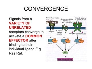
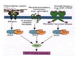
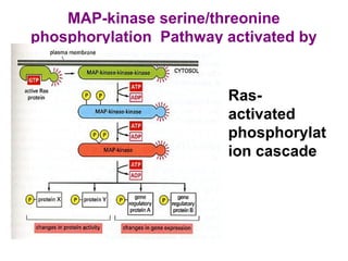
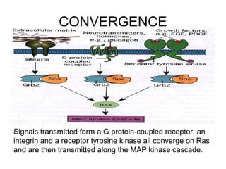
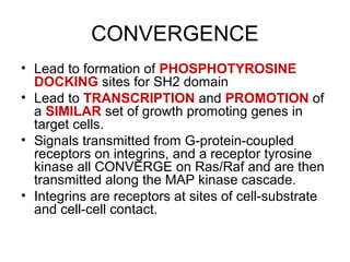
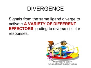
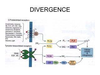
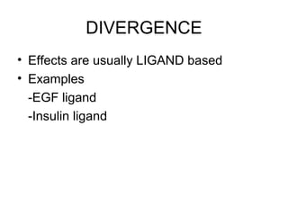
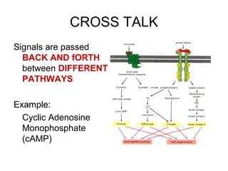
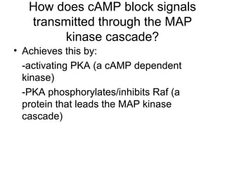
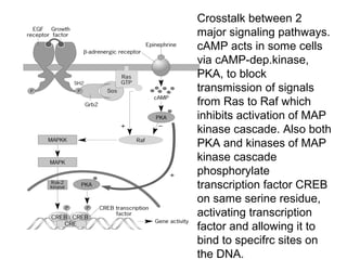
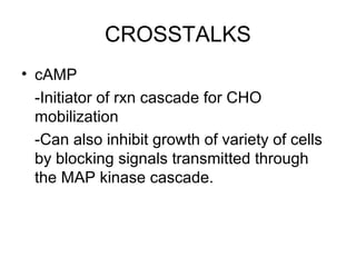
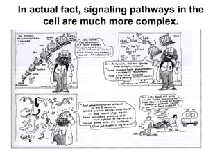
Convergence, Divergence and
Crosstalk
Among Different Signaling
Pathways
Herbert, Barnabas E. MB.Sc, Hse
Key Concept
• Convergence
• Divergence
• Crosstalk
• Signaling Pathways
Key Concept
• Convergence
• Divergence
• Crosstalk
• Signaling Pathways
CONVERGENCE
Signals from a
VARIETY OF
UNRELATED
receptors converge to
activate a COMMON
EFFECTOR after
binding to their
individual ligand.E.g
Ras Raf.
MAP-kinase serine/threonine
phosphorylation Pathway activated by
Ras
• Ras-
activated
phosphorylat
ion cascade
CONVERGENCE
• Signals usually from RECEPTORS
• Examples:
-G-protein coupled receptors
-Receptor tyrosine kinases
-Integrins
CONVERGENCE
Signals transmitted form a G protein-coupled receptor, an
integrin and a receptor tyrosine kinase all converge on Ras
and are then transmitted along the MAP kinase cascade.
CONVERGENCE
• Lead to formation of PHOSPHOTYROSINE
DOCKING sites for SH2 domain
• Lead to TRANSCRIPTION and PROMOTION of
a SIMILAR set of growth promoting genes in
target cells.
• Signals transmitted from G-protein-coupled
receptors on integrins, and a receptor tyrosine
kinase all CONVERGE on Ras/Raf and are then
transmitted along the MAP kinase cascade.
• Integrins are receptors at sites of cell-substrate
and cell-cell contact.
Key Concept
• Convergence
• Divergence
• Crosstalk
• Signaling Pathways
Key Concept
• Convergence
• Divergence
• Crosstalk
• Signaling Pathways
DIVERGENCE
Signals from the same ligand diverge to
activate A VARIETY OF DIFFERENT
EFFECTORS leading to diverse cellular
responses.
DIVERGENCE
DIVERGENCE
• Effects are usually LIGAND based
• Examples
-EGF ligand
-Insulin ligand
KEY CONCEPTS
• Convergence
• Divergence
• Crosstalk
• Signaling Pathways
KEY CONCEPTS
• Convergence
• Divergence
• Crosstalk
• Signaling Pathways
CROSS TALK
Signals are passed
BACK AND fORTH
between DIFFERENT
PATHWAYS
Example:
Cyclic Adenosine
Monophosphate
(cAMP)
How does cAMP block signals
transmitted through the MAP
kinase cascade?
• Achieves this by:
-activating PKA (a cAMP dependent
kinase)
-PKA phosphorylates/inhibits Raf (a
protein that leads the MAP kinase
cascade)
Crosstalk between 2
major signaling pathways.
cAMP acts in some cells
via cAMP-dep.kinase,
PKA, to block
transmission of signals
from Ras to Raf which
inhibits activation of MAP
kinase cascade. Also both
PKA and kinases of MAP
kinase cascade
phosphorylate
transcription factor CREB
on same serine residue,
activating transcription
factor and allowing it to
bind to specifrc sites on
the DNA.
CROSSTALKS
• cAMP
-Initiator of rxn cascade for CHO
mobilization
-Can also inhibit growth of variety of cells
by blocking signals transmitted through
the MAP kinase cascade.
• Convergence
• Divergence
• Crosstalk
• Signaling Pathways
• Convergence
• Divergence
• Crosstalk
• Signaling Pathways
SIGNALING PATHWAYS
• Provide a mechanism for
routing information through a
cell
• Comparable to the nervous
system:
-the cell receives information
about its environment
through the activation of
various surface receptors.
CELL SURFACE RECEPTORS
• Acts like sensors to detect extracellular
stimuli
• Can bind only to specific ligands
• Unaffected by the presence of a large
variety of UNRELATED molecules
Do not forget!
“A single cell may have dozens of different
receptors sending signals to the cell
interior simultaneously!”
What happens
when signals
are transmitted
into cells???
The signals are selectively routed along a
number of different signaling pathways
that may cause the cell to:
-Divide (Mitosis)
-Change shape
-Activate a specific metabolic pathway
-Apoptosis (Commit suicide)
CENTRAL IDEA
In this way, the cell integrates information
arriving from different sources and mounts
an appropriate and comprehensive
response
How are different
stimuli able to
evoke distinct
responses, even
though they utilize
similar pathways?
Contrasting cellular responses are due to
differences in the protein composition of
different cell types (Different cells have
different isoforms of these various
proteins)
A working theory, not a satisfactory
answer!
In actual fact, signaling pathways in the
cell are much more complex.
SURMARY
Signals from a variety of UNRELATED
RECEPTORS can CONVERGE to
activate a common effector, such as
Ras/Raf; signals fro the SAME LIGAND
can DIVERGE to activate a variety of
DIFFERENT EFFECTORS; and signals
can be passed BACK AND FORTH
between pathways (Cross talk).
• Paroxysmal vertigo
• Define the following and give
examples.
-Convergence
-Divergence
-Crosstalk
QUESTION ONE
What happens when signals
are transmitted into cells???
QUESTION TWO
INDISCRIMINATE FIRING ?
Concentration is the Key! Keep calm!! 
THE ROLE OF NITRIC OXIDE
AS AN INTRACELLULAR
MESSENGER
Herbert, Barnabas E.
History of Second Messengers
• Before 1980
-Organic compounds e.g cAMP
-Ions e.g Ca2+
• After 1980
-Inorganic gas -> Nitric Oxide (NO)
Nitric Oxide
- Formed from L-arginine (amino acid) in a
rxn catalyzed by the enz Nitric Oxide
Synthase (NOS)
- Discovered as second messenger by
accidental observation
Flash Back: Acetylcholine
- Known to act in the body to relax smooth
muscle cells of blood vessels.
- Response could not be duplicated in vitro
- Binds to receptors on the surface of
endothelial cells
- Leads to the production and release of an
AGENT that diffuse through the cell’s
plasma membrane
Acetylcholine
- Causes the muscle cells to relax
- The AGENT was later discovered to be
Nitric Oxide (NO)
NITRIC OXIDE: MOA
Step one:
Acetylcholine binds to the outer surface
of endothelial cell
Step two:
Causes a rise in cytosolic Ca2+
concentration
Step three:
Ca2+
activates NOS to synthesize NO
NITRIC OXIDE: MOA
Step four:
NO formed in endothelial cell diffuses
across the plasma membrane to
adjacent muscle cells
Step five:
Stimulates guanyl cyclase in smooth
muscle which synthesizes cGMP, a
2nd messenger similar in structure to
cAMP
NITRIC OXIDE: MOA
Step six:
cGMP leads to a decrease in cytosolic
Ca2+
concentration which leads to
smooth muscle cell relaxation
Conclusion/Discovery:
“NO acts as an activator of guanyl
cyclase”
Medical Relevance: Nitroglycerine
-used to treat the pain of angina that results
from an inadequate flow of blood to the heart.
-metabolized to NO which stimulates the
relaxation of the smooth muscles lining the
blood vessels of the heart
-leads to increase blood flow to the organ
Pharmacological Relevance: Sildenafil
- Aka Viagra
- Developed
following the
discovery of NO
Sildenafil: MOA
• During sexual act
-nerve endings in the penis release NO
-causes:
(a) relaxation of smooth muscle
cells in the lining of penile
blood vessels
(b) Engorgement of the organ
with blood
Sildenafil: MOA
• Viagra
-has no effect on the release of NO or the
activation of guanylyl cyclase
-(instead) inhibits cGMP
phosphodiesterase
Phosphodiesterase: MOA
• An enzyme
• Destroys cGMP
• Inhibition of this enzyme leads to:
(A) maintained, elevated levels of
cGMP ->
(B) promotes the development and
maintenance of an erection
Since Viagra acts to maintain
elevated levels of cGMP,
does it affect the heart as
well???
Viagra
-specific for one particular isoform of cGMP
phosphodiesterase (PDE5)
-version that acts in the penis
PDE3
-plays key role in the regulation of heart
muscle contraction (not inhibited by Viagra)
NO or N2O???
Do not forget!
Nitric Oxide (NO) should not be confused
with Nitrous Oxide (Laughing Gas)
Describe the steps
in the signaling
pathway by which
nitric oxide
mediates dilation
of blood vessels.
Since Viagra acts to
maintain elevated levels of
cGMP, does it affect the
heart as well??? Explain!
Information overload!!!
Five Minutes Break!!!
For everything there is a season, and a time
for every matter under heaven: A time to be
born, and a time to die…
Ecclesiastes 3: 1f
For every cell, there is a time to live and a
time to die…
Apoptosis
Apoptosis
…Programmed cell death
Discussed by:
HERBERT, B.
Objectives
• At the end of the discuss, students should
be able to:
– Describe the steps that occur between the
time that a TNF molecule binds to its receptor
and the eventual death of the cell.
– Describe the steps that occur between the
time a proapoptotic Bcl-2 membrane binds to
the outer mitochondrial membrane and the
death of the cell.
Cells die for two quite different
reasons
• Accidental death
• result of mechanical trauma or exposure to some
kind of toxic agent (necrosis)
• only type of death seen in unicellular organisms
• Deliberate death
• result of an built-in suicide mechanism known as
apoptosis or programmed cell death.
Necrosis
• When cells are injured
– ATP concentrations fall so low that the Na+
/K+
ATPase can no longer operate,
– ion concentrations are no longer controlled
– causes the cells to swell and then burst
– cell contents leak out
– causes the surrounding tissues to become
inflamed
Apoptosis
• A normal occurrence
• An orchestrated sequence of events
• Leads to death of a cell
• Eliminates cells with sustained irreparable
genomic damage
– Important because damage to genetic blue print can
result in unregulated cell division -> Cancer
• Etymology: John Kerr et al., (1972)
Apoptosis: Characteristics
• Shrinkage of cell volume and nucleus
• Loss of adhesion to neighbouring cells
• Formation of blebs at cell surface
• Dissection of chromatin into small
fragments
• Engulfment of the ‘corpse’ by
phagocytosis
Apoptosis: Working Examples
• Neurons (During embryonic development)
 grow out of CNS to innervate organs present in
the periphery of the body
 usually many more neurons grow out than are
needed for normal innervation
 Neurons that reach their destination receive
signal from the target tissue that allows them to
survive
 Neurons that fail to find their way to the target
tissue do not receive the survival signal and are
eliminated by apoptosis
Apoptosis: Working Example
• T Lymphocytes
– Cells of the immune system
– Recognize and kill abnormal or pathogen
infected cells
– Recognizes target cells via specific receptors
that are present on its surface
Apoptosis: T Lymphocytes
– Sometimes produced during embryonic
development with receptors capable of
binding tightly to proteins present on surface
of normal cells within the body.
– T Lymphocytes that have this dangerous
capability are eliminated by apoptosis.
Apoptosis: Medical Relevance
• Apoptosis is involved in neurodegenerative
diseases such as:
– Alzheimers’s
– Parkinson’s
– Huntington’s
Elimination of essential neurons during dz
progression gives rise to loss of memory or
decrease in motor coordination
Apoptosis: Triggers
Apoptosis can be triggered in three ways:
(a) binding of ligand to death domain
receptors,
(b) denial of growth factors, and
(c) cell stress.
Central Idea
“Apoptosis is important in maintaining
homeostasis in multicellular organisms
and failure to regulate apoptosis can
result in serious damage to an organism”
Apoptosis is a normal occurrence!
You can’t escape from it!
Apoptosis: A Worm’s Eye View!
• First revealed in studies on nematode
worm Caenorhabditis elegans
»Cells can be followed with absolute
precision during embryonic
development
»131 cells are normally destined to die
by apoptosis
»Worms carrying mutation in the CED-
3 gene proceed through development
without losing any of their cells to
apoptosis
Apoptosis: Caspases
• A homologous gene to CED-3 found in
humans
• Distinctive group of cysteine proteins
»i.e proteases with a key cysteine
residue in their catalytic site
»Activated at an early stage of
apoptosis
»Responsible for triggering changes
observed during cell death
MOA: Caspases
• Achieves apoptosis by cleaving a selected
group of proteins
• All the cells of our body contain caspases
• they are normally locked in an inactive
form by an integral inhibitory domain of
the protein
• Proteolysis cleaves the inhibitory domain
off, releasing the active caspases
Viruses: A Hostile Take Over!
• no protein synthesis is required to activate
the apoptotic pathway—all the
components are already present
• if a virus infects a cell and takes over all
protein synthesis, the cell can still commit
suicide and hence prevent viral replication
Caspases: Targets
• Focal Adhesion Kinase (FAK), PKB, and
Raf 1
– Inactivation of FAK disrupts cell adhession,
leading to detachment of apoptotic cell from
its neighbours
• Lamins
– Make up inner lining of nuclear envelope
– Cleavage of lamins leads to the disassembly
of nuclear lamina and shrinkage of the
nucleus
Caspases: Targets
• Proteins of the Cytoskeleton
– Such as those of the intermediate filaments,
actin, tubulin and gelsolin
– Cleavage and consequent inactivation of these
proteins lead to changes in cell shape
• Caspase activated Dnase (CAD)
– An endonuclease
– Activated following caspase cleavage of an
inhibitory protein
– Translocates from cytoplasm to nucleus
– Attacks DNA, severing it into fragments
Apoptosis: What activates it?
A. Internal stimuli (Intrinsic Pathway)
–Abnormalities in DNA
A. External stimuli (Extrinsic Pathway)
– Removal of growth factors from the
medium
The exTrinsic PaThway of aPoPTosis
Extrinsic Pathway of Apoptosis
– Removal of growth factors from the medium
– Epithelial cells of the prostate become
apoptotic when deprived of the male sex
hormone, testosterone
» Hence prostate cancer that has spread to other tissues
are often tx with drugs that interfere with testosterone
production
– Stimulis is carried by an extracellular
messenger pr called TNF
Tumor Necrosis Factor
• So called for its ability to kill tumor cells
• Produced by cells of the immune system
in response to adverse conditions, such
as:
• Exposure to ionizing radiation
• Elevated temperature
• Viral infection
• Toxic chemical agents such as those used in
cancer chemotherapy
TNF Receptor
• Present in plasma membrane as a
preassembled trimer
• Cytoplasmic domain of each receptor
subunit contains a segment of about 70
a.a called ‘death domain’ (mediates pr-pr
interactions)
TNF: MOA
• Evokes its response by binding to a
transmembrane receptor, TNFR1
• Member of family related to ‘death receptor’ that
mediates apoptosis
• TNF binds to the trimer receptor -> change
in conformation of the receptor’s death
domain -> recruitment of a number of pr
• Last pr to join complex are two
procaspases (8 molecules)
TNF: MOA
• Synthesis of caspases as proenzymes
protects the cell from accidental proteolytic
damage
• When two or more procaspases are held
in close association with one another, they
are capable of cleaving one another’s
polypeptide chain and converting the other
molwcule to the fully active caspase
Caspase 8
• Final mature enzyme
• Contains four polypeptide chains
• Derived from two procaspase precursors
• Described as an initiator caspase
• Initiates apoptosis by cleaving and
activating downstream or executioner
caspases.
Executioner Caspases
• Carry out the controlled self-destruction of
the cell
The inTrinsic PaThway of aPoPTosis
Examples of Internal Stimuli
• Irreparable genetic damage
• Extremely high concentrations of cytosolic
Ca2+
• Severe oxidative stress
• Lack of survival signals (Absence of
growth factors)
Bcl-2 Family of Proteins
• Regulates activation of the intrinsic
pathway
• Originally identified as a tumor-causing
oncogene
• Subdivided into two:-
– Proapoptotic : promotes apoptosis (e.g, Bad
and Bax)
– Antiapoptotic: protects cells from apoptosis (e.g
Bcl-XL, Bcl-w, and Bcl-2)
Don’t Forget!
“Bcl-2 acts as an oncogene by promoting
survival of potential cancer cells that would
otherwise die.”
MOA: Intrinsic Pathway
• Stressful stimuli:- activates proapoptotic
members of the Bcl-2 family (Bad/Bax)
Translocates from the cytosol to outer
mitochondrial membrane
Attaches to outer mitochondrial membrane
MOA: Intrinsic Pathway
Increases membrane permeability
Promotes release of cytochrome C (which
resides in the intermembrane space)
Moves to cytosol
Forms apoptosome (a multi protein complex
that includes procaspase-9)
Procaspase-9
• Activated by simply joining the multiprotein
complex
• Does not require proteolytic cleavage
• An initiator caspase; initiates executioner
caspases
Apoptosis
Do not forget!
“The external pathway is receptor-mediated
while the internal pathway is mitochondrial
mediated! They however CONVERGE by
activating the same executioner caspase,
which cleaves the same cellular targets.”
Finally!
• As cells execute the proapoptotic program
they lose contact with neighbors and start
to shrink
• Cell disintegrates into a condensed,
membrane-enclosed apoptotic body
• Apoptotic bodies are recognizd by the
presence of phosphatidylserine on their
surface
Phosphatidylserine
• A phospholipid that is normaly present
only on the inner leaflet of the plasma
membrane
During apoptosis, a phospholipid “scramblase”
moves phosphatidylserine molecules to the
outer leaflet of the plasma membrane where
they are recognized as an “eat me” signal by
specialized macrophages.
Convergence, divergence and crosstalk

Convergence, divergence and crosstalk

  • 2.
    Convergence, Divergence and Crosstalk AmongDifferent Signaling Pathways Herbert, Barnabas E. MB.Sc, Hse
  • 3.
    Key Concept • Convergence •Divergence • Crosstalk • Signaling Pathways
  • 4.
    Key Concept • Convergence •Divergence • Crosstalk • Signaling Pathways
  • 5.
    CONVERGENCE Signals from a VARIETYOF UNRELATED receptors converge to activate a COMMON EFFECTOR after binding to their individual ligand.E.g Ras Raf.
  • 7.
    MAP-kinase serine/threonine phosphorylation Pathwayactivated by Ras • Ras- activated phosphorylat ion cascade
  • 8.
    CONVERGENCE • Signals usuallyfrom RECEPTORS • Examples: -G-protein coupled receptors -Receptor tyrosine kinases -Integrins
  • 9.
    CONVERGENCE Signals transmitted forma G protein-coupled receptor, an integrin and a receptor tyrosine kinase all converge on Ras and are then transmitted along the MAP kinase cascade.
  • 10.
    CONVERGENCE • Lead toformation of PHOSPHOTYROSINE DOCKING sites for SH2 domain • Lead to TRANSCRIPTION and PROMOTION of a SIMILAR set of growth promoting genes in target cells. • Signals transmitted from G-protein-coupled receptors on integrins, and a receptor tyrosine kinase all CONVERGE on Ras/Raf and are then transmitted along the MAP kinase cascade. • Integrins are receptors at sites of cell-substrate and cell-cell contact.
  • 11.
    Key Concept • Convergence •Divergence • Crosstalk • Signaling Pathways
  • 12.
    Key Concept • Convergence •Divergence • Crosstalk • Signaling Pathways
  • 13.
    DIVERGENCE Signals from thesame ligand diverge to activate A VARIETY OF DIFFERENT EFFECTORS leading to diverse cellular responses.
  • 14.
  • 15.
    DIVERGENCE • Effects areusually LIGAND based • Examples -EGF ligand -Insulin ligand
  • 16.
    KEY CONCEPTS • Convergence •Divergence • Crosstalk • Signaling Pathways
  • 17.
    KEY CONCEPTS • Convergence •Divergence • Crosstalk • Signaling Pathways
  • 19.
    CROSS TALK Signals arepassed BACK AND fORTH between DIFFERENT PATHWAYS Example: Cyclic Adenosine Monophosphate (cAMP)
  • 20.
    How does cAMPblock signals transmitted through the MAP kinase cascade? • Achieves this by: -activating PKA (a cAMP dependent kinase) -PKA phosphorylates/inhibits Raf (a protein that leads the MAP kinase cascade)
  • 21.
    Crosstalk between 2 majorsignaling pathways. cAMP acts in some cells via cAMP-dep.kinase, PKA, to block transmission of signals from Ras to Raf which inhibits activation of MAP kinase cascade. Also both PKA and kinases of MAP kinase cascade phosphorylate transcription factor CREB on same serine residue, activating transcription factor and allowing it to bind to specifrc sites on the DNA.
  • 22.
    CROSSTALKS • cAMP -Initiator ofrxn cascade for CHO mobilization -Can also inhibit growth of variety of cells by blocking signals transmitted through the MAP kinase cascade.
  • 23.
    • Convergence • Divergence •Crosstalk • Signaling Pathways
  • 24.
    • Convergence • Divergence •Crosstalk • Signaling Pathways
  • 25.
    SIGNALING PATHWAYS • Providea mechanism for routing information through a cell • Comparable to the nervous system: -the cell receives information about its environment through the activation of various surface receptors.
  • 26.
    CELL SURFACE RECEPTORS •Acts like sensors to detect extracellular stimuli • Can bind only to specific ligands • Unaffected by the presence of a large variety of UNRELATED molecules
  • 27.
    Do not forget! “Asingle cell may have dozens of different receptors sending signals to the cell interior simultaneously!”
  • 28.
    What happens when signals aretransmitted into cells???
  • 29.
    The signals areselectively routed along a number of different signaling pathways that may cause the cell to: -Divide (Mitosis) -Change shape -Activate a specific metabolic pathway -Apoptosis (Commit suicide)
  • 30.
    CENTRAL IDEA In thisway, the cell integrates information arriving from different sources and mounts an appropriate and comprehensive response
  • 31.
    How are different stimuliable to evoke distinct responses, even though they utilize similar pathways?
  • 32.
    Contrasting cellular responsesare due to differences in the protein composition of different cell types (Different cells have different isoforms of these various proteins)
  • 33.
    A working theory,not a satisfactory answer!
  • 34.
    In actual fact,signaling pathways in the cell are much more complex.
  • 35.
    SURMARY Signals from avariety of UNRELATED RECEPTORS can CONVERGE to activate a common effector, such as Ras/Raf; signals fro the SAME LIGAND can DIVERGE to activate a variety of DIFFERENT EFFECTORS; and signals can be passed BACK AND FORTH between pathways (Cross talk).
  • 37.
    • Paroxysmal vertigo •Define the following and give examples. -Convergence -Divergence -Crosstalk QUESTION ONE
  • 38.
    What happens whensignals are transmitted into cells??? QUESTION TWO
  • 39.
  • 40.
    Concentration is theKey! Keep calm!! 
  • 41.
    THE ROLE OFNITRIC OXIDE AS AN INTRACELLULAR MESSENGER Herbert, Barnabas E.
  • 42.
    History of SecondMessengers • Before 1980 -Organic compounds e.g cAMP -Ions e.g Ca2+ • After 1980 -Inorganic gas -> Nitric Oxide (NO)
  • 43.
    Nitric Oxide - Formedfrom L-arginine (amino acid) in a rxn catalyzed by the enz Nitric Oxide Synthase (NOS) - Discovered as second messenger by accidental observation
  • 44.
    Flash Back: Acetylcholine -Known to act in the body to relax smooth muscle cells of blood vessels. - Response could not be duplicated in vitro - Binds to receptors on the surface of endothelial cells - Leads to the production and release of an AGENT that diffuse through the cell’s plasma membrane
  • 45.
    Acetylcholine - Causes themuscle cells to relax - The AGENT was later discovered to be Nitric Oxide (NO)
  • 46.
    NITRIC OXIDE: MOA Stepone: Acetylcholine binds to the outer surface of endothelial cell Step two: Causes a rise in cytosolic Ca2+ concentration Step three: Ca2+ activates NOS to synthesize NO
  • 47.
    NITRIC OXIDE: MOA Stepfour: NO formed in endothelial cell diffuses across the plasma membrane to adjacent muscle cells Step five: Stimulates guanyl cyclase in smooth muscle which synthesizes cGMP, a 2nd messenger similar in structure to cAMP
  • 48.
    NITRIC OXIDE: MOA Stepsix: cGMP leads to a decrease in cytosolic Ca2+ concentration which leads to smooth muscle cell relaxation Conclusion/Discovery: “NO acts as an activator of guanyl cyclase”
  • 49.
    Medical Relevance: Nitroglycerine -usedto treat the pain of angina that results from an inadequate flow of blood to the heart. -metabolized to NO which stimulates the relaxation of the smooth muscles lining the blood vessels of the heart -leads to increase blood flow to the organ
  • 50.
    Pharmacological Relevance: Sildenafil -Aka Viagra - Developed following the discovery of NO
  • 51.
    Sildenafil: MOA • Duringsexual act -nerve endings in the penis release NO -causes: (a) relaxation of smooth muscle cells in the lining of penile blood vessels (b) Engorgement of the organ with blood
  • 52.
    Sildenafil: MOA • Viagra -hasno effect on the release of NO or the activation of guanylyl cyclase -(instead) inhibits cGMP phosphodiesterase
  • 53.
    Phosphodiesterase: MOA • Anenzyme • Destroys cGMP • Inhibition of this enzyme leads to: (A) maintained, elevated levels of cGMP -> (B) promotes the development and maintenance of an erection
  • 54.
    Since Viagra actsto maintain elevated levels of cGMP, does it affect the heart as well???
  • 55.
    Viagra -specific for oneparticular isoform of cGMP phosphodiesterase (PDE5) -version that acts in the penis PDE3 -plays key role in the regulation of heart muscle contraction (not inhibited by Viagra)
  • 56.
  • 57.
    Do not forget! NitricOxide (NO) should not be confused with Nitrous Oxide (Laughing Gas)
  • 60.
    Describe the steps inthe signaling pathway by which nitric oxide mediates dilation of blood vessels.
  • 61.
    Since Viagra actsto maintain elevated levels of cGMP, does it affect the heart as well??? Explain!
  • 62.
  • 63.
  • 64.
    For everything thereis a season, and a time for every matter under heaven: A time to be born, and a time to die… Ecclesiastes 3: 1f
  • 65.
    For every cell,there is a time to live and a time to die…
  • 66.
  • 67.
  • 68.
    Objectives • At theend of the discuss, students should be able to: – Describe the steps that occur between the time that a TNF molecule binds to its receptor and the eventual death of the cell. – Describe the steps that occur between the time a proapoptotic Bcl-2 membrane binds to the outer mitochondrial membrane and the death of the cell.
  • 69.
    Cells die fortwo quite different reasons • Accidental death • result of mechanical trauma or exposure to some kind of toxic agent (necrosis) • only type of death seen in unicellular organisms • Deliberate death • result of an built-in suicide mechanism known as apoptosis or programmed cell death.
  • 70.
    Necrosis • When cellsare injured – ATP concentrations fall so low that the Na+ /K+ ATPase can no longer operate, – ion concentrations are no longer controlled – causes the cells to swell and then burst – cell contents leak out – causes the surrounding tissues to become inflamed
  • 71.
    Apoptosis • A normaloccurrence • An orchestrated sequence of events • Leads to death of a cell • Eliminates cells with sustained irreparable genomic damage – Important because damage to genetic blue print can result in unregulated cell division -> Cancer • Etymology: John Kerr et al., (1972)
  • 72.
    Apoptosis: Characteristics • Shrinkageof cell volume and nucleus • Loss of adhesion to neighbouring cells • Formation of blebs at cell surface • Dissection of chromatin into small fragments • Engulfment of the ‘corpse’ by phagocytosis
  • 73.
    Apoptosis: Working Examples •Neurons (During embryonic development)  grow out of CNS to innervate organs present in the periphery of the body  usually many more neurons grow out than are needed for normal innervation  Neurons that reach their destination receive signal from the target tissue that allows them to survive  Neurons that fail to find their way to the target tissue do not receive the survival signal and are eliminated by apoptosis
  • 74.
    Apoptosis: Working Example •T Lymphocytes – Cells of the immune system – Recognize and kill abnormal or pathogen infected cells – Recognizes target cells via specific receptors that are present on its surface
  • 75.
    Apoptosis: T Lymphocytes –Sometimes produced during embryonic development with receptors capable of binding tightly to proteins present on surface of normal cells within the body. – T Lymphocytes that have this dangerous capability are eliminated by apoptosis.
  • 76.
    Apoptosis: Medical Relevance •Apoptosis is involved in neurodegenerative diseases such as: – Alzheimers’s – Parkinson’s – Huntington’s Elimination of essential neurons during dz progression gives rise to loss of memory or decrease in motor coordination
  • 77.
    Apoptosis: Triggers Apoptosis canbe triggered in three ways: (a) binding of ligand to death domain receptors, (b) denial of growth factors, and (c) cell stress.
  • 78.
    Central Idea “Apoptosis isimportant in maintaining homeostasis in multicellular organisms and failure to regulate apoptosis can result in serious damage to an organism”
  • 79.
    Apoptosis is anormal occurrence! You can’t escape from it!
  • 80.
    Apoptosis: A Worm’sEye View! • First revealed in studies on nematode worm Caenorhabditis elegans »Cells can be followed with absolute precision during embryonic development »131 cells are normally destined to die by apoptosis »Worms carrying mutation in the CED- 3 gene proceed through development without losing any of their cells to apoptosis
  • 81.
    Apoptosis: Caspases • Ahomologous gene to CED-3 found in humans • Distinctive group of cysteine proteins »i.e proteases with a key cysteine residue in their catalytic site »Activated at an early stage of apoptosis »Responsible for triggering changes observed during cell death
  • 82.
    MOA: Caspases • Achievesapoptosis by cleaving a selected group of proteins • All the cells of our body contain caspases • they are normally locked in an inactive form by an integral inhibitory domain of the protein • Proteolysis cleaves the inhibitory domain off, releasing the active caspases
  • 83.
    Viruses: A HostileTake Over! • no protein synthesis is required to activate the apoptotic pathway—all the components are already present • if a virus infects a cell and takes over all protein synthesis, the cell can still commit suicide and hence prevent viral replication
  • 84.
    Caspases: Targets • FocalAdhesion Kinase (FAK), PKB, and Raf 1 – Inactivation of FAK disrupts cell adhession, leading to detachment of apoptotic cell from its neighbours • Lamins – Make up inner lining of nuclear envelope – Cleavage of lamins leads to the disassembly of nuclear lamina and shrinkage of the nucleus
  • 85.
    Caspases: Targets • Proteinsof the Cytoskeleton – Such as those of the intermediate filaments, actin, tubulin and gelsolin – Cleavage and consequent inactivation of these proteins lead to changes in cell shape • Caspase activated Dnase (CAD) – An endonuclease – Activated following caspase cleavage of an inhibitory protein – Translocates from cytoplasm to nucleus – Attacks DNA, severing it into fragments
  • 86.
    Apoptosis: What activatesit? A. Internal stimuli (Intrinsic Pathway) –Abnormalities in DNA A. External stimuli (Extrinsic Pathway) – Removal of growth factors from the medium
  • 87.
  • 88.
    Extrinsic Pathway ofApoptosis – Removal of growth factors from the medium – Epithelial cells of the prostate become apoptotic when deprived of the male sex hormone, testosterone » Hence prostate cancer that has spread to other tissues are often tx with drugs that interfere with testosterone production – Stimulis is carried by an extracellular messenger pr called TNF
  • 89.
    Tumor Necrosis Factor •So called for its ability to kill tumor cells • Produced by cells of the immune system in response to adverse conditions, such as: • Exposure to ionizing radiation • Elevated temperature • Viral infection • Toxic chemical agents such as those used in cancer chemotherapy
  • 90.
    TNF Receptor • Presentin plasma membrane as a preassembled trimer • Cytoplasmic domain of each receptor subunit contains a segment of about 70 a.a called ‘death domain’ (mediates pr-pr interactions)
  • 91.
    TNF: MOA • Evokesits response by binding to a transmembrane receptor, TNFR1 • Member of family related to ‘death receptor’ that mediates apoptosis • TNF binds to the trimer receptor -> change in conformation of the receptor’s death domain -> recruitment of a number of pr • Last pr to join complex are two procaspases (8 molecules)
  • 92.
    TNF: MOA • Synthesisof caspases as proenzymes protects the cell from accidental proteolytic damage • When two or more procaspases are held in close association with one another, they are capable of cleaving one another’s polypeptide chain and converting the other molwcule to the fully active caspase
  • 93.
    Caspase 8 • Finalmature enzyme • Contains four polypeptide chains • Derived from two procaspase precursors • Described as an initiator caspase • Initiates apoptosis by cleaving and activating downstream or executioner caspases.
  • 94.
    Executioner Caspases • Carryout the controlled self-destruction of the cell
  • 96.
  • 97.
    Examples of InternalStimuli • Irreparable genetic damage • Extremely high concentrations of cytosolic Ca2+ • Severe oxidative stress • Lack of survival signals (Absence of growth factors)
  • 98.
    Bcl-2 Family ofProteins • Regulates activation of the intrinsic pathway • Originally identified as a tumor-causing oncogene • Subdivided into two:- – Proapoptotic : promotes apoptosis (e.g, Bad and Bax) – Antiapoptotic: protects cells from apoptosis (e.g Bcl-XL, Bcl-w, and Bcl-2)
  • 99.
    Don’t Forget! “Bcl-2 actsas an oncogene by promoting survival of potential cancer cells that would otherwise die.”
  • 100.
    MOA: Intrinsic Pathway •Stressful stimuli:- activates proapoptotic members of the Bcl-2 family (Bad/Bax) Translocates from the cytosol to outer mitochondrial membrane Attaches to outer mitochondrial membrane
  • 101.
    MOA: Intrinsic Pathway Increasesmembrane permeability Promotes release of cytochrome C (which resides in the intermembrane space) Moves to cytosol Forms apoptosome (a multi protein complex that includes procaspase-9)
  • 102.
    Procaspase-9 • Activated bysimply joining the multiprotein complex • Does not require proteolytic cleavage • An initiator caspase; initiates executioner caspases Apoptosis
  • 103.
    Do not forget! “Theexternal pathway is receptor-mediated while the internal pathway is mitochondrial mediated! They however CONVERGE by activating the same executioner caspase, which cleaves the same cellular targets.”
  • 104.
    Finally! • As cellsexecute the proapoptotic program they lose contact with neighbors and start to shrink • Cell disintegrates into a condensed, membrane-enclosed apoptotic body • Apoptotic bodies are recognizd by the presence of phosphatidylserine on their surface
  • 105.
    Phosphatidylserine • A phospholipidthat is normaly present only on the inner leaflet of the plasma membrane
  • 106.
    During apoptosis, aphospholipid “scramblase” moves phosphatidylserine molecules to the outer leaflet of the plasma membrane where they are recognized as an “eat me” signal by specialized macrophages.