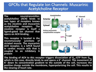
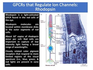
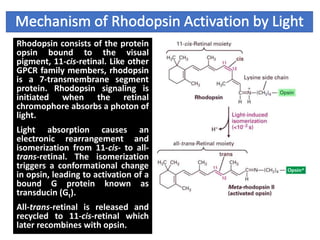
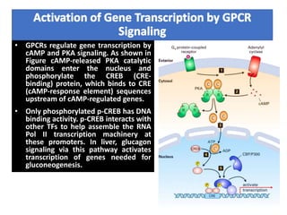
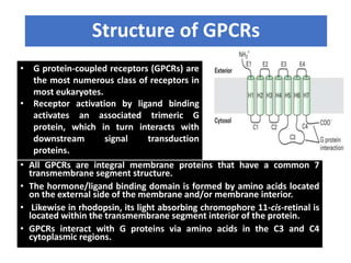
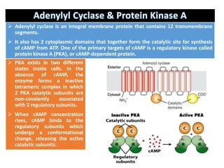
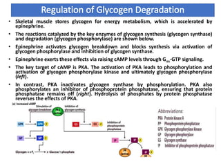
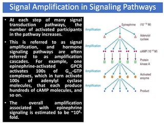
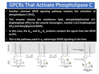
PRESENTED BY :-
DARSHANA SAHARAN
M.SC. ZOOLOGY SEM. 3RD
DEPARTMENT OF ZOOLOGY, UNIVERSITY OF
RAJASTHAN
• General principles of signal transduction
• G Protein-coupled Receptors (GPCRs):
Structure and Mechanism.
• GPCRs that Regulate Adenylyl Cyclase.
• GPCRs that Activate Phospholipase C.
• GPCRs that Regulate Ion Channels.
• GPCRs that Regulate Gene Transcription.
General Principles of Signal Transduction
• Signal transduction refers to the
overall process of converting
extracellular signals into intracellular
responses.
• Key players in signal transduction are
signaling molecules, receptors, signal
transduction proteins and second
messengers, and effector proteins.
• Cells respond to signals by changing
the activity of existing enzymes (fast)
and/or the levels of expression of
enzymes and cell components
(slower) by gene regulation (Steps 7a
& 7b).
• Receptors and signal transduction
systems have evolved to detect and
respond to hormones, growth
factors, neurotransmitters,
pheromones, oxygen, nutrients,
light, touch, heat, etc.
Structure of GPCRs
• G protein-coupled receptors (GPCRs) are
the most numerous class of receptors in
most eukaryotes.
• Receptor activation by ligand binding
activates an associated trimeric G
protein, which in turn interacts with
downstream signal transduction
proteins.
• All GPCRs are integral membrane proteins that have a common 7
transmembrane segment structure.
• The hormone/ligand binding domain is formed by amino acids located
on the external side of the membrane and/or membrane interior.
• Likewise in rhodopsin, its light absorbing chromophore 11-cis-retinal is
located within the transmembrane segment interior of the protein.
• GPCRs interact with G proteins via amino acids in the C3 and C4
cytoplasmic regions.
• The trimeric G protein cycle of activity in hormone-stimulated GPCR regulation of
effector proteins is summarized in next slide.
• Initially, the G protein complex is tethered to the inner leaflet of the cytoplasmic
membrane via lipid anchors attached to the Ga and Gg subunits.
• The trimeric GDP-bound form of the G protein is inactive in signaling.
• The binding of a hormone to the GPCR triggers a conformational change in the receptor
(Step 1) which promotes its binding to the trimeric G protein (Step 2).
• Binding to the activated GPCR triggers the dissociation of GDP (Step 3).
• Subsequent binding of GTP to the Ga subunit activates it, and causes its dissociation
from the receptor and the Gßg complex (Step 4).
• Ga-GTP then binds to the effector protein regulating its activity. The hormone
eventually dissociates from the receptor (Step 5).
• Over time (often less than 1 min), GTP is hydrolyzed to GDP and Ga becomes inactive.
• It then dissociates from the effector and recombines with Gßg (Step 6). A hormone-
bound GPCR activates multiple G proteins, until the hormone dissociates.
• Proteins known as regulators of G protein signaling (RGS) accelerate GTP hydrolysis by
Ga decreasing the time-period during which Ga is active (not shown).
G Protein Coupled
Receptors
• Adenylyl cyclase is an effector enzyme that synthesizes cAMP. Ga-GTP subunits bind to
the catalytic domains of the cyclase, regulating their activity. Gas-GTP activates the
catalytic domains, whereas Gai-GTP inhibits them. A given cell type can express
multiple types of GPCRs that all couple to adenylyl cyclase.
• The net activity of adenylyl cyclase thus depends on the combined level of G protein
signaling via the multiple GPCRs. In liver, GPCRs for epinephrine and glucagon both
activate the cyclase. In adipose tissue epinephrine, glucagon, and ACTH activate the
cyclase via Gas-GTP, while PGE1 and adenosine inactivate the cyclase via Gai-GTP.
 Adenylyl cyclase is an integral membrane protein that contains 12 transmembrane
segments.
 It also has 2 cytoplasmic domains that together form the catalytic site for synthesis
of cAMP from ATP. One of the primary targets of cAMP is a regulatory kinase called
protein kinase A (PKA), or cAMP-dependent protein.
 PKA exists in two different
states inside cells. In the
absence of cAMP, the
enzyme forms a inactive
tetrameric complex in which
2 PKA catalytic subunits are
non-covalently associated
with 2 regulatory subunits.
 When cAMP concentration
rises, cAMP binds to the
regulatory subunits which
undergo a conformational
change, releasing the active
catalytic subunits.
• Skeletal muscle stores glycogen for energy metabolism, which is accelerated by
epinephrine.
• The reactions catalyzed by the key enzymes of glycogen synthesis (glycogen synthase)
and degradation (glycogen phosphorylase) are shown below.
• Epinephrine activates glycogen breakdown and blocks synthesis via activation of
glycogen phosphorylase and inhibition of glycogen synthase.
• Epinephrine exerts these effects via raising cAMP levels through Gas-GTP signaling.
• The key target of cAMP is PKA. The activation of PKA leads to phosphorylation and
activation of glycogen phosphorylase kinase and ultimately glycogen phosphorylase
(left).
• In contrast, PKA inactivates glycogen synthase by phosphorylation. PKA also
phosphorylates an inhibitor of phosphoprotein phosphatase, ensuring that protein
phosphatase remains off (right). Hydrolysis of phosphates by protein phosphatase
reverses the effects of PKA.
• At each step of many signal
transduction pathways, the
number of activated participants
in the pathway increases.
• This is referred to as signal
amplification, and hormone
signaling pathways are often
referred to as amplification
cascades. For example, one
epinephrine-activated GPCR
activates 100s of Gas-GTP
complexes, which in turn activate
100s of adenylyl cyclase
molecules, that each produce
hundreds of cAMP molecules, and
so on.
• The overall amplification
associated with epinephrine
signaling is estimated to be ~108-
fold.
• Another common GPCR signaling pathway involves the activation of
phospholipase C (PLC).
• This enzyme cleaves the membrane lipid, phosphatidylinositol 4,5-
bisphosphate (PIP2) to the second messengers, inositol 1,4,5-trisphosphate
(IP3) and diacylglycerol (DAG).
• In this case, the Gao and Gaq Ga proteins conduct the signal from the GPCR
to PLC.
• This is the pathway used in a1-adrenergic GPCR signaling in the liver.
*
The steps downstream of PLC that make up the IP3/DAG signaling pathway are
illustrated in given picture.
IP3 diffuses from the cytoplasmic membrane to the ER where it binds to and triggers the
opening of IP3-gated Ca2+ channels (Steps 3 & 4).
Another kinase, protein kinase C (PKC) binds to DAG in the cytoplasmic membrane and is
activated (Step 6).
In liver, the rise in cytoplasmic [Ca2+] activates enzymes such as glycogen phosphorylase
kinase, which phosphorylates and activates glycogen phosphorylase. Glycogen
phosphorylase kinase is activated by Ca2+-calmodulin. In addition, PKC phosphorylates
and inactivates glycogen synthase.
• A related signaling pathway involving phospholipase C operates in vascular
endothelial cells and causes adjacent smooth muscle cells to relax in
response to circulating acetylcholine.
• In the NO/cGMP signaling pathway, the downstream target of
Ca2+/calmodulin is nitric oxide synthase, which synthesizes the gas NO from
arginine. NO diffuses into smooth muscle cells and causes relaxation by
activating guanylyl cyclase and increasing [cGMP].
• As a result arteries in tissues such as the heart dilate, increasing blood supply
to the tissue. NO also is produced from the drug nitroglycerin which is given
to heart attack patients and patients being treated for angina.
The neurotransmitter,
acetylcholine (ACH) binds to
two types of receptors known
as the nicotinic and muscarinic
acetylcholine receptors. The
nicotinic receptor is itself a
ligand-gated ion channel that
opens on ACH binding.
This receptor is located in the
neuromuscular junctions of
striated muscle. The muscarinic
ACH receptor, is a GPCR found
in cardiac muscle cells that is
coupled to an inhibitory G
protein
The binding of ACH to this receptor triggers dissociation of Gai-GTP from Gßg,
which in this case, directly binds to and opens a K+ channel. The movement of
K+ down its concentration gradient to the outside of the cell, increases the
positive charge outside the membrane, hyperpolarizing the cell. This results in
the slowing of heart rate.
Rhodopsin is a light-activated
GPCR found in the rod cells of
the eye.
Rhodopsin molecules are
located within membrane disks
in the outer segments of rod
cells.
About 107 copies of rhodopsin
occur per cell. Rod cells are
important in capture of low
intensity light having a broad
range of wavelengths.
Closely related color pigment
receptors that respond to more
limited regions of the visual
spectrum (i.e., blue, green, &
red light) are present in cone
cells.
Rhodopsin consists of the protein
opsin bound to the visual
pigment, 11-cis-retinal. Like other
GPCR family members, rhodopsin
is a 7-transmembrane segment
protein. Rhodopsin signaling is
initiated when the retinal
chromophore absorbs a photon of
light.
Light absorption causes an
electronic rearrangement and
isomerization from 11-cis- to all-
trans-retinal. The isomerization
triggers a conformational change
in opsin, leading to activation of a
bound G protein known as
transducin (Gt).
All-trans-retinal is released and
recycled to 11-cis-retinal which
later recombines with opsin.
The rhodopsin signal transduction pathway is shown in. Light
absorption by rhodopsin triggers GTP/GDP exchange on the
transducin Gat subunit, and dissociation of this trimeric G protein
(Steps 1 & 2).
Gat-GTP binds to and activates a cGMP phosphodiesterase,
reducing intracellular cGMP level (Steps 3 & 4).
This indirectly results in the closing of non-selective Na+/Ca2+ ion
channels in the cytoplasmic membrane and hyperpolarization of
the membrane potential (Step 6).
This results in decreased release of neurotransmitter from the
cells. Thus, light is perceived by the brain due to a decrease in
nerve impulses coming from rod cells. Studies have shown that
only 5 photons must be absorbed per human rod cell to transmit a
signal.
A single activated molecule of rhodopsin activates ~500 transducin
molecules in a classic example of signal amplification.
• GPCRs regulate gene transcription by
cAMP and PKA signaling. As shown in
Figure cAMP-released PKA catalytic
domains enter the nucleus and
phosphorylate the CREB (CRE-
binding) protein, which binds to CRE
(cAMP-response element) sequences
upstream of cAMP-regulated genes.
• Only phosphorylated p-CREB has DNA
binding activity. p-CREB interacts with
other TFs to help assemble the RNA
Pol II transcription machinery at
these promoters. In liver, glucagon
signaling via this pathway activates
transcription of genes needed for
gluconeogenesis.
Activation of the Tubby transcription
factor following ligand binding to receptors coupled to
Go or Gq.
In resting cells, Tubby is bound
tightly to PIP2 in the plasma
membrane. Receptor stimulation
(not shown) leads to activation
of phospholipase C, hydrolysis of
PIP2, and release of Tubby into
the cytosol ( 1 ). Directed by two
functional nuclear localization
sequences (NLS) in its N-terminal
domain, Tubby translocates into
the nucleus ( 2 ) and activates
transcription of target genes ( 3 ). It
is not known whether IP3 remains
bound to Tubby.
• A number of events contribute to the termination of
signaling by a GPCR.
• These include dissociation of the hormone from the
receptor, hydrolysis of GTP by Ga, hydrolysis of cAMP via
cAMP phosphodiesterase, and phosphorylation and
“desensitization” of receptors by kinases such as PKA.
Gpcr signalling

Gpcr signalling

  • 1.
    PRESENTED BY :- DARSHANASAHARAN M.SC. ZOOLOGY SEM. 3RD DEPARTMENT OF ZOOLOGY, UNIVERSITY OF RAJASTHAN
  • 2.
    • General principlesof signal transduction • G Protein-coupled Receptors (GPCRs): Structure and Mechanism. • GPCRs that Regulate Adenylyl Cyclase. • GPCRs that Activate Phospholipase C. • GPCRs that Regulate Ion Channels. • GPCRs that Regulate Gene Transcription.
  • 3.
    General Principles ofSignal Transduction • Signal transduction refers to the overall process of converting extracellular signals into intracellular responses. • Key players in signal transduction are signaling molecules, receptors, signal transduction proteins and second messengers, and effector proteins. • Cells respond to signals by changing the activity of existing enzymes (fast) and/or the levels of expression of enzymes and cell components (slower) by gene regulation (Steps 7a & 7b). • Receptors and signal transduction systems have evolved to detect and respond to hormones, growth factors, neurotransmitters, pheromones, oxygen, nutrients, light, touch, heat, etc.
  • 4.
    Structure of GPCRs •G protein-coupled receptors (GPCRs) are the most numerous class of receptors in most eukaryotes. • Receptor activation by ligand binding activates an associated trimeric G protein, which in turn interacts with downstream signal transduction proteins. • All GPCRs are integral membrane proteins that have a common 7 transmembrane segment structure. • The hormone/ligand binding domain is formed by amino acids located on the external side of the membrane and/or membrane interior. • Likewise in rhodopsin, its light absorbing chromophore 11-cis-retinal is located within the transmembrane segment interior of the protein. • GPCRs interact with G proteins via amino acids in the C3 and C4 cytoplasmic regions.
  • 5.
    • The trimericG protein cycle of activity in hormone-stimulated GPCR regulation of effector proteins is summarized in next slide. • Initially, the G protein complex is tethered to the inner leaflet of the cytoplasmic membrane via lipid anchors attached to the Ga and Gg subunits. • The trimeric GDP-bound form of the G protein is inactive in signaling. • The binding of a hormone to the GPCR triggers a conformational change in the receptor (Step 1) which promotes its binding to the trimeric G protein (Step 2). • Binding to the activated GPCR triggers the dissociation of GDP (Step 3). • Subsequent binding of GTP to the Ga subunit activates it, and causes its dissociation from the receptor and the Gßg complex (Step 4). • Ga-GTP then binds to the effector protein regulating its activity. The hormone eventually dissociates from the receptor (Step 5). • Over time (often less than 1 min), GTP is hydrolyzed to GDP and Ga becomes inactive. • It then dissociates from the effector and recombines with Gßg (Step 6). A hormone- bound GPCR activates multiple G proteins, until the hormone dissociates. • Proteins known as regulators of G protein signaling (RGS) accelerate GTP hydrolysis by Ga decreasing the time-period during which Ga is active (not shown).
  • 6.
  • 8.
    • Adenylyl cyclaseis an effector enzyme that synthesizes cAMP. Ga-GTP subunits bind to the catalytic domains of the cyclase, regulating their activity. Gas-GTP activates the catalytic domains, whereas Gai-GTP inhibits them. A given cell type can express multiple types of GPCRs that all couple to adenylyl cyclase. • The net activity of adenylyl cyclase thus depends on the combined level of G protein signaling via the multiple GPCRs. In liver, GPCRs for epinephrine and glucagon both activate the cyclase. In adipose tissue epinephrine, glucagon, and ACTH activate the cyclase via Gas-GTP, while PGE1 and adenosine inactivate the cyclase via Gai-GTP.
  • 9.
     Adenylyl cyclaseis an integral membrane protein that contains 12 transmembrane segments.  It also has 2 cytoplasmic domains that together form the catalytic site for synthesis of cAMP from ATP. One of the primary targets of cAMP is a regulatory kinase called protein kinase A (PKA), or cAMP-dependent protein.  PKA exists in two different states inside cells. In the absence of cAMP, the enzyme forms a inactive tetrameric complex in which 2 PKA catalytic subunits are non-covalently associated with 2 regulatory subunits.  When cAMP concentration rises, cAMP binds to the regulatory subunits which undergo a conformational change, releasing the active catalytic subunits.
  • 10.
    • Skeletal musclestores glycogen for energy metabolism, which is accelerated by epinephrine. • The reactions catalyzed by the key enzymes of glycogen synthesis (glycogen synthase) and degradation (glycogen phosphorylase) are shown below. • Epinephrine activates glycogen breakdown and blocks synthesis via activation of glycogen phosphorylase and inhibition of glycogen synthase. • Epinephrine exerts these effects via raising cAMP levels through Gas-GTP signaling. • The key target of cAMP is PKA. The activation of PKA leads to phosphorylation and activation of glycogen phosphorylase kinase and ultimately glycogen phosphorylase (left). • In contrast, PKA inactivates glycogen synthase by phosphorylation. PKA also phosphorylates an inhibitor of phosphoprotein phosphatase, ensuring that protein phosphatase remains off (right). Hydrolysis of phosphates by protein phosphatase reverses the effects of PKA.
  • 11.
    • At eachstep of many signal transduction pathways, the number of activated participants in the pathway increases. • This is referred to as signal amplification, and hormone signaling pathways are often referred to as amplification cascades. For example, one epinephrine-activated GPCR activates 100s of Gas-GTP complexes, which in turn activate 100s of adenylyl cyclase molecules, that each produce hundreds of cAMP molecules, and so on. • The overall amplification associated with epinephrine signaling is estimated to be ~108- fold.
  • 12.
    • Another commonGPCR signaling pathway involves the activation of phospholipase C (PLC). • This enzyme cleaves the membrane lipid, phosphatidylinositol 4,5- bisphosphate (PIP2) to the second messengers, inositol 1,4,5-trisphosphate (IP3) and diacylglycerol (DAG). • In this case, the Gao and Gaq Ga proteins conduct the signal from the GPCR to PLC. • This is the pathway used in a1-adrenergic GPCR signaling in the liver. *
  • 13.
    The steps downstreamof PLC that make up the IP3/DAG signaling pathway are illustrated in given picture. IP3 diffuses from the cytoplasmic membrane to the ER where it binds to and triggers the opening of IP3-gated Ca2+ channels (Steps 3 & 4). Another kinase, protein kinase C (PKC) binds to DAG in the cytoplasmic membrane and is activated (Step 6). In liver, the rise in cytoplasmic [Ca2+] activates enzymes such as glycogen phosphorylase kinase, which phosphorylates and activates glycogen phosphorylase. Glycogen phosphorylase kinase is activated by Ca2+-calmodulin. In addition, PKC phosphorylates and inactivates glycogen synthase.
  • 14.
    • A relatedsignaling pathway involving phospholipase C operates in vascular endothelial cells and causes adjacent smooth muscle cells to relax in response to circulating acetylcholine. • In the NO/cGMP signaling pathway, the downstream target of Ca2+/calmodulin is nitric oxide synthase, which synthesizes the gas NO from arginine. NO diffuses into smooth muscle cells and causes relaxation by activating guanylyl cyclase and increasing [cGMP]. • As a result arteries in tissues such as the heart dilate, increasing blood supply to the tissue. NO also is produced from the drug nitroglycerin which is given to heart attack patients and patients being treated for angina.
  • 15.
    The neurotransmitter, acetylcholine (ACH)binds to two types of receptors known as the nicotinic and muscarinic acetylcholine receptors. The nicotinic receptor is itself a ligand-gated ion channel that opens on ACH binding. This receptor is located in the neuromuscular junctions of striated muscle. The muscarinic ACH receptor, is a GPCR found in cardiac muscle cells that is coupled to an inhibitory G protein The binding of ACH to this receptor triggers dissociation of Gai-GTP from Gßg, which in this case, directly binds to and opens a K+ channel. The movement of K+ down its concentration gradient to the outside of the cell, increases the positive charge outside the membrane, hyperpolarizing the cell. This results in the slowing of heart rate.
  • 16.
    Rhodopsin is alight-activated GPCR found in the rod cells of the eye. Rhodopsin molecules are located within membrane disks in the outer segments of rod cells. About 107 copies of rhodopsin occur per cell. Rod cells are important in capture of low intensity light having a broad range of wavelengths. Closely related color pigment receptors that respond to more limited regions of the visual spectrum (i.e., blue, green, & red light) are present in cone cells.
  • 17.
    Rhodopsin consists ofthe protein opsin bound to the visual pigment, 11-cis-retinal. Like other GPCR family members, rhodopsin is a 7-transmembrane segment protein. Rhodopsin signaling is initiated when the retinal chromophore absorbs a photon of light. Light absorption causes an electronic rearrangement and isomerization from 11-cis- to all- trans-retinal. The isomerization triggers a conformational change in opsin, leading to activation of a bound G protein known as transducin (Gt). All-trans-retinal is released and recycled to 11-cis-retinal which later recombines with opsin.
  • 18.
    The rhodopsin signaltransduction pathway is shown in. Light absorption by rhodopsin triggers GTP/GDP exchange on the transducin Gat subunit, and dissociation of this trimeric G protein (Steps 1 & 2). Gat-GTP binds to and activates a cGMP phosphodiesterase, reducing intracellular cGMP level (Steps 3 & 4). This indirectly results in the closing of non-selective Na+/Ca2+ ion channels in the cytoplasmic membrane and hyperpolarization of the membrane potential (Step 6). This results in decreased release of neurotransmitter from the cells. Thus, light is perceived by the brain due to a decrease in nerve impulses coming from rod cells. Studies have shown that only 5 photons must be absorbed per human rod cell to transmit a signal. A single activated molecule of rhodopsin activates ~500 transducin molecules in a classic example of signal amplification.
  • 21.
    • GPCRs regulategene transcription by cAMP and PKA signaling. As shown in Figure cAMP-released PKA catalytic domains enter the nucleus and phosphorylate the CREB (CRE- binding) protein, which binds to CRE (cAMP-response element) sequences upstream of cAMP-regulated genes. • Only phosphorylated p-CREB has DNA binding activity. p-CREB interacts with other TFs to help assemble the RNA Pol II transcription machinery at these promoters. In liver, glucagon signaling via this pathway activates transcription of genes needed for gluconeogenesis.
  • 22.
    Activation of theTubby transcription factor following ligand binding to receptors coupled to Go or Gq. In resting cells, Tubby is bound tightly to PIP2 in the plasma membrane. Receptor stimulation (not shown) leads to activation of phospholipase C, hydrolysis of PIP2, and release of Tubby into the cytosol ( 1 ). Directed by two functional nuclear localization sequences (NLS) in its N-terminal domain, Tubby translocates into the nucleus ( 2 ) and activates transcription of target genes ( 3 ). It is not known whether IP3 remains bound to Tubby.
  • 23.
    • A numberof events contribute to the termination of signaling by a GPCR. • These include dissociation of the hormone from the receptor, hydrolysis of GTP by Ga, hydrolysis of cAMP via cAMP phosphodiesterase, and phosphorylation and “desensitization” of receptors by kinases such as PKA.