Downloaded 170 times
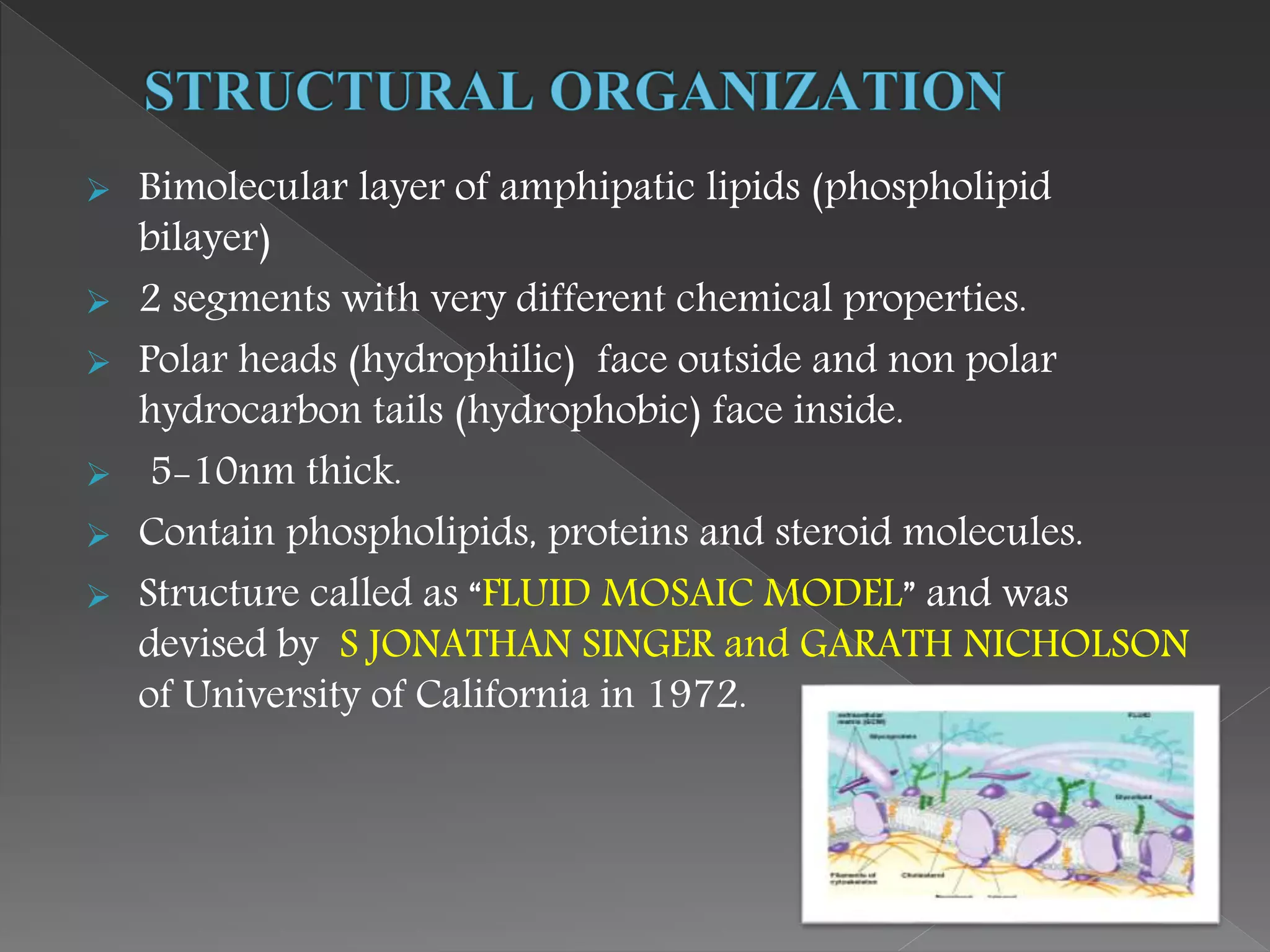
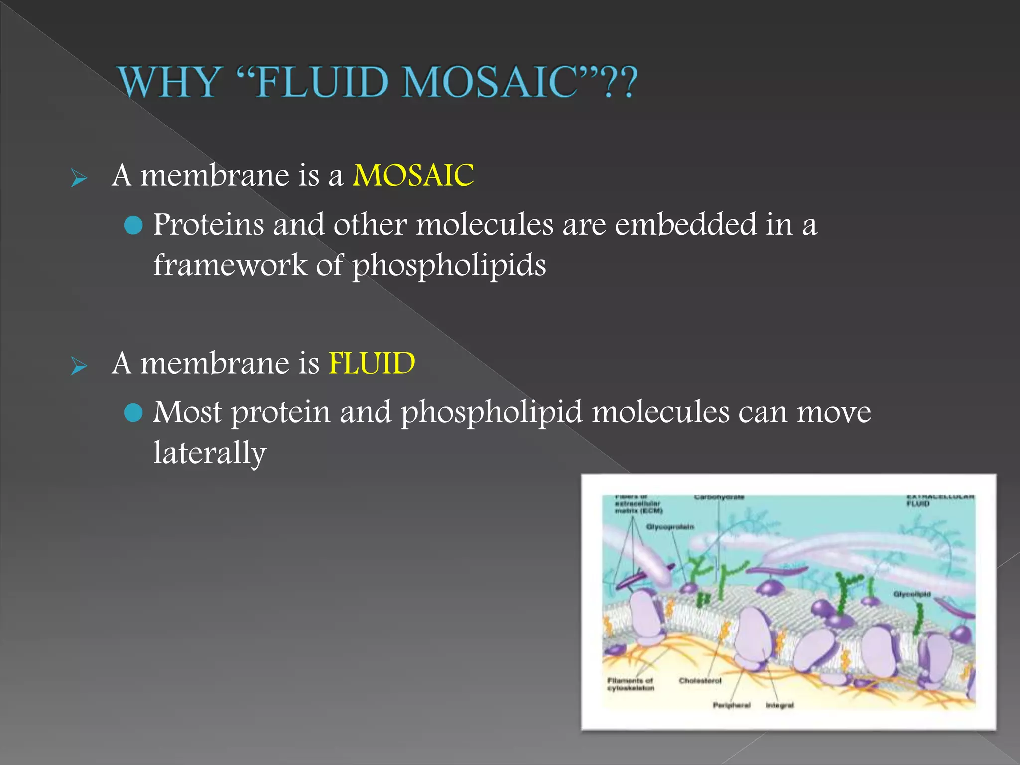

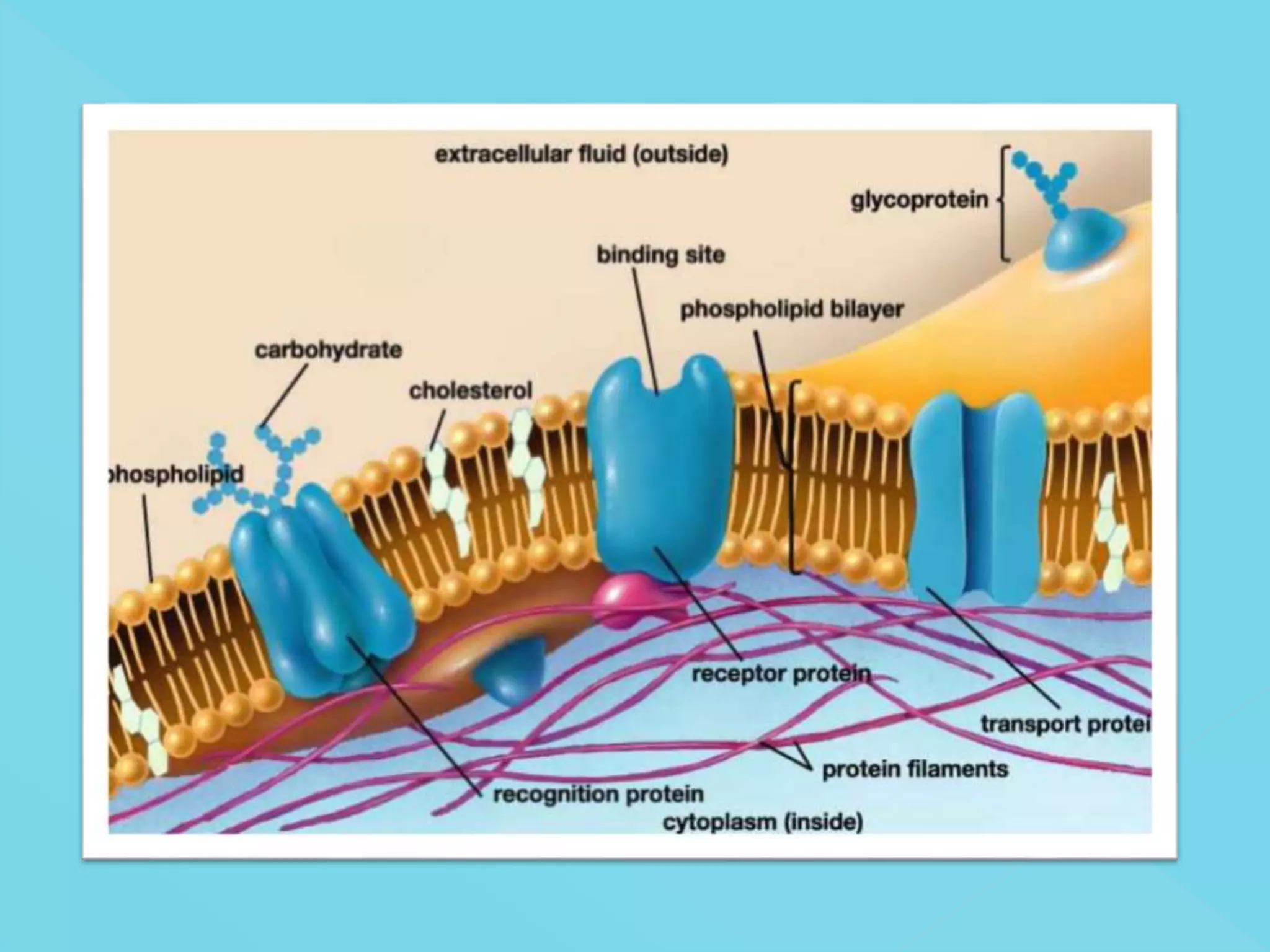
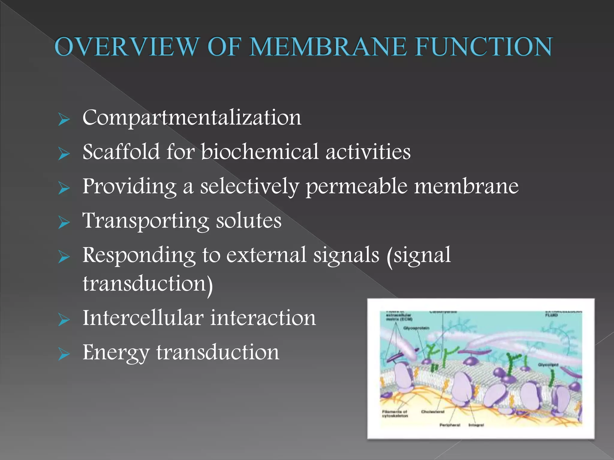
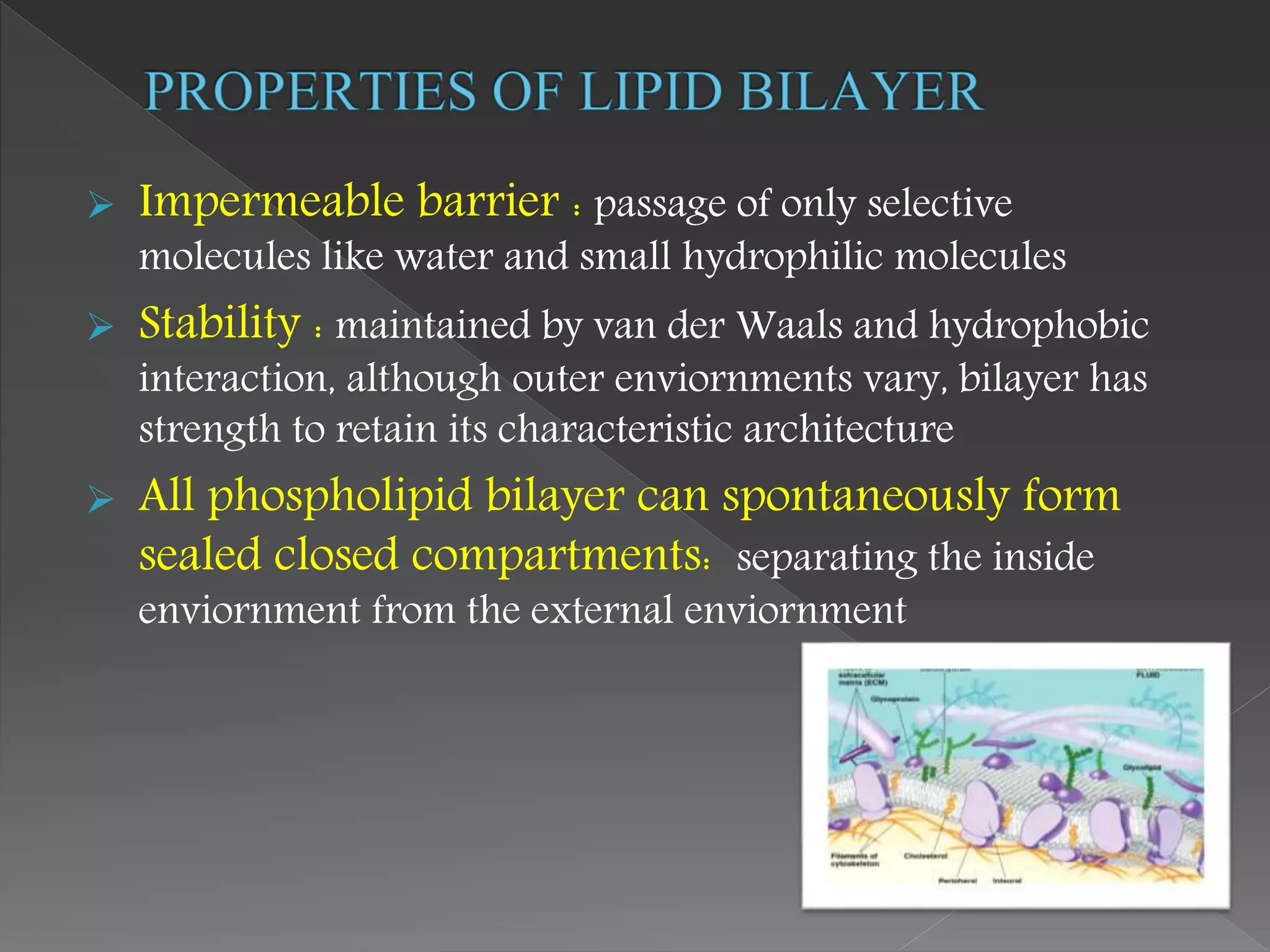
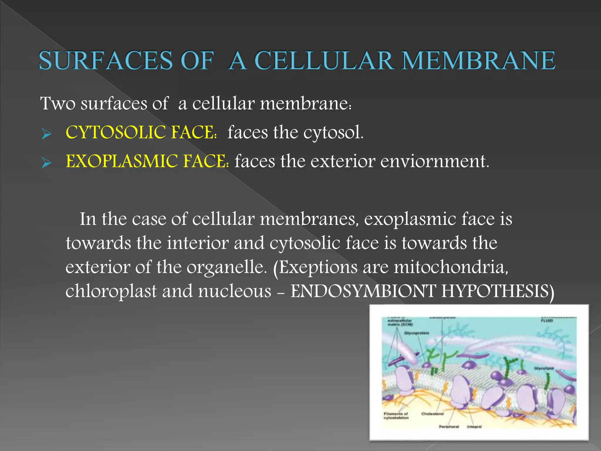
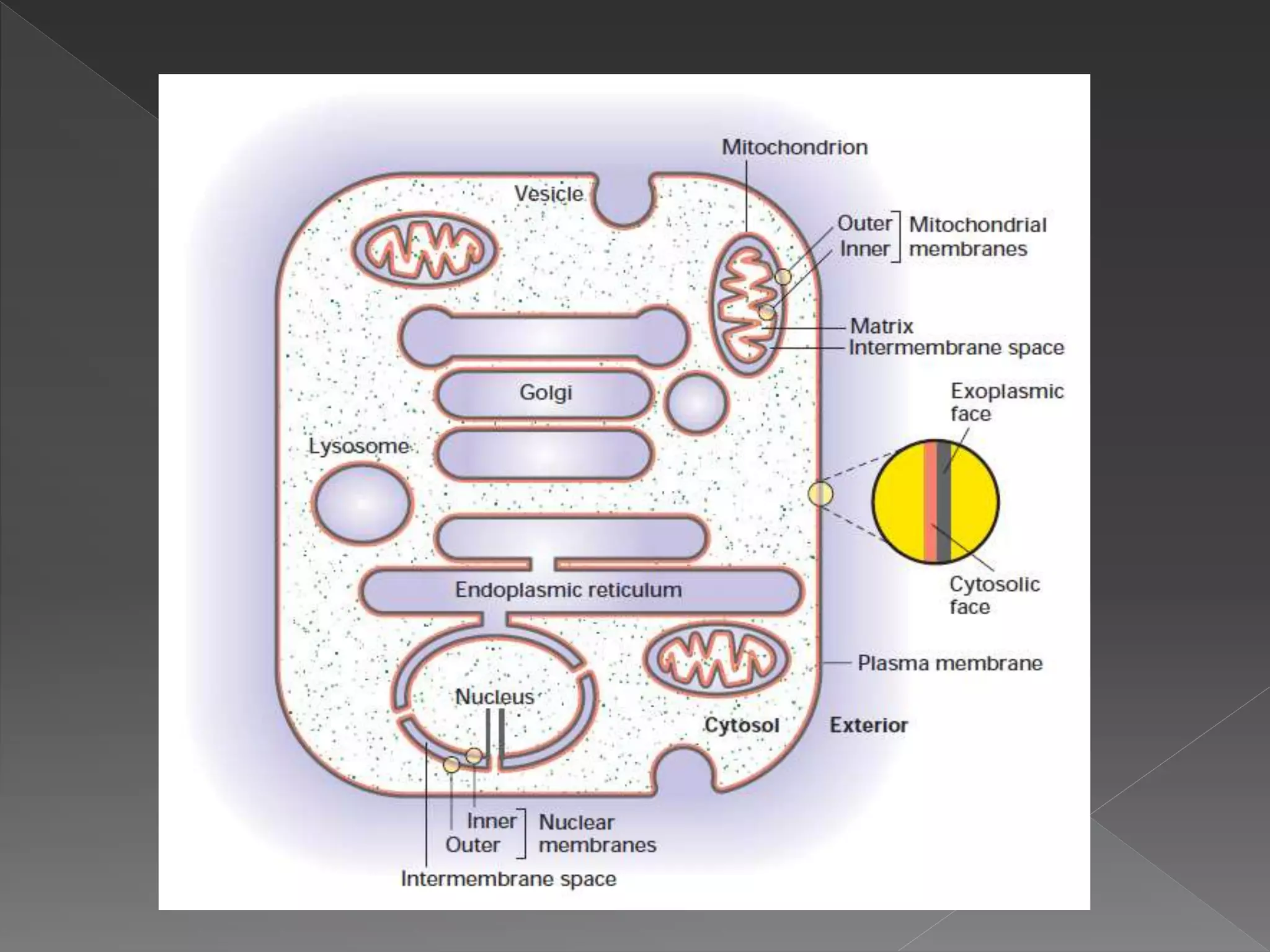
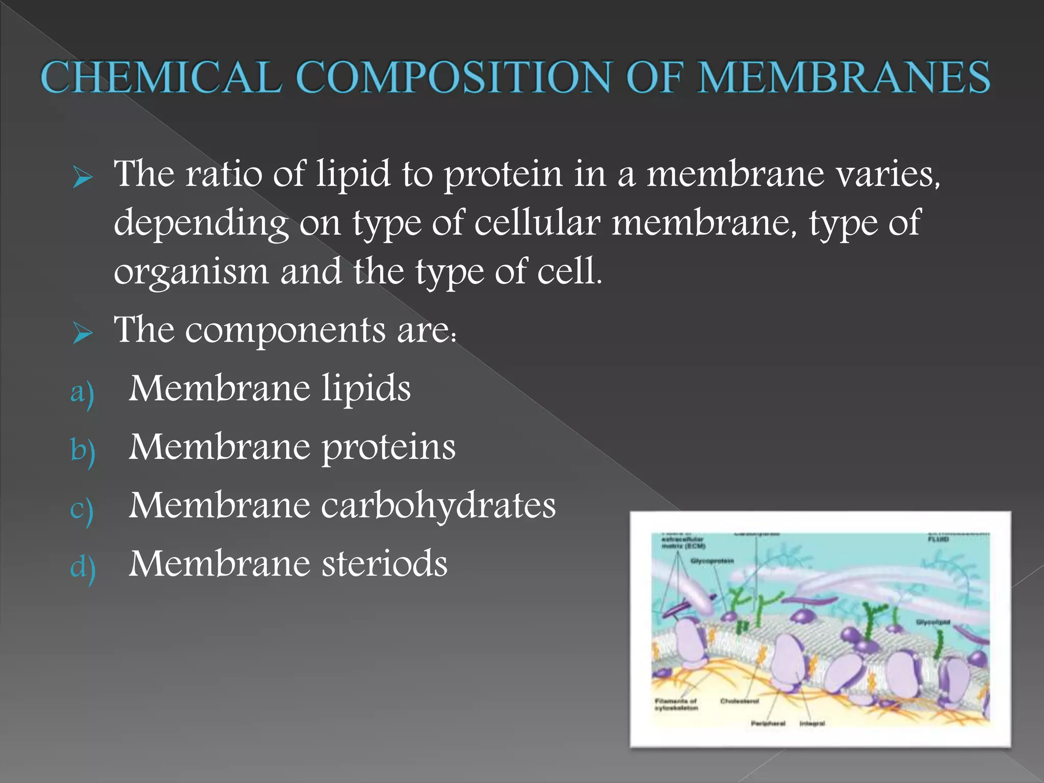
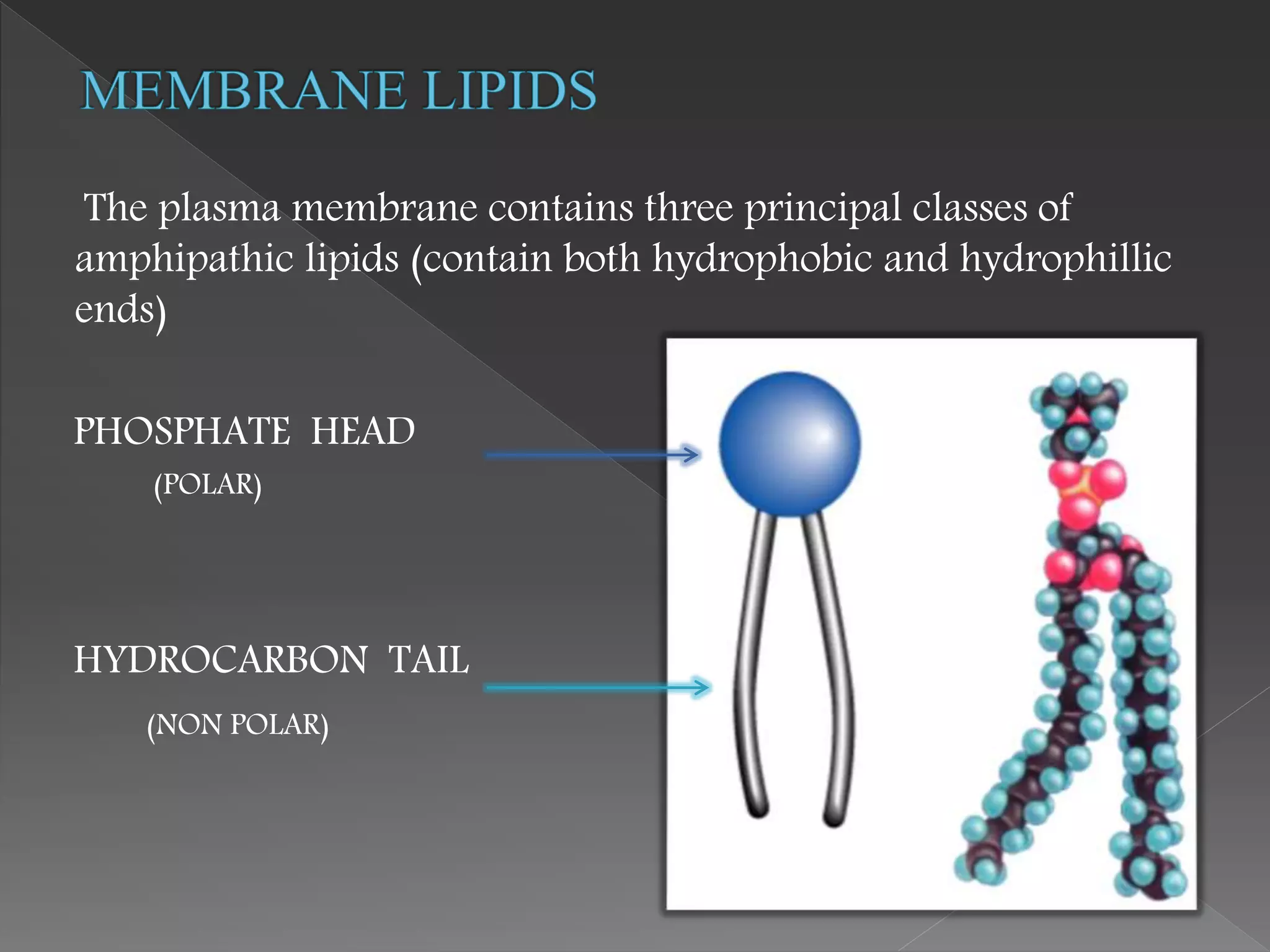
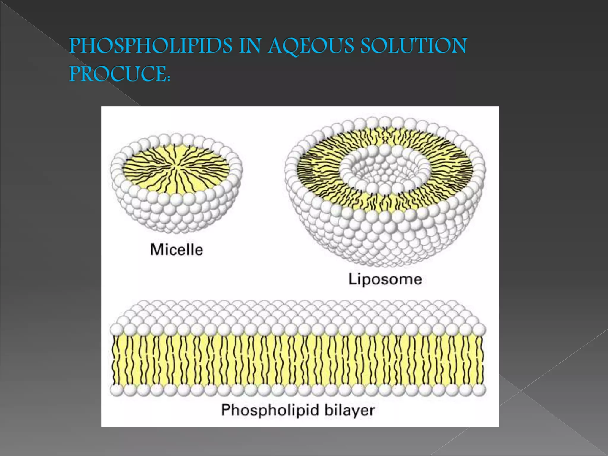

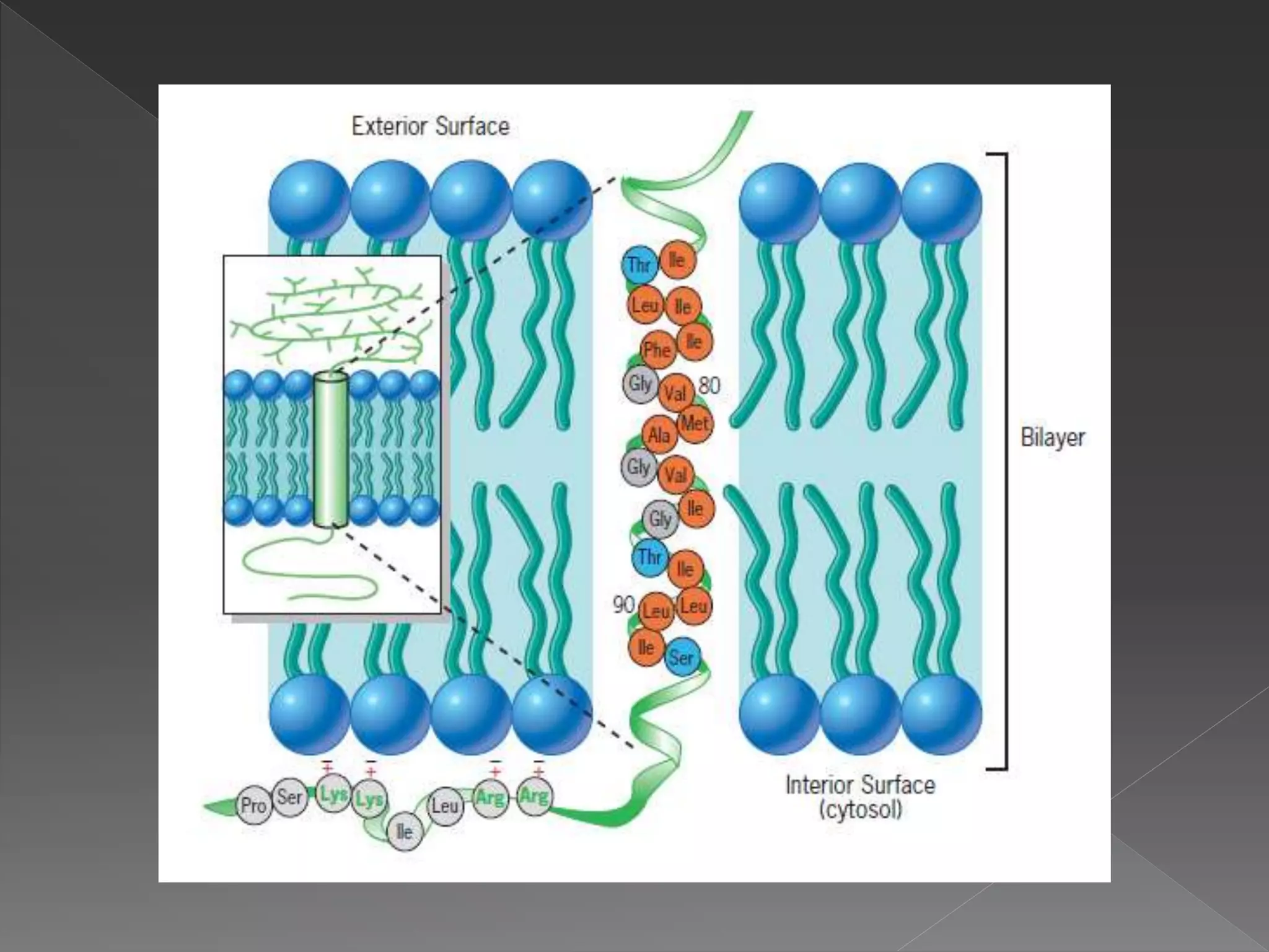
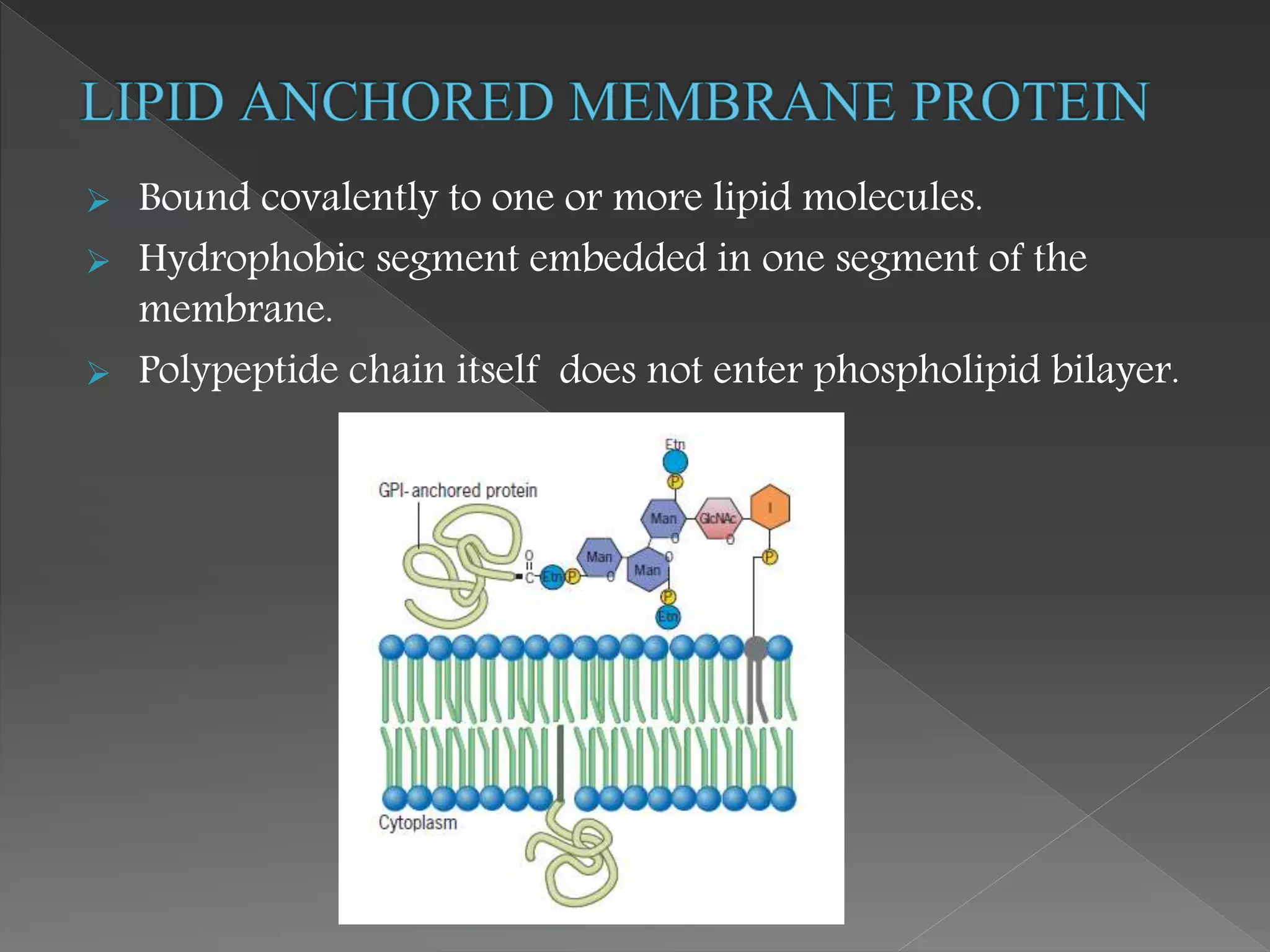
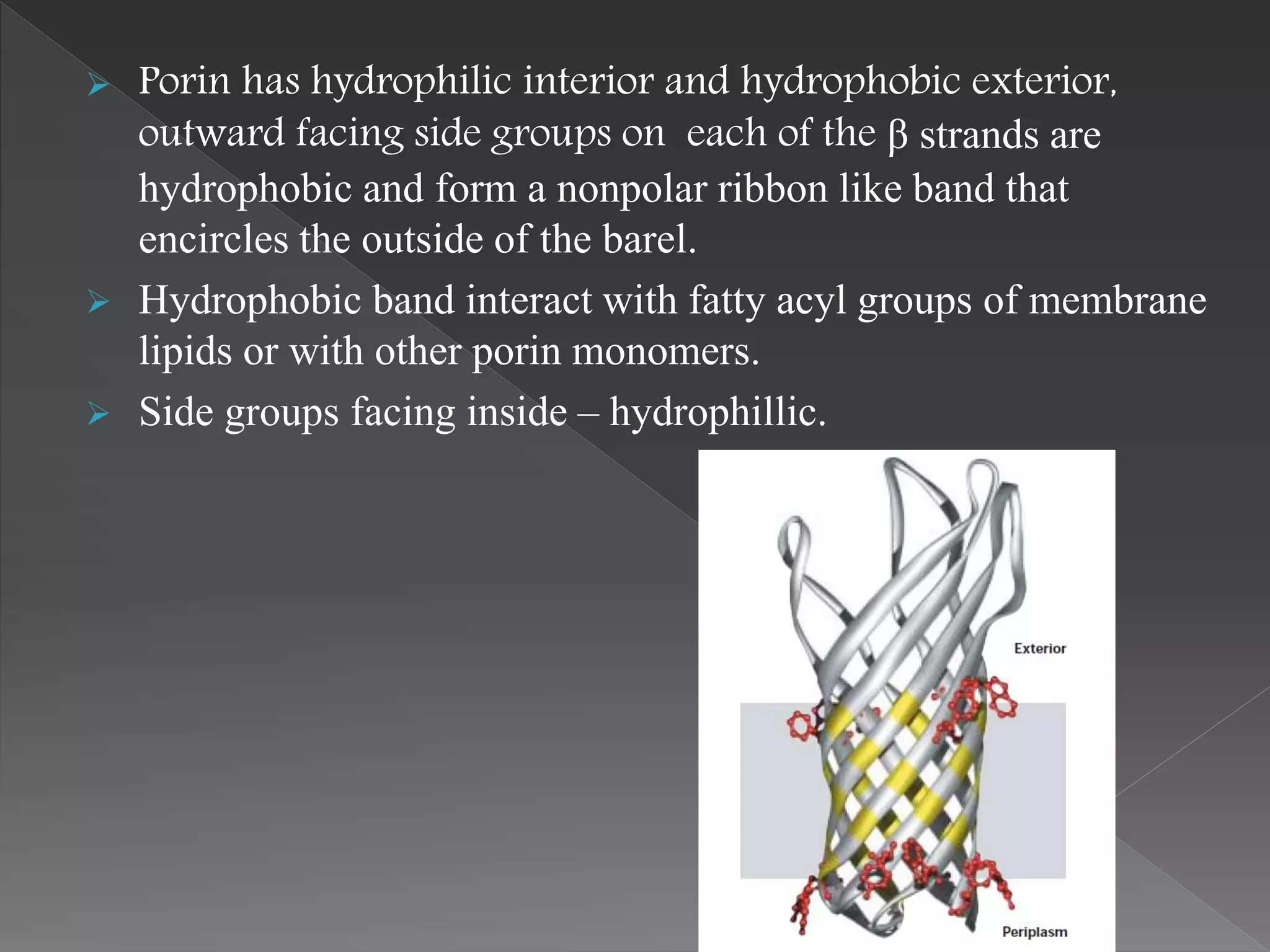


The document provides a detailed overview of the structure and functions of cellular membranes, emphasizing the phospholipid bilayer's selective permeability, fluid mosaic model, and the roles of various proteins and lipids. It discusses the organization and composition of membranes, including integral and peripheral proteins, lipid rafts, and the importance of carbohydrates in cell interaction. Additionally, it highlights membrane fluidity, thickness, and curvature, key to cellular functions such as signal transduction, energy transduction, and maintaining cellular architecture.


