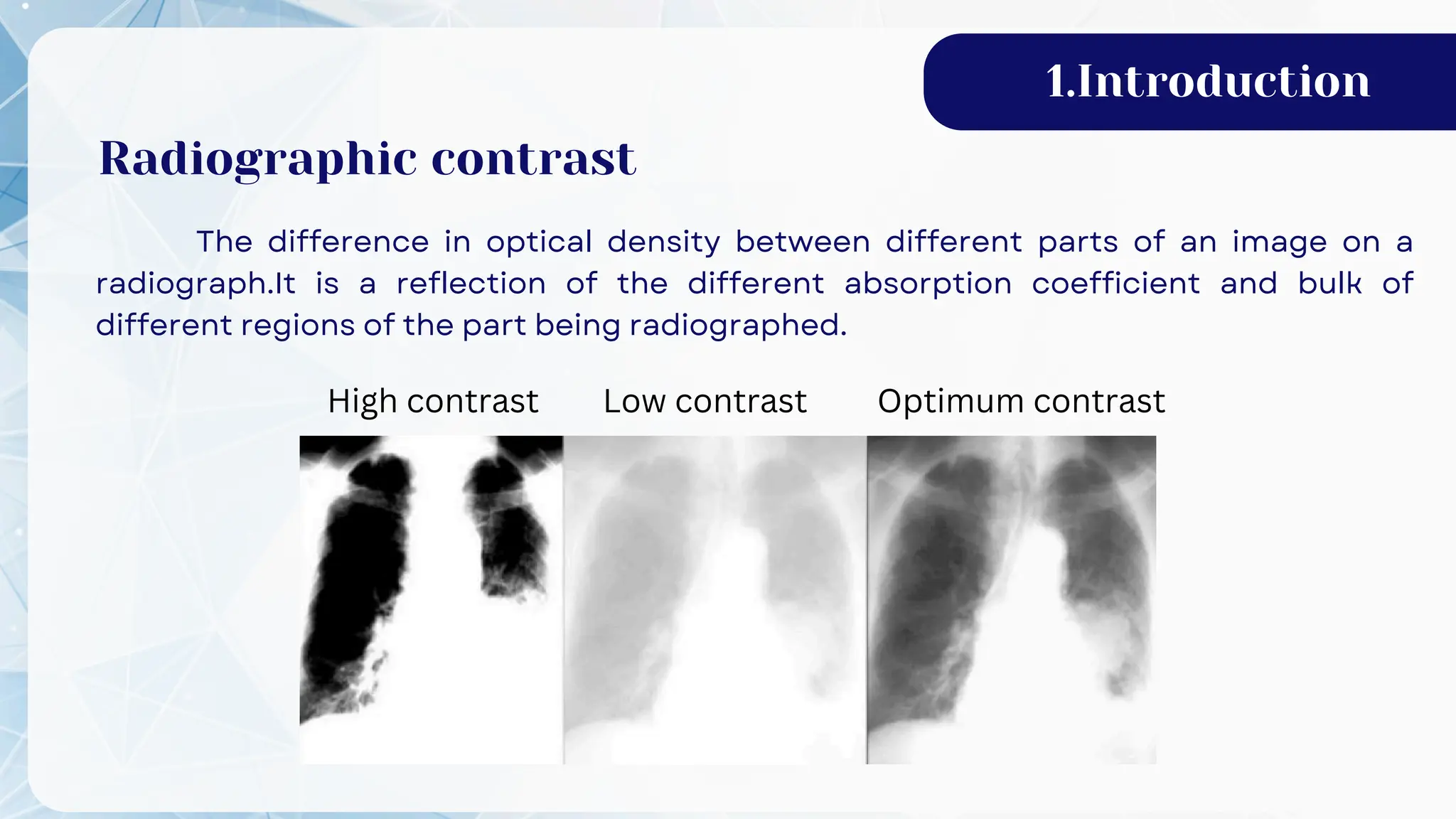
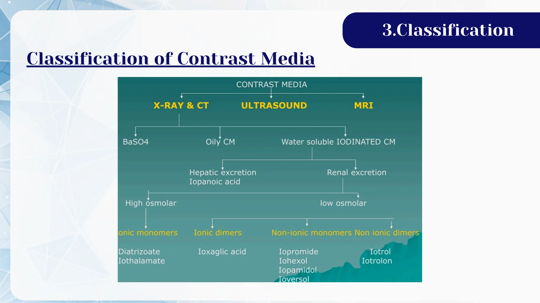
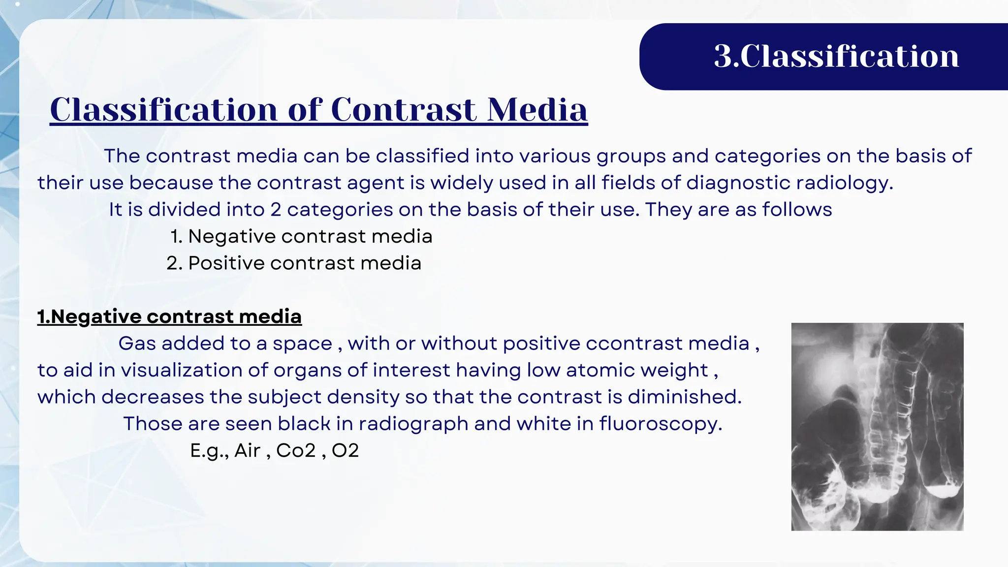
Contrast Media
BY
POOVARASU
And
their adverse reactions
01 Introduction 02 Route of administration
Table of content
03 Classification
05 Contrast media in MRI
07
04 Contast media in XRAY & CT
06
08 Responsibilities of radiographer
Contast media in USG
Reactions of contrast media
1.Introduction
Radiographic contrast
The difference in optical density between different parts of an image on a
radiograph.It is a reflection of the different absorption coefficient and bulk of
different regions of the part being radiographed.
High contrast Low contrast Optimum contrast
1.Introduction
Contrast Media
Contrast media are any external agent that can be introduced into the sub
human body which increases or decreases the subject density in order to increase
the image contrast.
The human body consists of soft tissues and bony structures. As among the soft
tissues itself,there are very little differences in density to provide enough contrast to
visualize the anatomic details. It is necessary to differentiate the density of the organ to
provide desired contrast.
Contrast media is used because soft tissue is more difficult to demonstrate than
bony structure ( usually used for blood vessels and gastrointestinal tract ).
1.Introduction
Properties of Contrast Media
It should be radio-opaque to x-ray and another form of radiation.
Relatively high or low atomic weight in comparison to that human tissue.
Inert i.e. shouldn't react with the body.
Absorbable by the body.
Easily excreted for the body.
Cheap and easily available.
2.Route of administration
Modes of administration of
Contrast Media
Barium - Sulfate contrast materials that are swallowed or administrated
by mouth (orally) are used to enhance x-ray and CT images of the
gastrointestinal (GI) tract,including Pharynx , Esophagus , Stomach , Small
intestine , Large intestine.
Preoral :
rectal :
Barium - Sulfate contrast materials that are administrated by enema
(rectally) are used to enhance x-ray and CT images of the lower
gastrointestinal(GI) tract including Colon and Rectum.
2.Route of administration
Contrast materials injected into a vein (intravenously) are used to
enhance x-ray and CT images. Gadolinium is injected into vein is used
to enhance MRI images.
It is used to enhance internal organs , gastrointestinal tract ,
arteries and veins , soft tissue , brain , breast.
intravenous :
intra artial :
Contrast media is injected through the artery.
Used in Angiography.
intra articular :
Contrast materials injected in joint space.
Used in Arthogram.
intra thecal:
Contrast media is injected through sub-arachnoid space.
Used in Myelography.
3.Classification
Classification of Contrast Media
The contrast media can be classified into various groups and categories on the basis of
their use because the contrast agent is widely used in all fields of diagnostic radiology.
It is divided into 2 categories on the basis of their use. They are as follows
1. Negative contrast media
2. Positive contrast media
1.Negative contrast media
Gas added to a space , with or without positive ccontrast media ,
to aid in visualization of organs of interest having low atomic weight ,
which decreases the subject density so that the contrast is diminished.
Those are seen black in radiograph and white in fluoroscopy.
E.g., Air , Co2 , O2
3.Classification
Classification of Contrast Media
3.Classification
Advantages :
✯ Eliminated by one pass through the lungs.
✯ Lacks allergic potential and renal toxicity.
✯ Low viscosity ( 1/400 that of iodinated contrast ).
✯ Easy delivery through micro catheter or between catheter and wire.
✯ Flows readily into bleeding sites.
✯ Allows superior opacification of portal vein via wedge portal venography or parenchymal
injecton.
Advice :
✧ Uses closed system and never connect Co2 cylinder directly.
✧ Flush regularly with Co2 , not saline.
✧ Wait 2-5 minutes between injections to minimize risk of ischemia ( restriction in blood
supply to tissues ).
✧ Change patient position and use vasodilators
3.Classification
Disadvantage :
✭ Potential neurotoxicity and cardiac ischemia.
✭ Contraindicated in cerebral and coronary circulations , thoracic aorta , brachial artery or if
the patient has a significant AV shunt.
✭ Invisible
*Potential undetected air contamniation ; use sealed delivery system.
✭ Buoyant
*Raises to non-dependent portion of a larger diameter vessels ; potential to overestimate
stenoses and may not fill dependent vessels.
*Prone position and head elevated position contraindicated.
✭ Compressible
*Potential excessive volumes or explosive delivery ( uses of finite volume plastic bag
delivery system is recommended ).
Note:
Highly soluble gases are less likely to cause problems.Gases which are highly soluble in blood include Co2 and N2O. Room
air contains nitrogen , which is not very soluble. Oxygen diffuses too rapidly to be useful. Infusion of negative contrast
agents , as with positive contrast agents, should be infused carefully to prevent over-inflation . Never attach a gas source
directly to the catheter used for infusion. Always load a syringe with the gas , and then attach the syringe to the catheter.
2.Positive contrast media
Those contrast media which has high atomic weight , which increases the subject
density so that the contrast is enhanced . Cannot be easily penetrated by X-rays.
They are seen white in radiograph and black in fluoroscopy.
E.g., Barium sulphate
Omnipaque ( Iohexol ) etc...
IN CASE OF CT AND X-RAY
We use certain contrast agent which has higher densities . They are described as
follows:
1. Water soluble contrast media.
2. Water insolube contrast media
3. Oily contrast media.
3.Classification
4.Contast media in XRAY & CT
1. Water-soluble contrast media
or Organic Iodine compounds
Those contrast media which are soluble in water is known as water soluble contrast
media or organic iodine compound.
History
In early age NaI as OSBON was used for treatment of SYPHYLIS.
When the same patient was radiographed then his radiograph showed positive contrast
with NaI.
But it was found as toxic.
So later iodine is mixed with organic compounds & started using as contrast agents
Properties of contrast media
❃ Have complex molecular structure
❃ Contains iodine which is radio opaque
❃ Water soluble & absorbable
❃ Reacts less within body
❃ In absorption of these compounds is excreted by kidneys
❃ But , carrier factor may remain & form a glomerular residue which is patiently harmful
FURTHER, ORGANIC IONIC COMPOUNDS ARE
CLASSIFIED INTO 2 GROUPS
They are :
1 - Ionic contrast media
2 - Non ionic contrast media
Ionic contast media
Iodinated contrast is a form of intravenous radio contrast ( radiographic dye ) containing
iodine , which enhances the visibility of vascular structures and organs during radiographic
procedures.
Most often they are used in intravenously but can be administrated intra-arterially , intra-
thecally , and intra - abdominally.
4.Contast media in XRAY & CT
Properties:
⍟ It produces ions when dissolve in water and have more charge particles.
⍟ It has got high osmolarity.
⍟ It is cheap but toxic in nature.
⍟ It has less hydroxyl group.
⍟ Ionic contrast media has more viscosity.
TYPES OF IONIC CONTRAST MEDIA:
a. Ionic monomer
Most toxic water soluble contrast media due to high osmolarity
✧Used where there is no contrast of blood
✧Some monomers are conrey, Urogrphin, Angiographin
b. Ionic dimer
Linked tri- Iodinated benzene rings in which atleast 1 carboxylate containing group is
substituted on atleast 1 benzene ring
✧ They have two organic molecule and other iodine
✧They are toxic and used where there is no direct contact with blood
✧ Some dimers are Inorganic acid ( Hexabrix )
4.Contast media in XRAY & CT
2.Non ionic contrast media
The non ionic form decreases the risk, but is much more expensive. The non ionic contrast
media is much more widely used today.
Properties
✾ It produces less ion
✾ It has got high osmolarity
✾ It is expensive but not toxic to the body
✾ Such contrast media has less viscosity.
E. g., Omnipaque, Ultravist
Types of non ionic contrast media
1. Non ionic monomer
2. Non ionic dimer
4.Contast media in XRAY & CT
a.Non ionic monomer
Single tri- iodinated benzene ring without a carboxylate containing benzene substituent.
★ These contrast have one organic molecule at molecule level
★ They are less toxic that can be used in blood stream
★ Some non ionic monomers are:
Iohexal (omnipaque)
Lopamidol (lopamiro, isovue, niopam, solutrast)
Iopromide(Ultrasvist)
Iopentol ( Imagopaque)
Ioversol ( Optoray)
b.Non ionic dimer
Linked tri iodinated benzene ring that do not contain a carboxylate functional group within
any benzene substituent.
★ They contain two organic compounds at molecular level.
★ They are less toxic so are called iso osmolar contrast media.
4.Contast media in XRAY & CT
★ Some non ionic dimers are
Iotral
Iotrolan ( Isovist)
Iodixanol
4.Contast media in XRAY & CT
CONTRAST TERMINOLOGY
HOCM
❈ Tri iodinated benzoic acid ( Ionic contrast, therefore charged particle)
❈ High Osmolality
1500-2300 mOsm/kg water ( monomers)
690 mOsm/kg water(dimers)
c/w Plasma Osmolality 280-303 mOsm/kg water
❈ 1:40, 000 Mortality
❈ Higher incidence of adverse reactions
❈ 5% Allergy
❈ Higher incidence of contrast induced nephropathy (CIN)
❈ Not for use intravascularly
LOCM
❉ Tri iodinated substituted ring compound
❉ Side chains make molecule highly hydrophilic to increase solubility without dissociation.
4.Contast media in XRAY & CT
❉ Low Osmolality
580 mOsm/kg water ( monomer) ( Ultravist / Niopam / Optiray / Omnipaque)
272 mOsm/kg (dimers) (Visipaque) (Saline added to increase osmolality) also called
isomolar or ICM500-2300 mOsm/kg water ( monomers)
❉ 1:1,70, 000 mortality
❉ 4:10, 000 severe or life threatening reactions
❉ Intravascular use : 80-90% intravenous, 10-20% intra arteria
OILY CONTRAST MEDIA
✣They are especially a combination of iodine & poppy seed oil
✣It may be linked with those organic compounds which cannot be
injected intravenously.
✣It can neither be injected nor passed into body by a physiological
process.
✣Therefore used only for direct introduction into spaces & tracts
✣Used for the study of Myelography, Bronchography, Lymphography,
Pleural cavity & ducts like salivary ducts, bladder, biliary ducts etc..
E.g., Lipidol used for Bronchography , Lymphography
Myodil used for Myelography
4.Contast media in XRAY & CT
WATER INSOLUBLE CONTRAST MEDIA :
Those contrast media which is insoluble in water are used for diagnosis of the body parts
i.e., Barium
BARIUM
✥Barium has atomic number of 56 , therefore it is highly radiopaque
✥Insoluble in water like as lipid
✥ It is non absorbable so non toxic. If body absorbs barium it is toxic.
✥It is inert to tissue i.e., it has no capacity to change the anatomy of tissue.
✥It can be used for double contrast study
✥Used generally in GIT studies
✥Barium sulphate salts are commercially available in solid state as in suspension
state.
✥Commercially available are micropaque, baritop.
✥95% weight by volume is available
For single contrast 50% wt by vol.
Double contrast 250% wt by vol.
✥Nowadays barium suspension is available in different flavors like vanilla,
strawberry, chocolate etc..
4.Contast media in XRAY & CT
ALTERNATIVE TO BA CONTRAST
GASTROGRAFFIN OR HYPAQUE ( IODINE)
⋆ High atomic - Close to Iodine
⋆ Similar usage as Barium
⋆ Recommended for use where there is suspected leakage into pleural or
peritoneal cavities.
⋆ Water soluble, safe in abdominal cavity
-Safe to use if perforation is suspected.
⋆ Very harmful to the lungs tissue
-do not use if aspiration is possible.
⋆ Particularly useful in neonates & paediatrics.
⋆ Also useful in CT to distinguish bowel from other structures
( 15ml in 1 litre)
4.Contast media in XRAY & CT
MRI CONTRAST MEDIA
These are paramagnetic substance which are combined with DTPA ( Diethylene
Triamine Penta Acetic acid) Most commonly used compounds for contrast enhancement are
Gadolinium - based.
Mechanism of action
⭒ They affect T1 & T2 relaxation time in tissue
⭒ They maximize the difference of signal intensity between two times.
⭒ They either increase or decrease the signal intensity of a tissue relative to another
5.Contrast media in MRI
TYPES OF MRI CONTRAST MEDIA
1. Parenteral contrast agents
2. Oral contrast agents
1. PARENTERAL CONTRAST AGENTS:
A. Depending on relativity:
a) Positive contrast agents (T1 agent)
b) Negative contrast agents (T2 agent)
a. Positive contrast agent:
• This affects the T1 relaxation time of tissue.
• It reduced T1 relaxation time and increase the signal intensity of the T1 weighted image
E.g., Gadolinium, Mn – DPDP.
5.Contrast media in MRI
b. Negative contrast agents:
• They affect the T2 relaxation time of the tissue results, reduced in the signal intensity
of T2 weighted image.
E.g., Iron oxide, Gadolinium (high dose)
5.Contrast media in MRI
B. Depending on susceptibility
a) Paramagnetic agents
b) Super paramagnetic agents
a. Paramagnetic agents :
• Gadolinium is a paramagnetic agent.
• They are usually positive contrast agent but at higher dose
administration can cause T2 shortening resulting into
decreased signal on T2 weighted image.
• This effect used in perfusion studies.
b. Super paramagnetic agents:
• They are negative contrast agents.
• They cause photon dephasing faster leading to T2 shortening and signal loss.
E.g., Iron oxide (Fe3O4) and super paramagnetic iron oxide
2. ORAL CONTRAST AGENTS:
a) Positive contrast agent
b) Negative contrast agent
a. Positive contrast agent:
Manganese chloride, Gd – DTPA increases the resolution and contrast in bowel
investigation.
5.Contrast media in MRI
b. Negative contrast agents:
• They decrease signal from and reduce the motion of bowel.
• They are also used in MRCP.
E.g., Super paramagnetic iron oxide
OTHER MR CONTRAST AGENTS :
Hepatobiliary contrast agents
a. Iron oxide (Fe3O4):
• It is a super paramagnetic agent.
• It is phagocytosed by reticuloendothelial system (RES) with prominent uptake in liver
and spleen.
• Normal liver tissue takes up iron oxide and becomes dark on T2 weighted images.
• Focal lesions such as metastases do not have RES cells within them, so they remain same
and appear relatively bright.
b. Mn – DPDP (Mangafodipir trisodium):
• This is hepatobiliary specific contrast agent.
• In this contrast, 50% excreted by kidney and another 50% excreted by the biliary system.
• This contrast takes up by the hepatocytes and becomes positive contrast enhancement
(brighter) on a T1 weighted image.
• The lesions without hepatocytes like metastasis will not show enhancement and remain
darker.
5.Contrast media in MRI
ULTRASOUND CONTRAST MEDIA
❃ Artifacts is used here as contrast.
❃ Micro air bubbles (<7mu m) suspended in galactose is used.
❃ These bubbles last for 5mins
❃ Microbubbles have a high degree of echogenicity, and ability to reflect the ultrasound
waves.
❃ The echogenicity difference between the gas in the microbubbles and the soft tissue
surrounding, which produce a high echogenicity difference image
❃ These air bubbles produces artifacts & those artifacts used in diagnostic purpose.
❃ Not much in practice.
❃ Very expensive.
❃ Commercially it is available as Echovist, Laevosist
❃ Palmitic acid also used in Laevosist to reduce surface tension of the bubbles..
6.Contrast media in USG
Working
• Ultrasound contrast agents are injected intravenously into the systemic circulation, which
remains in the systemic circulation for a certain period.
• During that time, ultrasound waves are directed on the area of the interest.
• The microbubbles reflect unique echo from the surrounding tissue.
• The ultrasound system converts the strong echogenicity into a contrast enhanced image of
the area of interest.
6.Contrast media in USG
DIFFERENT TYPES OF ULTRASOUND CONTRAST AGENT:
A. Tissue enhancing ultrasound contrast agents:
It improves the assessment of certain organs like liver, kidney, pancreas, prostate and
ovary by improving the acoustic difference between normal and abnormal portion of
organs. They are as follows:
1. Levovist
2. Sonovist (Schering)
3. Sonozoid (hycomed – amershan)
6.Contrast media in USG
B. Vascular enhancing ultrasound contrast agent:
• This are gas microbubbles with a diameter less than 5 to 10 micrometer, to pass through
lung capillaries and into the systemic circulations.
• These are mostly used to imaging of malignant tumors in liver, kidney, ovary, pancreas
and prostate.
• It also used in cardiac evaluation. E.g.,Albunex, infosan.
• Albunex has been developed by MALLINCNODT – INC. The shell stabilizer is albumin and
the gas used is air.
ADVERSE REACTIONS TO CONTRAST MEDIA
Based on Severity
Mild , Moderate , Severe.
7.Reactions of contrast media
Nausea
Vomiting
Altered taste
Sweats
Cough
Shaking
Rash, hives
Itching
Swelling : eyes, face
Anxiety
Headache
Flushing
Tachycardia/bradycardia
Wheezing
Hypotension
Bronchospasm
Hypertension
Dyspnea
Laryngeal edema
Pulmonary edema
Abdominal cramps
Laryngeal edema
(severe or progressive)
Profound hypotension
Convulsions
Unresponsive
Clinically manifest
Arrhythmia
Cardiopulmonary arrest
MILD MODERATE SEVERE
Types of contrast reaction
1. Anaphylactoid
2. Non anaphylactoid
3. Combined
7.Reactions of contrast media
1. Anaphylactoid reaction
It occurs unexpectedly and the specific cause is uncertain. Therefore it is often
referred as 'Idiosyncratic'
✦ Allergic asthma
✦ Drug allergies
✦ Food allergies
✦ Prior reaction to contrast
7.Reactions of contrast media
2. Non-Anaphylactoid reaction
Other reactions relating to osmotic, chemotoxic, direct organ toxicity or vasomotor
effects are more predictable and better understood. These are referred as non
anaphylactoid reaction
✦ Chemotoxic
✦ Vasovagal
✦ Idiopathic
A) Chemotoxic
The clinical effects of contrast agents not only result from high
osmolality, but also from their own specific pharmacology, which
mediates chemotoxic effects. The chemotoxic effects of the new
nonionic agent, iohexol, are compared with those of standard ionic
and other low osmolality contrast agents, ionic and nonionic.
Organ specific
*Nephrotoxicity
*Cardiovascular toxicity
*Neurotoxicity
7.Reactions of contrast media
Nephrotoxicity
Contrast-medium nephropathy is usually defined as impairment of renal function
occurring within 48 hours after administration of contrast media.
It is manifested by an absolute increase in the serum creatinine level of at least 44
μmol/L,or by a relative increase of at least 25% over the baseline value in the absence of
another cause.
Cardiovascular toxicity
The injection of hypertonic contrast medium causes significant fluid and ion shifts.
Immediately after injection there is significant increase in serum osmolality.This
causes an influx of water from interstitial space into vascular compartment , an increase
in blood volume , an increase in cardiac output and a brief increase systemic blood
pressure , peripheral dilation causes more prolonged fall of blood pressure.
Reactions include hypotension,tachycardia,and arrhythmias.Uncommon reactions
include congestive heart failure , pulmonary edema and cardiac arrest
7.Reactions of contrast media
Neurotoxicity
Contrast media delived to brain via artial route may cross blood brain
barrior.Neurotoxicity is caused by both osmolality and chemotoxic effect and IV contrast
medium may rarely provoke in patient with epilepsy or cerebral tumors.
These risks are reduced when low or iso-osmolar agents are used.
Potential reacctions include headache , confusion , seizure , altered consciousness
, visual disturbance and dizziness.
B. Vasovagal reactions
It results of increased vagal tone on the heart and blood vessels.
Results bradycardia and hypotension and may be accompanied by apprehension ,
confusion , sweating , unresponsiveness , and loss of bowel or bladder control signals
C. Idiopathic
Of unknown cause . Any disease that is of uncertain or unknown origin may be
termed as Idiopathic.
3. Combined
Both anaphylactoid and non anaphylactoid occurs simultaneouosly.
ADVERSE REACTIONS TO MRI CONTRAST MEDIA
A. Minor effects
Pain and burning at the injection site, low blood pressure (hypotension), minor skin rash,
mild headache, lightheadedness and nausea.
B. Allergic reactions
Urticaria (hives), swelling of the face, rashes, itching, sweating, watery or itchy eyes and
shortness of breath.
C.Nephrogenic systemic fibrosis (NSF)
Patients with renal failure and kidney diseases can’t filter the chemical contrast quickly
enough and it stays in the body.
There it causes a serious medical condition called nephrogenic systemic fibrosis (NSF).
NSF symptoms include hardening skin with red patches and are most commonly found
in the limbs
7.Reactions of contrast media
Responsibilities of Radiographer during contrast studies
1. Should check patient history
Clinical complains
Food or drug allergies
Previous contrast media reactions
Ashma, hay fever or hives
2. Selection and peroration of contrast media
Read labels 3 times
Empty container available (venipuncture)
3. Preparation for possible reaction
Fully stocked emergency chart ( epinephrine available - emergency medical treatment to
treat life threatening allergic reaction)
Cardiopulmonary resuscitation equipment
Oxygen and suction available
4. Radiographer responsibility during mild and moderate reaction include
Call for medical assistance
Become familiar with work environment - Emergency assistance protocol (how to help)
Know where your crash cart and drug trays are located and how to call for help?
Monitor and comfort patient
Document patient reaction
Use emergency drugs and equipments according to the reaction
8.Responsibilities of Radiographers
✪ ADRENALINE - FOR ALLERGIC REACTIONS
✪ ATROPINE -ANTI-ARYTHMATIC
✪ BUSCOPAN - FOR PAIN RELIVING
✪ HYDROCORTISONE -FOR ALLERGIC REACTIONS
✪ DOPAMINE -TO INCREASE BLOOD PRESSURE
✪ SODIUM BICARBONATE -FOR ELECTROLYTE IMBALANCE
✪ DIAZEPAM -FOR SEDATION
✪ AVILAS ANTI-ALLERGIC AGENT
EMERGENCY DRUGS USED IN RADIOLOGY
• Whenever there is sudden onset of allergic reactions after the injection of contrast,
crash cart is rushed into for management.
• The crash cart contains various emergency drugs and equipment for management
for those crucial events which includes:
8.Responsibilities of Radiographers
Thank You

Contrast media and their adverse reactions

  • 1.
  • 2.
    01 Introduction 02Route of administration Table of content 03 Classification 05 Contrast media in MRI 07 04 Contast media in XRAY & CT 06 08 Responsibilities of radiographer Contast media in USG Reactions of contrast media
  • 3.
    1.Introduction Radiographic contrast The differencein optical density between different parts of an image on a radiograph.It is a reflection of the different absorption coefficient and bulk of different regions of the part being radiographed. High contrast Low contrast Optimum contrast
  • 4.
    1.Introduction Contrast Media Contrast mediaare any external agent that can be introduced into the sub human body which increases or decreases the subject density in order to increase the image contrast. The human body consists of soft tissues and bony structures. As among the soft tissues itself,there are very little differences in density to provide enough contrast to visualize the anatomic details. It is necessary to differentiate the density of the organ to provide desired contrast. Contrast media is used because soft tissue is more difficult to demonstrate than bony structure ( usually used for blood vessels and gastrointestinal tract ).
  • 5.
    1.Introduction Properties of ContrastMedia It should be radio-opaque to x-ray and another form of radiation. Relatively high or low atomic weight in comparison to that human tissue. Inert i.e. shouldn't react with the body. Absorbable by the body. Easily excreted for the body. Cheap and easily available.
  • 6.
    2.Route of administration Modesof administration of Contrast Media Barium - Sulfate contrast materials that are swallowed or administrated by mouth (orally) are used to enhance x-ray and CT images of the gastrointestinal (GI) tract,including Pharynx , Esophagus , Stomach , Small intestine , Large intestine. Preoral : rectal : Barium - Sulfate contrast materials that are administrated by enema (rectally) are used to enhance x-ray and CT images of the lower gastrointestinal(GI) tract including Colon and Rectum.
  • 7.
    2.Route of administration Contrastmaterials injected into a vein (intravenously) are used to enhance x-ray and CT images. Gadolinium is injected into vein is used to enhance MRI images. It is used to enhance internal organs , gastrointestinal tract , arteries and veins , soft tissue , brain , breast. intravenous : intra artial : Contrast media is injected through the artery. Used in Angiography. intra articular : Contrast materials injected in joint space. Used in Arthogram. intra thecal: Contrast media is injected through sub-arachnoid space. Used in Myelography.
  • 8.
  • 9.
    The contrast mediacan be classified into various groups and categories on the basis of their use because the contrast agent is widely used in all fields of diagnostic radiology. It is divided into 2 categories on the basis of their use. They are as follows 1. Negative contrast media 2. Positive contrast media 1.Negative contrast media Gas added to a space , with or without positive ccontrast media , to aid in visualization of organs of interest having low atomic weight , which decreases the subject density so that the contrast is diminished. Those are seen black in radiograph and white in fluoroscopy. E.g., Air , Co2 , O2 3.Classification Classification of Contrast Media
  • 10.
    3.Classification Advantages : ✯ Eliminatedby one pass through the lungs. ✯ Lacks allergic potential and renal toxicity. ✯ Low viscosity ( 1/400 that of iodinated contrast ). ✯ Easy delivery through micro catheter or between catheter and wire. ✯ Flows readily into bleeding sites. ✯ Allows superior opacification of portal vein via wedge portal venography or parenchymal injecton. Advice : ✧ Uses closed system and never connect Co2 cylinder directly. ✧ Flush regularly with Co2 , not saline. ✧ Wait 2-5 minutes between injections to minimize risk of ischemia ( restriction in blood supply to tissues ). ✧ Change patient position and use vasodilators
  • 11.
    3.Classification Disadvantage : ✭ Potentialneurotoxicity and cardiac ischemia. ✭ Contraindicated in cerebral and coronary circulations , thoracic aorta , brachial artery or if the patient has a significant AV shunt. ✭ Invisible *Potential undetected air contamniation ; use sealed delivery system. ✭ Buoyant *Raises to non-dependent portion of a larger diameter vessels ; potential to overestimate stenoses and may not fill dependent vessels. *Prone position and head elevated position contraindicated. ✭ Compressible *Potential excessive volumes or explosive delivery ( uses of finite volume plastic bag delivery system is recommended ). Note: Highly soluble gases are less likely to cause problems.Gases which are highly soluble in blood include Co2 and N2O. Room air contains nitrogen , which is not very soluble. Oxygen diffuses too rapidly to be useful. Infusion of negative contrast agents , as with positive contrast agents, should be infused carefully to prevent over-inflation . Never attach a gas source directly to the catheter used for infusion. Always load a syringe with the gas , and then attach the syringe to the catheter.
  • 12.
    2.Positive contrast media Thosecontrast media which has high atomic weight , which increases the subject density so that the contrast is enhanced . Cannot be easily penetrated by X-rays. They are seen white in radiograph and black in fluoroscopy. E.g., Barium sulphate Omnipaque ( Iohexol ) etc... IN CASE OF CT AND X-RAY We use certain contrast agent which has higher densities . They are described as follows: 1. Water soluble contrast media. 2. Water insolube contrast media 3. Oily contrast media. 3.Classification
  • 13.
    4.Contast media inXRAY & CT 1. Water-soluble contrast media or Organic Iodine compounds Those contrast media which are soluble in water is known as water soluble contrast media or organic iodine compound. History In early age NaI as OSBON was used for treatment of SYPHYLIS. When the same patient was radiographed then his radiograph showed positive contrast with NaI. But it was found as toxic. So later iodine is mixed with organic compounds & started using as contrast agents Properties of contrast media ❃ Have complex molecular structure ❃ Contains iodine which is radio opaque ❃ Water soluble & absorbable ❃ Reacts less within body ❃ In absorption of these compounds is excreted by kidneys ❃ But , carrier factor may remain & form a glomerular residue which is patiently harmful
  • 14.
    FURTHER, ORGANIC IONICCOMPOUNDS ARE CLASSIFIED INTO 2 GROUPS They are : 1 - Ionic contrast media 2 - Non ionic contrast media Ionic contast media Iodinated contrast is a form of intravenous radio contrast ( radiographic dye ) containing iodine , which enhances the visibility of vascular structures and organs during radiographic procedures. Most often they are used in intravenously but can be administrated intra-arterially , intra- thecally , and intra - abdominally. 4.Contast media in XRAY & CT
  • 15.
    Properties: ⍟ It producesions when dissolve in water and have more charge particles. ⍟ It has got high osmolarity. ⍟ It is cheap but toxic in nature. ⍟ It has less hydroxyl group. ⍟ Ionic contrast media has more viscosity. TYPES OF IONIC CONTRAST MEDIA: a. Ionic monomer Most toxic water soluble contrast media due to high osmolarity ✧Used where there is no contrast of blood ✧Some monomers are conrey, Urogrphin, Angiographin b. Ionic dimer Linked tri- Iodinated benzene rings in which atleast 1 carboxylate containing group is substituted on atleast 1 benzene ring ✧ They have two organic molecule and other iodine ✧They are toxic and used where there is no direct contact with blood ✧ Some dimers are Inorganic acid ( Hexabrix ) 4.Contast media in XRAY & CT
  • 16.
    2.Non ionic contrastmedia The non ionic form decreases the risk, but is much more expensive. The non ionic contrast media is much more widely used today. Properties ✾ It produces less ion ✾ It has got high osmolarity ✾ It is expensive but not toxic to the body ✾ Such contrast media has less viscosity. E. g., Omnipaque, Ultravist Types of non ionic contrast media 1. Non ionic monomer 2. Non ionic dimer 4.Contast media in XRAY & CT
  • 17.
    a.Non ionic monomer Singletri- iodinated benzene ring without a carboxylate containing benzene substituent. ★ These contrast have one organic molecule at molecule level ★ They are less toxic that can be used in blood stream ★ Some non ionic monomers are: Iohexal (omnipaque) Lopamidol (lopamiro, isovue, niopam, solutrast) Iopromide(Ultrasvist) Iopentol ( Imagopaque) Ioversol ( Optoray) b.Non ionic dimer Linked tri iodinated benzene ring that do not contain a carboxylate functional group within any benzene substituent. ★ They contain two organic compounds at molecular level. ★ They are less toxic so are called iso osmolar contrast media. 4.Contast media in XRAY & CT
  • 18.
    ★ Some nonionic dimers are Iotral Iotrolan ( Isovist) Iodixanol 4.Contast media in XRAY & CT
  • 19.
    CONTRAST TERMINOLOGY HOCM ❈ Triiodinated benzoic acid ( Ionic contrast, therefore charged particle) ❈ High Osmolality 1500-2300 mOsm/kg water ( monomers) 690 mOsm/kg water(dimers) c/w Plasma Osmolality 280-303 mOsm/kg water ❈ 1:40, 000 Mortality ❈ Higher incidence of adverse reactions ❈ 5% Allergy ❈ Higher incidence of contrast induced nephropathy (CIN) ❈ Not for use intravascularly LOCM ❉ Tri iodinated substituted ring compound ❉ Side chains make molecule highly hydrophilic to increase solubility without dissociation. 4.Contast media in XRAY & CT
  • 20.
    ❉ Low Osmolality 580mOsm/kg water ( monomer) ( Ultravist / Niopam / Optiray / Omnipaque) 272 mOsm/kg (dimers) (Visipaque) (Saline added to increase osmolality) also called isomolar or ICM500-2300 mOsm/kg water ( monomers) ❉ 1:1,70, 000 mortality ❉ 4:10, 000 severe or life threatening reactions ❉ Intravascular use : 80-90% intravenous, 10-20% intra arteria OILY CONTRAST MEDIA ✣They are especially a combination of iodine & poppy seed oil ✣It may be linked with those organic compounds which cannot be injected intravenously. ✣It can neither be injected nor passed into body by a physiological process. ✣Therefore used only for direct introduction into spaces & tracts ✣Used for the study of Myelography, Bronchography, Lymphography, Pleural cavity & ducts like salivary ducts, bladder, biliary ducts etc.. E.g., Lipidol used for Bronchography , Lymphography Myodil used for Myelography 4.Contast media in XRAY & CT
  • 21.
    WATER INSOLUBLE CONTRASTMEDIA : Those contrast media which is insoluble in water are used for diagnosis of the body parts i.e., Barium BARIUM ✥Barium has atomic number of 56 , therefore it is highly radiopaque ✥Insoluble in water like as lipid ✥ It is non absorbable so non toxic. If body absorbs barium it is toxic. ✥It is inert to tissue i.e., it has no capacity to change the anatomy of tissue. ✥It can be used for double contrast study ✥Used generally in GIT studies ✥Barium sulphate salts are commercially available in solid state as in suspension state. ✥Commercially available are micropaque, baritop. ✥95% weight by volume is available For single contrast 50% wt by vol. Double contrast 250% wt by vol. ✥Nowadays barium suspension is available in different flavors like vanilla, strawberry, chocolate etc.. 4.Contast media in XRAY & CT
  • 22.
    ALTERNATIVE TO BACONTRAST GASTROGRAFFIN OR HYPAQUE ( IODINE) ⋆ High atomic - Close to Iodine ⋆ Similar usage as Barium ⋆ Recommended for use where there is suspected leakage into pleural or peritoneal cavities. ⋆ Water soluble, safe in abdominal cavity -Safe to use if perforation is suspected. ⋆ Very harmful to the lungs tissue -do not use if aspiration is possible. ⋆ Particularly useful in neonates & paediatrics. ⋆ Also useful in CT to distinguish bowel from other structures ( 15ml in 1 litre) 4.Contast media in XRAY & CT
  • 23.
    MRI CONTRAST MEDIA Theseare paramagnetic substance which are combined with DTPA ( Diethylene Triamine Penta Acetic acid) Most commonly used compounds for contrast enhancement are Gadolinium - based. Mechanism of action ⭒ They affect T1 & T2 relaxation time in tissue ⭒ They maximize the difference of signal intensity between two times. ⭒ They either increase or decrease the signal intensity of a tissue relative to another 5.Contrast media in MRI TYPES OF MRI CONTRAST MEDIA 1. Parenteral contrast agents 2. Oral contrast agents
  • 24.
    1. PARENTERAL CONTRASTAGENTS: A. Depending on relativity: a) Positive contrast agents (T1 agent) b) Negative contrast agents (T2 agent) a. Positive contrast agent: • This affects the T1 relaxation time of tissue. • It reduced T1 relaxation time and increase the signal intensity of the T1 weighted image E.g., Gadolinium, Mn – DPDP. 5.Contrast media in MRI b. Negative contrast agents: • They affect the T2 relaxation time of the tissue results, reduced in the signal intensity of T2 weighted image. E.g., Iron oxide, Gadolinium (high dose)
  • 25.
    5.Contrast media inMRI B. Depending on susceptibility a) Paramagnetic agents b) Super paramagnetic agents a. Paramagnetic agents : • Gadolinium is a paramagnetic agent. • They are usually positive contrast agent but at higher dose administration can cause T2 shortening resulting into decreased signal on T2 weighted image. • This effect used in perfusion studies. b. Super paramagnetic agents: • They are negative contrast agents. • They cause photon dephasing faster leading to T2 shortening and signal loss. E.g., Iron oxide (Fe3O4) and super paramagnetic iron oxide
  • 26.
    2. ORAL CONTRASTAGENTS: a) Positive contrast agent b) Negative contrast agent a. Positive contrast agent: Manganese chloride, Gd – DTPA increases the resolution and contrast in bowel investigation. 5.Contrast media in MRI b. Negative contrast agents: • They decrease signal from and reduce the motion of bowel. • They are also used in MRCP. E.g., Super paramagnetic iron oxide
  • 27.
    OTHER MR CONTRASTAGENTS : Hepatobiliary contrast agents a. Iron oxide (Fe3O4): • It is a super paramagnetic agent. • It is phagocytosed by reticuloendothelial system (RES) with prominent uptake in liver and spleen. • Normal liver tissue takes up iron oxide and becomes dark on T2 weighted images. • Focal lesions such as metastases do not have RES cells within them, so they remain same and appear relatively bright. b. Mn – DPDP (Mangafodipir trisodium): • This is hepatobiliary specific contrast agent. • In this contrast, 50% excreted by kidney and another 50% excreted by the biliary system. • This contrast takes up by the hepatocytes and becomes positive contrast enhancement (brighter) on a T1 weighted image. • The lesions without hepatocytes like metastasis will not show enhancement and remain darker. 5.Contrast media in MRI
  • 28.
  • 29.
    ULTRASOUND CONTRAST MEDIA ❃Artifacts is used here as contrast. ❃ Micro air bubbles (<7mu m) suspended in galactose is used. ❃ These bubbles last for 5mins ❃ Microbubbles have a high degree of echogenicity, and ability to reflect the ultrasound waves. ❃ The echogenicity difference between the gas in the microbubbles and the soft tissue surrounding, which produce a high echogenicity difference image ❃ These air bubbles produces artifacts & those artifacts used in diagnostic purpose. ❃ Not much in practice. ❃ Very expensive. ❃ Commercially it is available as Echovist, Laevosist ❃ Palmitic acid also used in Laevosist to reduce surface tension of the bubbles.. 6.Contrast media in USG
  • 30.
    Working • Ultrasound contrastagents are injected intravenously into the systemic circulation, which remains in the systemic circulation for a certain period. • During that time, ultrasound waves are directed on the area of the interest. • The microbubbles reflect unique echo from the surrounding tissue. • The ultrasound system converts the strong echogenicity into a contrast enhanced image of the area of interest. 6.Contrast media in USG
  • 31.
    DIFFERENT TYPES OFULTRASOUND CONTRAST AGENT: A. Tissue enhancing ultrasound contrast agents: It improves the assessment of certain organs like liver, kidney, pancreas, prostate and ovary by improving the acoustic difference between normal and abnormal portion of organs. They are as follows: 1. Levovist 2. Sonovist (Schering) 3. Sonozoid (hycomed – amershan) 6.Contrast media in USG B. Vascular enhancing ultrasound contrast agent: • This are gas microbubbles with a diameter less than 5 to 10 micrometer, to pass through lung capillaries and into the systemic circulations. • These are mostly used to imaging of malignant tumors in liver, kidney, ovary, pancreas and prostate. • It also used in cardiac evaluation. E.g.,Albunex, infosan. • Albunex has been developed by MALLINCNODT – INC. The shell stabilizer is albumin and the gas used is air.
  • 32.
    ADVERSE REACTIONS TOCONTRAST MEDIA Based on Severity Mild , Moderate , Severe. 7.Reactions of contrast media Nausea Vomiting Altered taste Sweats Cough Shaking Rash, hives Itching Swelling : eyes, face Anxiety Headache Flushing Tachycardia/bradycardia Wheezing Hypotension Bronchospasm Hypertension Dyspnea Laryngeal edema Pulmonary edema Abdominal cramps Laryngeal edema (severe or progressive) Profound hypotension Convulsions Unresponsive Clinically manifest Arrhythmia Cardiopulmonary arrest MILD MODERATE SEVERE
  • 33.
    Types of contrastreaction 1. Anaphylactoid 2. Non anaphylactoid 3. Combined 7.Reactions of contrast media 1. Anaphylactoid reaction It occurs unexpectedly and the specific cause is uncertain. Therefore it is often referred as 'Idiosyncratic' ✦ Allergic asthma ✦ Drug allergies ✦ Food allergies ✦ Prior reaction to contrast
  • 34.
    7.Reactions of contrastmedia 2. Non-Anaphylactoid reaction Other reactions relating to osmotic, chemotoxic, direct organ toxicity or vasomotor effects are more predictable and better understood. These are referred as non anaphylactoid reaction ✦ Chemotoxic ✦ Vasovagal ✦ Idiopathic A) Chemotoxic The clinical effects of contrast agents not only result from high osmolality, but also from their own specific pharmacology, which mediates chemotoxic effects. The chemotoxic effects of the new nonionic agent, iohexol, are compared with those of standard ionic and other low osmolality contrast agents, ionic and nonionic. Organ specific *Nephrotoxicity *Cardiovascular toxicity *Neurotoxicity
  • 35.
    7.Reactions of contrastmedia Nephrotoxicity Contrast-medium nephropathy is usually defined as impairment of renal function occurring within 48 hours after administration of contrast media. It is manifested by an absolute increase in the serum creatinine level of at least 44 μmol/L,or by a relative increase of at least 25% over the baseline value in the absence of another cause. Cardiovascular toxicity The injection of hypertonic contrast medium causes significant fluid and ion shifts. Immediately after injection there is significant increase in serum osmolality.This causes an influx of water from interstitial space into vascular compartment , an increase in blood volume , an increase in cardiac output and a brief increase systemic blood pressure , peripheral dilation causes more prolonged fall of blood pressure. Reactions include hypotension,tachycardia,and arrhythmias.Uncommon reactions include congestive heart failure , pulmonary edema and cardiac arrest
  • 36.
    7.Reactions of contrastmedia Neurotoxicity Contrast media delived to brain via artial route may cross blood brain barrior.Neurotoxicity is caused by both osmolality and chemotoxic effect and IV contrast medium may rarely provoke in patient with epilepsy or cerebral tumors. These risks are reduced when low or iso-osmolar agents are used. Potential reacctions include headache , confusion , seizure , altered consciousness , visual disturbance and dizziness. B. Vasovagal reactions It results of increased vagal tone on the heart and blood vessels. Results bradycardia and hypotension and may be accompanied by apprehension , confusion , sweating , unresponsiveness , and loss of bowel or bladder control signals C. Idiopathic Of unknown cause . Any disease that is of uncertain or unknown origin may be termed as Idiopathic. 3. Combined Both anaphylactoid and non anaphylactoid occurs simultaneouosly.
  • 37.
    ADVERSE REACTIONS TOMRI CONTRAST MEDIA A. Minor effects Pain and burning at the injection site, low blood pressure (hypotension), minor skin rash, mild headache, lightheadedness and nausea. B. Allergic reactions Urticaria (hives), swelling of the face, rashes, itching, sweating, watery or itchy eyes and shortness of breath. C.Nephrogenic systemic fibrosis (NSF) Patients with renal failure and kidney diseases can’t filter the chemical contrast quickly enough and it stays in the body. There it causes a serious medical condition called nephrogenic systemic fibrosis (NSF). NSF symptoms include hardening skin with red patches and are most commonly found in the limbs 7.Reactions of contrast media
  • 38.
    Responsibilities of Radiographerduring contrast studies 1. Should check patient history Clinical complains Food or drug allergies Previous contrast media reactions Ashma, hay fever or hives 2. Selection and peroration of contrast media Read labels 3 times Empty container available (venipuncture) 3. Preparation for possible reaction Fully stocked emergency chart ( epinephrine available - emergency medical treatment to treat life threatening allergic reaction) Cardiopulmonary resuscitation equipment Oxygen and suction available 4. Radiographer responsibility during mild and moderate reaction include Call for medical assistance Become familiar with work environment - Emergency assistance protocol (how to help) Know where your crash cart and drug trays are located and how to call for help? Monitor and comfort patient Document patient reaction Use emergency drugs and equipments according to the reaction 8.Responsibilities of Radiographers
  • 39.
    ✪ ADRENALINE -FOR ALLERGIC REACTIONS ✪ ATROPINE -ANTI-ARYTHMATIC ✪ BUSCOPAN - FOR PAIN RELIVING ✪ HYDROCORTISONE -FOR ALLERGIC REACTIONS ✪ DOPAMINE -TO INCREASE BLOOD PRESSURE ✪ SODIUM BICARBONATE -FOR ELECTROLYTE IMBALANCE ✪ DIAZEPAM -FOR SEDATION ✪ AVILAS ANTI-ALLERGIC AGENT EMERGENCY DRUGS USED IN RADIOLOGY • Whenever there is sudden onset of allergic reactions after the injection of contrast, crash cart is rushed into for management. • The crash cart contains various emergency drugs and equipment for management for those crucial events which includes: 8.Responsibilities of Radiographers
  • 40.