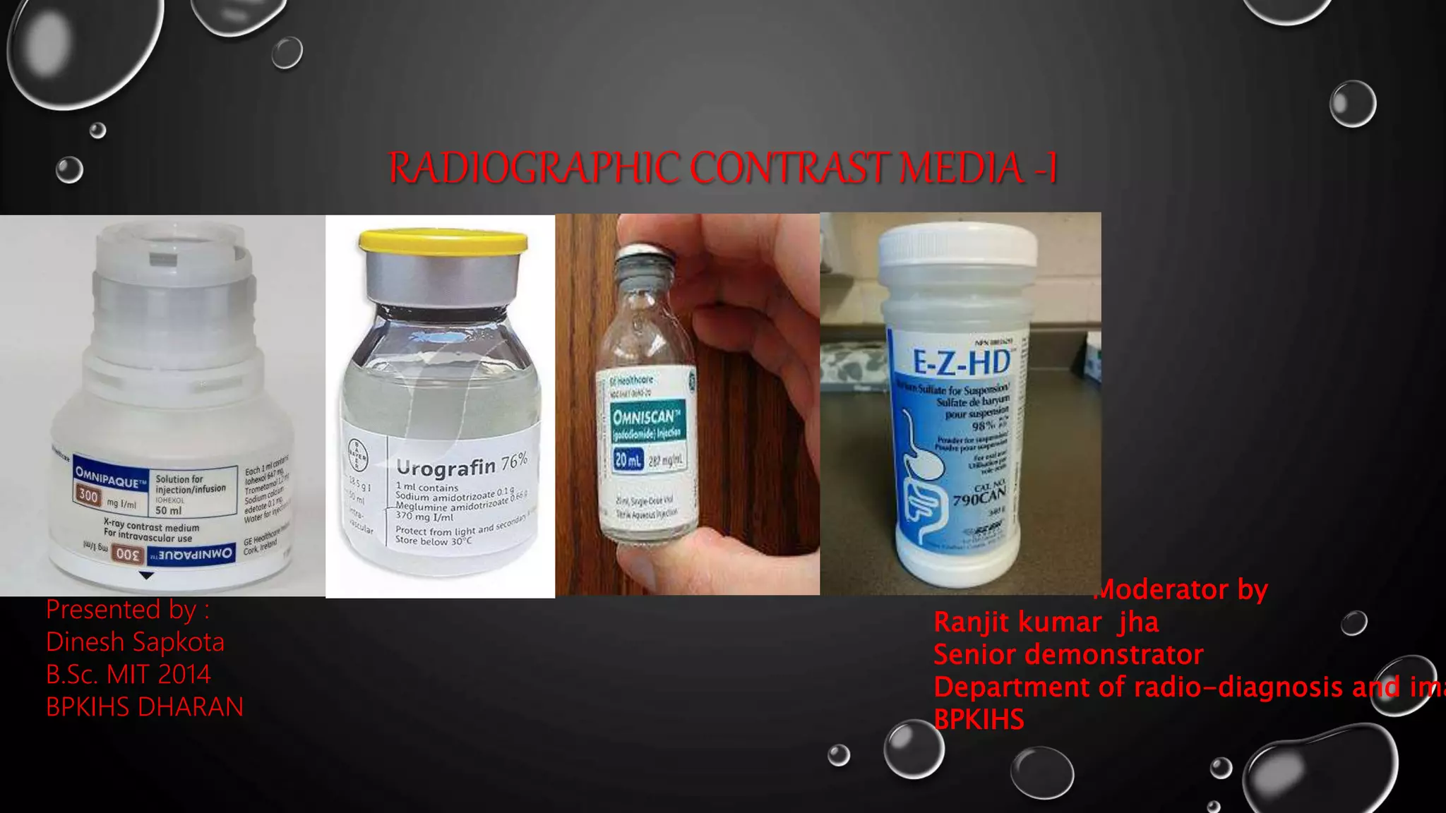
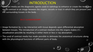
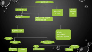
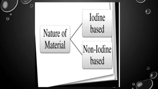
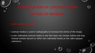
Presented by :
Dinesh Sapkota
B.Sc. MIT 2014
BPKIHS DHARAN
RADIOGRAPHIC CONTRAST MEDIA -I
Moderator by
Ranjit kumar jha
Senior demonstrator
Department of radio-diagnosis and ima
BPKIHS
INTRODUCTION
• Contrast media are the diagnostic agents used in radiology to enhance or create the necessary
visual contrast in an image between the organs, vessels or tract in which they are present and
the surrounding tissues in the body .
IN CT & RADIOGRAPH
• Image formation by x-ray interaction with tissue depends upon differential absorption
between tissues. The introduction of a contrast medium into the organs makes it’s
visualization possible by resulting in either more or less x-ray absorption.
• The used of contrast media has made possible to delineate the anatomical structures along
with the physiological functions of different parts of body.
PROPERTIES OF AN
IDEAL CONTRAST
MEDIA
• Should provide maximum opacity to x-
ray.
• The k edge of contrast media should
be closed to the mean energy of the
diagnostic x-ray .*
• Should have rapid eliminated .
• Its should be non-toxic or non-
poisons.
• It must be safe both locally where as
administer.
• Viscosity , easily available and low
• CONTRAST MEDIA IS NEEDED BECAUSE:
 soft tissues has a low absorption/
interaction
• ABSORPTION DEPEND UPON :
thickness, density & atomic number
• CONTRAST MEDIA AVAILABLE ARE :
 gas, iodine & barium
VISCOSITY
• Viscosity described the
thickness or resistance to flow
of a contrast agents
• The thickness of the contrast
media is related to the
concentration and size of
molecules
• Viscosity affects the rate of
injection
OSMOLALITY
• The osmolality of a solution is
the measurements of the
number of molecules and
particles in a solution per kg
of water.
HISTORY OF CONTRAST MEDIA
• IN 1920S, THE FIRST RADIOGRAPHIC CONTRAST MEDIUM INTRODUCED, SODIUM
IODIDE.
• FIRST MAJOR BREAKTHROUGH - IODINE WAS BOUND TO ORGANIC MOLECULES
[ UROSELECTAN (IOPAX), UROSELECTAN-B (NEOIOPAX) ].
• 1960S, MAJORITY OF WATER SOLUBLE CONTRAST MEDIA WERE SALTS OF IODINATED
FULLY SUBSTITUTED BENZOIC ACID DERIVATIVES [TRIIODINATED BENZOIC ACID].
• 1970S, INTRODUCTION OF LOW OSMOLAR CONTRAST MEDIA (LOCM) - [ IOHEXOL,
IOVERSOL,IOPAMIDOL AND IOBITRIDOL ].
• 1980S AND 1990S, ONGOING DEVELOPMENT OF NON-IONIC ISOTONIC DIMERS.
CONTRAST MEDIA FOR X-RAY
AND CTPOSITIVE CONTRAST
• CONTRAST MATERIAL IS RADIOPAQUE.
• HIGH ATOMIC NUMBER MATERIAL
• WHITE ON FILM
• EXAMPLE:
• 1) BARIUM SULFATE USE: GI STUDIES.
• 2) IODINE COMPOUNDS. USE:
• ANGIOGRAPHY,
• INTRAVENOUS AND RETROGRADE UROGRAPHY
• HYSTEROSALPHINGOGRAPHY
• SIALOGRAPHY
• MYELOGRAPHY
• CHOLANGIOGRAPHY
NEGATIVE CONTRAST
• CONTRAST MATERIAL
THAT IS NOT
• RADIOPAQUE
• LOW ATOMIC NUMBER
MATERIAL
• BLACK ON FILM
• EXAMPLE:
• 1) WATER, AIR AND
CARBON DIOXIDE
X-ray & CT
MRI Gd-
DTPA
USG
Echovi
st
Monom
er Dimer
Non-ionicIonic
NON-
IODINATED
Barium, others
IODINATED
POSITIVE
MEDIA NEGATIVE MEDIA
AIR,CO2,O2
Contrast
Monomer Dimer
1stge
2nd Newe
st
CLASSIFICATION OF CONTRAST MEDIA
NATURE OF MATERIAL
1. NON-IODINATED BASED :
• contrast media is used in radiography to increase the clarity of the image.
• a non-iodinated contrast media is one that does not contain iodine and may
instead contain barium or other non-iodinated media as the radio opaque
substance.
BARIUM SULPHATE
• Barium suspension is made up of pure barium
sulphate (BASO4 ).
• 0.1-3micrometer particles size
• Atomic number: 56
• PH: 5.3 which makes it stable in gastric acid
• Non absorbable and non toxic
• Barium is an ideal radiographic contrast, as its k-
edge of 37 kev approximately, is closed to mean
energy of x-ray used in diagnostic radiography.
BARIUM SULPHATE
DIS. ADVANTAGE
• Subsequent abdominal CT &
USG rendered difficult to
interpret
• High morbidity associated
with Ba in peritoneal cavity
ADVANTAGE
• Excellent mucosal coating
which allowed the
demonstration of normal &
abnormal mucosal patterns
• Lower cost
BARIUM SULPHATE
CONTRAINDICATION
• In case of suspected perforation it may
caused peritonitis
• Suspected perforation
• Suspected fistula
• Suspected partial or complete stenosis
• Paralytic ileus
• Haemorrhage in the gastrointestinal tract
• Toxic megacolon
• Prior to surgery or endoscopy
• If the patient has had a recent
gastrointestinal wide bore biopsy (usually
within 3–5 days) or a recent anastomosis
COMPLICATION
• Barium may lead to barium
peritonitis
• Ba if aspirated may lead to
pneumonitis and granuloma
formation
• Ba if intra vasation may lead
to pulmonary embolus
• Conversion of partial
obstruction into complete
bowel obstruction
DILUTION OF BARIUM SULPHATE
WEIGHT BY WEIGHT
• E.g. 30%w/w suspension is to
weight 30g of barium
sulphate and add 70 g of
water to if for a total wt. of
100g
• 1gmof water = 1ml of water
WEIGHT BY VOLUME
• E.g. 80% w/v suspension is to
weight 80gm barium sulphate
and to add enough water to
make the total volume up to
100ml suspension
IODINATED BASED
• Iodine is the basic component of all the currently used intravascular contrast
media.
• Atomic no.: 53
• Atomic weight: 150
• Total iodine content in the body is 50mg
• It’s preferred because:
a. High contrast density due to high atomic number
b. Allows firm binding to highly variable benzene ring
c. Low toxicity
d. Rapidly execrated 90% by glomerular filtration with in 12 hours
• It’s not suitable for MRI
• Higher atomic no: and k-shell electron binding energy of 34kev which is lower
than but closed to mean energy used in diagnostic x-rays and thus maximizing
the photo-electric effect.( 63–77 kvp is the optimal range)
CHEMICAL STRUCTURE OF IODINATED
CONTRAST AGENTS
OIL SOLUBLE CONTRAST AGENTS
PROPERTIES
• made from fatty acids
• iodine added to Easter groups
• insoluble in water
• long persistence in body
• infrequently used except for
specific exam
• can not be used with plastic
syringes
• high viscosity
• high surface tension
• slow absorption & execration
• chances of embolism
 It was first introduced in myelography by
sicard as first generation contrast media in
1921 then it was used for Bronchography ,
pyelography & lymphography
 It is rarely used now days in some special
cases like COPD & pulmonary hypertension.
EXAMPLES
• MYODIL IS AN OILY ORGANIC IODINE CONTAINING CONTRAST MATERIAL
WHICH WAS USED FOR MYELOGRAPHY ,
• RESIDUAL CONTRAST REMAINS FOR YEARS CAUSED BY ARACHNOIDITIS
• ETHIODIZED: USED BY HSG & LYMPHANGIOGRAPHY
• PANTOPAQUE : 1994 , USED IN MYELOGRAM, LOCM , 6-17 ML DOSE
• LIPIODOL: 1925 HEUSER WAS FIRST REPORT ON THE USED OF LIPIODAL IN
HSGS, LOCM, LOW VISCOSITY , LESS TOXIC, BECAME WIDELY ACCEPTED
• DINOSIL: USED IN BRONCHOGRAPHY
• OIL EMBOLISM MAY OCCUR BY ACCIDENTAL INTRAVASCULAR INJECTION
WATER SOLUBLE CONTRAST AGENTS
NON-IONIC CONTRAST
AGENTS
• Non –ionic contrast media do not dissolve into charged particles when it
enters a solution.
• More expensive with high safer then ionic contrast media
• Non-ionic contrast media substitute the sodium & meglumine side chains
with non-ionizing radicals.
• The solubility of non-ionic contrast agents in water is due to the hydrogen
bonds formed between the side chains &water molecules.
IONIC CONTRAST AGENTS
• An ionic contrast agents dissociated into charge particles when it enters a
solution.
• Ionic contrast media break down into cations and anions charge particles.
• Ionic contrast agents has approximately five time the osmolarity of human
plasma(hyperosmolar).
• The solubility of ionic agents in water is due to dissociated of the molecule
in solution into diatrizoate ion & sodium or maglumine ion. These two
particles are generated in the solution.
Molecule
s
Catio
n
+
Anion
-
IONIC CONTRAST AGENTS
IONIC MONOMERS
(HOCM)
• Ionic monomer are derivatives of
benzene ring in which three iodine
atoms are present at position of 2,4 ,6 &
position of 3,5 are substituted by
organic molecules and position 1 contain
a carboxyl group.
• Iodine to particles ratio is 3:2
• Osmolality (1500–2000 mosm/kg h2o
compared with 300 mosm/kg h2o for
plasma)
• examples :
Urografin
Angiograffin
Iothalmic acids
IONIC CONTRAST AGENTS
IONIC DIMERS (LOCM)
• Two tri-iodinated benzoic acid groups are joined
by a linking bridge resulting in a dimeric acid E.g.
hexabrig
• Iodine to particles ratio is 6:2 or 3:1
• Hexabrix = sodium & maglumine salts mixed
(ioxaglate)
• Osmolality (600 mosmol/kg H2O)
NON- IONIC CONTRAST AGENTS
NON-IONIC MONOMERS
(LOCM)
• Ionic agents replacing carboxyl group by a d- glucose group which provides many hydrophilic
hydroxyl group
• Making them more tolerable and safer to use than ionic contrast. For every three iodine
molecules in a non-ionic solution, one neutral molecule is produced.
• Non-ionic contrast media are therefore referred to as 3 : 1 compounds.
• They substitute the sodium and meglumine side chains with non-ionising radicals (OH)N. two
major advantages arise through the change in chemical structure:
• the first is that the negative carboxyl group is eliminated, thereby reducing the neurotoxicity;
• the second is that the elimination of the positive ion reduces osmolality to 600–700 mosm/kg
h2o.
• non-ionic locm is recommended for intrathecal and vascular radiological procedures.
NON-IONIC MONOMERS
EXAMPLES :
OSMOLALITY (470 MOSM/KG H2O)
• Iohexol (omnipaqu)
• Ioversol (optiray)
• Iopromide (ultravist )
• Iopamidol(niopam)
NON-IONIC DIMERS (ISO-
OSMOLAR)
• These agents contains 2 benzene rings and 6 iodine atoms
• To maintain the solubility a larger no. Of hydrophilic
groups are replaced around the molecules
• High viscosity & low diffusibility of these agents
• Osmolality (300mosm/kg H2O)
• Ratio: 6:1
• These compounds represent a gold standard water-soluble
iodine contrast medium.
• E.G iotrolan, iodixanol
MRI CONTRAST MEDIA
• The first MR compatible contrast media used was introduced in the year 1981 using ferric
chloride in the GIT.
• Gadolinium compound was first introduced in 1984 which is still being used.
• The contrast agents used in mri are based on the shortening of t1 and t2 relaxation times of
the tissues that lead to hyper-intense and hypo-intense in the mr images respectively.
• The elements used in the mr contrast should have large magnetic moments so that when
inserted in the body will caused fluctuation in the main magnetic field leading to shortening
of t1 & t2 times of the tissues.
TYPES OF MRI CONTRAST AGENTS
POSITIVE CONTRAST
AGENTS
• The contrast that
predominantly affect T1
relaxation is referred to as
positive contrast agents as is
caused increased in signal by
shortening T1 relaxation.
• Hyperintense image is formed
• E.G. Gadolinium chelates
• Dose: 0.1 -0.2mmol/kg body
NEGATIVE CONTRAST
AGENTS
• The contrast that predominantly
affects T2 relaxation is referred
to as negative contrast agents as
is caused signal loss by
shortening T2 relaxation
• Hypo-intense image is formed.
• E.G. Superparamagnetic iron
oxide(spio)
• Dose: 0.56mmol/kg body weight
GADOLINIUM
 Gadolinium is a rare earth metal “heavy
metal”
 Gadolinium is chelated to DTPA
(magnevist)
 By binding DTPA to the gadolinium sites,
only one “free” gadolinium site is
available to attach to water molecules
 Gadolinium chelates are of small
molecular weight
 Diffuse freely & excreted by kidneys
 Typical adult dose = 0.2ml/kg (20ml
max)
GADOLINIUM-CONTAINING CONTRAST
AGENTS APPROVED FOR HUMAN USE
IRON OXIDE: SUPERPARAMAGNETIC
• Two types of iron oxide contrast agents exist:
• Superparamagnetic iron oxide (SPIO)
• And ultra small superparamagnetic iron oxide (USPIO).
• These contrast agents consist of suspended colloids of iron oxide
• Nanoparticles and when injected during imaging reduce the T2/
• T2* signals of absorbing tissues.
• Spio and uspio contrast agents have been used successfully in
• Some instances for liver tumor enhancement
INDICATION SIDE EFFECT OF MRI CONTRAST
• Gadolinium containing contrast agents usually
have no effect On blood chemistries and
hematologic studies except transient Elevation
of serum iron and bilirubin levels.
Risks:-
systemic nephrogenic fibrosis contraindicated
when gfr<30ml/min
• CNS
• Demyelinating disease
• More accurate delineation of
tumour margins from oedema
• Discrimination of tumour
recurrence from post up fibrosis
• Cardiac and aortic imaging
ULTRASOUND CONTRAST MEDIA
• Contrast-enhanced ultrasound (CEUS) involves the administration of intravenous
contrast agents containing microbubbles of perfluorocarbon or nitrogen gas.
• Ultrasound contrast agents rely on the different ways in which sound waves are
reflected from interfaces between substances.
• This may be the surface of a small air bubble or a more complex structure.
• Commercially available contrast media are gas-filled microbubbles that are
administered intravenously to the systemic circulation .
• Microbubbles have a high degree of echogenicity (the ability of an object to reflect
ultrasound waves). There is a great difference in echogenicity between the gas in the
microbubbles and the soft tissue surroundings of the body.
MICROBUBBLE
• Microbubble shell material determines how easily the microbubble is taken
up by the immune system.
• The material for microbubble determines its time in circulation and
elasticity.
• Microbubble shells are composed of albumin, galactose, lipid, or polymers .
• Microbubble gas core is the most important part because it determines the
echogenicity.
• Size of microbubble is around 1 - 4 μm. (Upto7mm)
• The microbubble is nearly around the size of RBCs as it should not cross
the vascular endothelium.
Fourth
generation
• Diagnostic and
therapeutic
• Navigate to the region
of interest with the
help of adhesive
ligands
IDEAL ULTRASOUND
CONTRAST AGENT
1) be injectable by a peripheral vein
2) be non toxic
3) small enough to pass through pulmonary,
cardiac & capillary systems
4) stable enough to undergo the shear forces,
hydrostatic pressure changes
& Diameter changes
5) half life should be sufficient to allow
complete examination
6) should require little preparation
APPLICATIONS
1) Evaluating normal, increased or decreased
vascularity.
2) detecting vascular stenosis & occlusions.
3) improving neoplasm detection.
4) analysing & characterizing tumour neo vascularity.
5) differentiating normal variants such as renal
column of bertin from Neoplasm.
6) echocardiography – cardiac cavities, valves,
coronary artery & myocardia Viability
 contrast medium

contrast medium

  • 1.
    Presented by : DineshSapkota B.Sc. MIT 2014 BPKIHS DHARAN RADIOGRAPHIC CONTRAST MEDIA -I Moderator by Ranjit kumar jha Senior demonstrator Department of radio-diagnosis and ima BPKIHS
  • 2.
    INTRODUCTION • Contrast mediaare the diagnostic agents used in radiology to enhance or create the necessary visual contrast in an image between the organs, vessels or tract in which they are present and the surrounding tissues in the body . IN CT & RADIOGRAPH • Image formation by x-ray interaction with tissue depends upon differential absorption between tissues. The introduction of a contrast medium into the organs makes it’s visualization possible by resulting in either more or less x-ray absorption. • The used of contrast media has made possible to delineate the anatomical structures along with the physiological functions of different parts of body.
  • 3.
    PROPERTIES OF AN IDEALCONTRAST MEDIA • Should provide maximum opacity to x- ray. • The k edge of contrast media should be closed to the mean energy of the diagnostic x-ray .* • Should have rapid eliminated . • Its should be non-toxic or non- poisons. • It must be safe both locally where as administer. • Viscosity , easily available and low • CONTRAST MEDIA IS NEEDED BECAUSE:  soft tissues has a low absorption/ interaction • ABSORPTION DEPEND UPON : thickness, density & atomic number • CONTRAST MEDIA AVAILABLE ARE :  gas, iodine & barium
  • 4.
    VISCOSITY • Viscosity describedthe thickness or resistance to flow of a contrast agents • The thickness of the contrast media is related to the concentration and size of molecules • Viscosity affects the rate of injection OSMOLALITY • The osmolality of a solution is the measurements of the number of molecules and particles in a solution per kg of water.
  • 5.
    HISTORY OF CONTRASTMEDIA • IN 1920S, THE FIRST RADIOGRAPHIC CONTRAST MEDIUM INTRODUCED, SODIUM IODIDE. • FIRST MAJOR BREAKTHROUGH - IODINE WAS BOUND TO ORGANIC MOLECULES [ UROSELECTAN (IOPAX), UROSELECTAN-B (NEOIOPAX) ]. • 1960S, MAJORITY OF WATER SOLUBLE CONTRAST MEDIA WERE SALTS OF IODINATED FULLY SUBSTITUTED BENZOIC ACID DERIVATIVES [TRIIODINATED BENZOIC ACID]. • 1970S, INTRODUCTION OF LOW OSMOLAR CONTRAST MEDIA (LOCM) - [ IOHEXOL, IOVERSOL,IOPAMIDOL AND IOBITRIDOL ]. • 1980S AND 1990S, ONGOING DEVELOPMENT OF NON-IONIC ISOTONIC DIMERS.
  • 6.
    CONTRAST MEDIA FORX-RAY AND CTPOSITIVE CONTRAST • CONTRAST MATERIAL IS RADIOPAQUE. • HIGH ATOMIC NUMBER MATERIAL • WHITE ON FILM • EXAMPLE: • 1) BARIUM SULFATE USE: GI STUDIES. • 2) IODINE COMPOUNDS. USE: • ANGIOGRAPHY, • INTRAVENOUS AND RETROGRADE UROGRAPHY • HYSTEROSALPHINGOGRAPHY • SIALOGRAPHY • MYELOGRAPHY • CHOLANGIOGRAPHY NEGATIVE CONTRAST • CONTRAST MATERIAL THAT IS NOT • RADIOPAQUE • LOW ATOMIC NUMBER MATERIAL • BLACK ON FILM • EXAMPLE: • 1) WATER, AIR AND CARBON DIOXIDE
  • 7.
    X-ray & CT MRIGd- DTPA USG Echovi st Monom er Dimer Non-ionicIonic NON- IODINATED Barium, others IODINATED POSITIVE MEDIA NEGATIVE MEDIA AIR,CO2,O2 Contrast Monomer Dimer 1stge 2nd Newe st
  • 9.
    CLASSIFICATION OF CONTRASTMEDIA NATURE OF MATERIAL 1. NON-IODINATED BASED : • contrast media is used in radiography to increase the clarity of the image. • a non-iodinated contrast media is one that does not contain iodine and may instead contain barium or other non-iodinated media as the radio opaque substance.
  • 10.
    BARIUM SULPHATE • Bariumsuspension is made up of pure barium sulphate (BASO4 ). • 0.1-3micrometer particles size • Atomic number: 56 • PH: 5.3 which makes it stable in gastric acid • Non absorbable and non toxic • Barium is an ideal radiographic contrast, as its k- edge of 37 kev approximately, is closed to mean energy of x-ray used in diagnostic radiography.
  • 11.
    BARIUM SULPHATE DIS. ADVANTAGE •Subsequent abdominal CT & USG rendered difficult to interpret • High morbidity associated with Ba in peritoneal cavity ADVANTAGE • Excellent mucosal coating which allowed the demonstration of normal & abnormal mucosal patterns • Lower cost
  • 12.
    BARIUM SULPHATE CONTRAINDICATION • Incase of suspected perforation it may caused peritonitis • Suspected perforation • Suspected fistula • Suspected partial or complete stenosis • Paralytic ileus • Haemorrhage in the gastrointestinal tract • Toxic megacolon • Prior to surgery or endoscopy • If the patient has had a recent gastrointestinal wide bore biopsy (usually within 3–5 days) or a recent anastomosis COMPLICATION • Barium may lead to barium peritonitis • Ba if aspirated may lead to pneumonitis and granuloma formation • Ba if intra vasation may lead to pulmonary embolus • Conversion of partial obstruction into complete bowel obstruction
  • 13.
    DILUTION OF BARIUMSULPHATE WEIGHT BY WEIGHT • E.g. 30%w/w suspension is to weight 30g of barium sulphate and add 70 g of water to if for a total wt. of 100g • 1gmof water = 1ml of water WEIGHT BY VOLUME • E.g. 80% w/v suspension is to weight 80gm barium sulphate and to add enough water to make the total volume up to 100ml suspension
  • 14.
    IODINATED BASED • Iodineis the basic component of all the currently used intravascular contrast media. • Atomic no.: 53 • Atomic weight: 150 • Total iodine content in the body is 50mg • It’s preferred because: a. High contrast density due to high atomic number b. Allows firm binding to highly variable benzene ring c. Low toxicity d. Rapidly execrated 90% by glomerular filtration with in 12 hours • It’s not suitable for MRI • Higher atomic no: and k-shell electron binding energy of 34kev which is lower than but closed to mean energy used in diagnostic x-rays and thus maximizing the photo-electric effect.( 63–77 kvp is the optimal range)
  • 15.
    CHEMICAL STRUCTURE OFIODINATED CONTRAST AGENTS
  • 17.
    OIL SOLUBLE CONTRASTAGENTS PROPERTIES • made from fatty acids • iodine added to Easter groups • insoluble in water • long persistence in body • infrequently used except for specific exam • can not be used with plastic syringes • high viscosity • high surface tension • slow absorption & execration • chances of embolism  It was first introduced in myelography by sicard as first generation contrast media in 1921 then it was used for Bronchography , pyelography & lymphography  It is rarely used now days in some special cases like COPD & pulmonary hypertension.
  • 18.
    EXAMPLES • MYODIL ISAN OILY ORGANIC IODINE CONTAINING CONTRAST MATERIAL WHICH WAS USED FOR MYELOGRAPHY , • RESIDUAL CONTRAST REMAINS FOR YEARS CAUSED BY ARACHNOIDITIS • ETHIODIZED: USED BY HSG & LYMPHANGIOGRAPHY • PANTOPAQUE : 1994 , USED IN MYELOGRAM, LOCM , 6-17 ML DOSE • LIPIODOL: 1925 HEUSER WAS FIRST REPORT ON THE USED OF LIPIODAL IN HSGS, LOCM, LOW VISCOSITY , LESS TOXIC, BECAME WIDELY ACCEPTED • DINOSIL: USED IN BRONCHOGRAPHY • OIL EMBOLISM MAY OCCUR BY ACCIDENTAL INTRAVASCULAR INJECTION
  • 19.
    WATER SOLUBLE CONTRASTAGENTS NON-IONIC CONTRAST AGENTS • Non –ionic contrast media do not dissolve into charged particles when it enters a solution. • More expensive with high safer then ionic contrast media • Non-ionic contrast media substitute the sodium & meglumine side chains with non-ionizing radicals. • The solubility of non-ionic contrast agents in water is due to the hydrogen bonds formed between the side chains &water molecules.
  • 20.
    IONIC CONTRAST AGENTS •An ionic contrast agents dissociated into charge particles when it enters a solution. • Ionic contrast media break down into cations and anions charge particles. • Ionic contrast agents has approximately five time the osmolarity of human plasma(hyperosmolar). • The solubility of ionic agents in water is due to dissociated of the molecule in solution into diatrizoate ion & sodium or maglumine ion. These two particles are generated in the solution. Molecule s Catio n + Anion -
  • 21.
    IONIC CONTRAST AGENTS IONICMONOMERS (HOCM) • Ionic monomer are derivatives of benzene ring in which three iodine atoms are present at position of 2,4 ,6 & position of 3,5 are substituted by organic molecules and position 1 contain a carboxyl group. • Iodine to particles ratio is 3:2 • Osmolality (1500–2000 mosm/kg h2o compared with 300 mosm/kg h2o for plasma) • examples : Urografin Angiograffin Iothalmic acids
  • 22.
    IONIC CONTRAST AGENTS IONICDIMERS (LOCM) • Two tri-iodinated benzoic acid groups are joined by a linking bridge resulting in a dimeric acid E.g. hexabrig • Iodine to particles ratio is 6:2 or 3:1 • Hexabrix = sodium & maglumine salts mixed (ioxaglate) • Osmolality (600 mosmol/kg H2O)
  • 23.
    NON- IONIC CONTRASTAGENTS NON-IONIC MONOMERS (LOCM) • Ionic agents replacing carboxyl group by a d- glucose group which provides many hydrophilic hydroxyl group • Making them more tolerable and safer to use than ionic contrast. For every three iodine molecules in a non-ionic solution, one neutral molecule is produced. • Non-ionic contrast media are therefore referred to as 3 : 1 compounds. • They substitute the sodium and meglumine side chains with non-ionising radicals (OH)N. two major advantages arise through the change in chemical structure: • the first is that the negative carboxyl group is eliminated, thereby reducing the neurotoxicity; • the second is that the elimination of the positive ion reduces osmolality to 600–700 mosm/kg h2o. • non-ionic locm is recommended for intrathecal and vascular radiological procedures.
  • 24.
    NON-IONIC MONOMERS EXAMPLES : OSMOLALITY(470 MOSM/KG H2O) • Iohexol (omnipaqu) • Ioversol (optiray) • Iopromide (ultravist ) • Iopamidol(niopam)
  • 25.
    NON-IONIC DIMERS (ISO- OSMOLAR) •These agents contains 2 benzene rings and 6 iodine atoms • To maintain the solubility a larger no. Of hydrophilic groups are replaced around the molecules • High viscosity & low diffusibility of these agents • Osmolality (300mosm/kg H2O) • Ratio: 6:1 • These compounds represent a gold standard water-soluble iodine contrast medium. • E.G iotrolan, iodixanol
  • 26.
    MRI CONTRAST MEDIA •The first MR compatible contrast media used was introduced in the year 1981 using ferric chloride in the GIT. • Gadolinium compound was first introduced in 1984 which is still being used. • The contrast agents used in mri are based on the shortening of t1 and t2 relaxation times of the tissues that lead to hyper-intense and hypo-intense in the mr images respectively. • The elements used in the mr contrast should have large magnetic moments so that when inserted in the body will caused fluctuation in the main magnetic field leading to shortening of t1 & t2 times of the tissues.
  • 27.
    TYPES OF MRICONTRAST AGENTS POSITIVE CONTRAST AGENTS • The contrast that predominantly affect T1 relaxation is referred to as positive contrast agents as is caused increased in signal by shortening T1 relaxation. • Hyperintense image is formed • E.G. Gadolinium chelates • Dose: 0.1 -0.2mmol/kg body NEGATIVE CONTRAST AGENTS • The contrast that predominantly affects T2 relaxation is referred to as negative contrast agents as is caused signal loss by shortening T2 relaxation • Hypo-intense image is formed. • E.G. Superparamagnetic iron oxide(spio) • Dose: 0.56mmol/kg body weight
  • 28.
    GADOLINIUM  Gadolinium isa rare earth metal “heavy metal”  Gadolinium is chelated to DTPA (magnevist)  By binding DTPA to the gadolinium sites, only one “free” gadolinium site is available to attach to water molecules  Gadolinium chelates are of small molecular weight  Diffuse freely & excreted by kidneys  Typical adult dose = 0.2ml/kg (20ml max)
  • 29.
  • 30.
    IRON OXIDE: SUPERPARAMAGNETIC •Two types of iron oxide contrast agents exist: • Superparamagnetic iron oxide (SPIO) • And ultra small superparamagnetic iron oxide (USPIO). • These contrast agents consist of suspended colloids of iron oxide • Nanoparticles and when injected during imaging reduce the T2/ • T2* signals of absorbing tissues. • Spio and uspio contrast agents have been used successfully in • Some instances for liver tumor enhancement
  • 31.
    INDICATION SIDE EFFECTOF MRI CONTRAST • Gadolinium containing contrast agents usually have no effect On blood chemistries and hematologic studies except transient Elevation of serum iron and bilirubin levels. Risks:- systemic nephrogenic fibrosis contraindicated when gfr<30ml/min • CNS • Demyelinating disease • More accurate delineation of tumour margins from oedema • Discrimination of tumour recurrence from post up fibrosis • Cardiac and aortic imaging
  • 32.
    ULTRASOUND CONTRAST MEDIA •Contrast-enhanced ultrasound (CEUS) involves the administration of intravenous contrast agents containing microbubbles of perfluorocarbon or nitrogen gas. • Ultrasound contrast agents rely on the different ways in which sound waves are reflected from interfaces between substances. • This may be the surface of a small air bubble or a more complex structure. • Commercially available contrast media are gas-filled microbubbles that are administered intravenously to the systemic circulation . • Microbubbles have a high degree of echogenicity (the ability of an object to reflect ultrasound waves). There is a great difference in echogenicity between the gas in the microbubbles and the soft tissue surroundings of the body.
  • 33.
    MICROBUBBLE • Microbubble shellmaterial determines how easily the microbubble is taken up by the immune system. • The material for microbubble determines its time in circulation and elasticity. • Microbubble shells are composed of albumin, galactose, lipid, or polymers . • Microbubble gas core is the most important part because it determines the echogenicity. • Size of microbubble is around 1 - 4 μm. (Upto7mm) • The microbubble is nearly around the size of RBCs as it should not cross the vascular endothelium.
  • 35.
    Fourth generation • Diagnostic and therapeutic •Navigate to the region of interest with the help of adhesive ligands
  • 36.
    IDEAL ULTRASOUND CONTRAST AGENT 1)be injectable by a peripheral vein 2) be non toxic 3) small enough to pass through pulmonary, cardiac & capillary systems 4) stable enough to undergo the shear forces, hydrostatic pressure changes & Diameter changes 5) half life should be sufficient to allow complete examination 6) should require little preparation APPLICATIONS 1) Evaluating normal, increased or decreased vascularity. 2) detecting vascular stenosis & occlusions. 3) improving neoplasm detection. 4) analysing & characterizing tumour neo vascularity. 5) differentiating normal variants such as renal column of bertin from Neoplasm. 6) echocardiography – cardiac cavities, valves, coronary artery & myocardia Viability