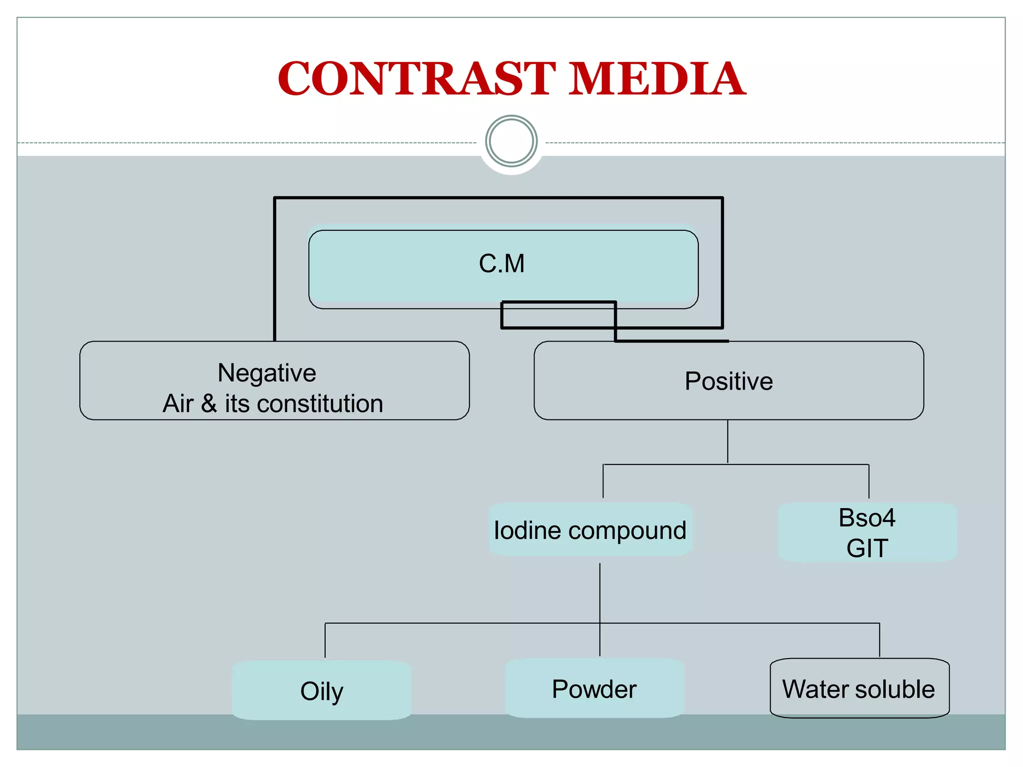
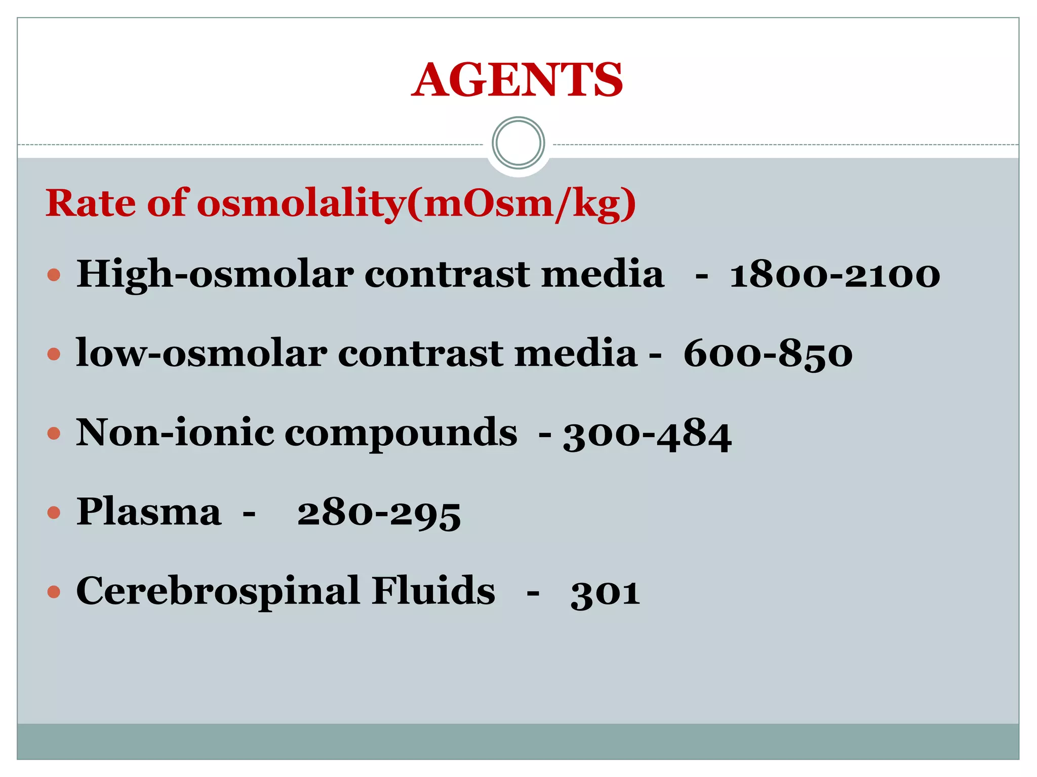
M A A J I D M O H I U D D I N M A L I K
L E C T U R E R C O P M S A D E S H U N I V E R S I T Y
B A T H I N D A P U N J A B
CONTRAST MEDIA
INTRODUCTION
In the field of radiology, some investigation or
procedure on a patient in need of contrast into the
patient’s body through a vein, artery, mouth or
anus, which is located in the body. Since the
invention of the era of the 1950s, iodine contrast
media (ICM) is apparently more secure have been
used extensively, almost daily use and is a widely
used drug in medical history.
CONTINUE
Various forms and types of contrast media was
used for the development of medical imaging.
Most current diagnostic imaging contrast agents
depends on this. Such as pharmaceutical drugs
most others, this contrast agent was almost no
negative effects even with a good safety record and
proven medicine/drug is safe for use.
CONTINUE
If seen on x-ray film, there are usually pictures of
the anatomy such as bone, and there also appears
a white colored gas that surrounds around the
organs. The real aim is the use of contrast media to
increase the density of the organ is usually not
visible on radiographs, such as passage of urine or
blood vessels, which originally did not appear at
regular chest x-ray contrast media are not used.
CONTINUE
After application of contrast media, the
resulting image will be white showing a hidden
organ. Contrast media is composed of two
types of ionic and non ionic contrast media is
divided into two categories
 Positive contrast media
 Negative contrast media
CONTRAST MEDIA
It is a chemical substance of very high or very
low atomic number or weight, there for it
increase or decrease the density of the organ
under examination.
CONTRAST MEDIA
C.M
Negative
Air & its constitution
Positive
Iodine compound Bso4
GIT
Oily Powder Water soluble
POSITIVE CONTRAST MEDIA
Positive contrast agents are agents that have
the atomic mass and a high rate of obstruction
(attenuation) high. So these agents are indeed
radiopaque material. Positive contrast media
are as iodine, bromine, and barium.
CONTINUE
Typically, these materials will be incorporated into the
body (according to appropriate procedures) and
contrast agents will meet the target organ. Effects
resulting radiopaque would explain the situation and
structure of the organ / tissue targeted at radiographic
images. Generally, a positive contrast agent is the key
to ensuring that the patient has allergies or not for the
selection of the contrast media can be tailored to the
patient to avoid the effects of anaphylactic shock.
NEGATIVE CONTRAST MEDIA
One of x-ray absorption is the weight of the atomic
mass of a substance. If the atomic mass is low, the
effect of obstacles (attenuate) is low and the
reverse occurs when the atomic mass increases.
When the mass of the atom is high, it will produce
an effect of density / contrast radiography of the
image because the image will become more
permeable and dark. This feature is useful when
an anatomical structure is shown with a
background of very permeable and the large
difference in contrast between the image and the
background (without producing blur the
boundaries between image and background).
CONTINUE
Contrast media such as air (oxygen, carbon
dioxide, etc.) are good examples as negative
contrast agents. Negative contrast agents often
used in conjunction with contrast agents and
one common procedures often incorporate the
use of these agents were negative double
barium enema.
NEGATIVE CONTRAST AGENTS
Carbon dioxide:
Produced in the gastrointestinal tract (GIT) with
barium sulfate to demonstrate mucosal pattern.
Double contrast barium meal for example. Carbon
dioxide can also be produced by the colon when
given double contrast media, as it reduces pain
and reducing stomach discomfort immediately.
CONTINUE
Oxygen:
Produced in the body cavities such as in the knee
when examining arteriogram to demonstrate the
knee joint.
Air:
Produced by the patient during the radiographic
examination, for example, took a deep breath and
hold during chest examination.
BASIC IODINE CONTRAST MEDIA
Several investigations in the radiology needs of this
contrast into the patient’s body through a artery or a
vein. A frequent example is the intravenous urogram.
Usually only the bones and the wind will be visible on
x-ray images. To distinguish the path of blood in the
urine or blood vessels, contrast media containing
iodine to be added to increase the intensity of blood or
urine. Then, this will cause the structure to look ‘white’
in films such as the bones of patients in x-ray images.
CLASSIFICATION OF CONTRAST MEDIA
There are two parts: the ionic and non ionic.
Ionic compound is divided into two parts; high
level of osmolar contrast media(hocm), and low
level of osmolar contrast media(locm). This
classification is based on the iodine concentration
of particles (generally as elements of contrast
media is frequently used in the procedure IVU)
absorbed / dissolved into the blood.
CONTINUE
If the rate of absorption of iodine is too high, it
can slow the blood flow into the central
nervous system (hyperosmolality). This
situation is a major factor many negative
reactions to patients due to contrast media
that are not appropriate.
AGENTS
Rate of osmolality(mOsm/kg)
 High-osmolar contrast media - 1800-2100
 low-osmolar contrast media - 600-850
 Non-ionic compounds - 300-484
 Plasma - 280-295
 Cerebrospinal Fluids - 301
IONIC:
Content molecules tightly to the anion and cation
(positive and negative) in the blood causing high
osmolality – toxic / reaction high. Properties of
molecules containing benzene rings, for example –
salt compound meglumine diatrizoate and
diatrizoate sodium (Renografin 60 and Hypaque
76 – osmolality high)
NON-IONIC:
Molecules are not resolved in the fluid / blood.
For example, a compound of sodium
diatrizoate (iopamidol, isovue, Iohexol /
omnipaque)
MEASURES OF CARE
 Can not exceed the dose.
 Stay away from the sun.
 Stored at room temperature.
CONTRAST MEDIA NEEDED FOR RADIOLOGICAL
EXAMINATION
To distinguish the path of blood in the urine or
blood vessel, usually just the bones and the air
will be visible on x-ray and to improve the
density of urine or blood.
ADVANTAGES OF CONTRAST MEDIA
 Easily dissolved.
 Inert
 Provide better mucosal detail
 There is no osmotic effect, so the radio
density is long-lasting.
 Cheap
 Relatively delicious
DISADVANTAGES OF CONTRAST MEDIA
 Aspiration pneumonia if inhaled.
 If got leakage into the cavity or organs of the
body. It can’t survive indefinitely and may
cause granulomatous reactions.
RADIOGRAPHY CONTRAST MEDIA.pdf

RADIOGRAPHY CONTRAST MEDIA.pdf

  • 1.
    M A AJ I D M O H I U D D I N M A L I K L E C T U R E R C O P M S A D E S H U N I V E R S I T Y B A T H I N D A P U N J A B CONTRAST MEDIA
  • 2.
    INTRODUCTION In the fieldof radiology, some investigation or procedure on a patient in need of contrast into the patient’s body through a vein, artery, mouth or anus, which is located in the body. Since the invention of the era of the 1950s, iodine contrast media (ICM) is apparently more secure have been used extensively, almost daily use and is a widely used drug in medical history.
  • 3.
    CONTINUE Various forms andtypes of contrast media was used for the development of medical imaging. Most current diagnostic imaging contrast agents depends on this. Such as pharmaceutical drugs most others, this contrast agent was almost no negative effects even with a good safety record and proven medicine/drug is safe for use.
  • 4.
    CONTINUE If seen onx-ray film, there are usually pictures of the anatomy such as bone, and there also appears a white colored gas that surrounds around the organs. The real aim is the use of contrast media to increase the density of the organ is usually not visible on radiographs, such as passage of urine or blood vessels, which originally did not appear at regular chest x-ray contrast media are not used.
  • 5.
    CONTINUE After application ofcontrast media, the resulting image will be white showing a hidden organ. Contrast media is composed of two types of ionic and non ionic contrast media is divided into two categories  Positive contrast media  Negative contrast media
  • 6.
    CONTRAST MEDIA It isa chemical substance of very high or very low atomic number or weight, there for it increase or decrease the density of the organ under examination.
  • 7.
    CONTRAST MEDIA C.M Negative Air &its constitution Positive Iodine compound Bso4 GIT Oily Powder Water soluble
  • 8.
    POSITIVE CONTRAST MEDIA Positivecontrast agents are agents that have the atomic mass and a high rate of obstruction (attenuation) high. So these agents are indeed radiopaque material. Positive contrast media are as iodine, bromine, and barium.
  • 9.
    CONTINUE Typically, these materialswill be incorporated into the body (according to appropriate procedures) and contrast agents will meet the target organ. Effects resulting radiopaque would explain the situation and structure of the organ / tissue targeted at radiographic images. Generally, a positive contrast agent is the key to ensuring that the patient has allergies or not for the selection of the contrast media can be tailored to the patient to avoid the effects of anaphylactic shock.
  • 10.
    NEGATIVE CONTRAST MEDIA Oneof x-ray absorption is the weight of the atomic mass of a substance. If the atomic mass is low, the effect of obstacles (attenuate) is low and the reverse occurs when the atomic mass increases. When the mass of the atom is high, it will produce an effect of density / contrast radiography of the image because the image will become more permeable and dark. This feature is useful when an anatomical structure is shown with a background of very permeable and the large difference in contrast between the image and the background (without producing blur the boundaries between image and background).
  • 11.
    CONTINUE Contrast media suchas air (oxygen, carbon dioxide, etc.) are good examples as negative contrast agents. Negative contrast agents often used in conjunction with contrast agents and one common procedures often incorporate the use of these agents were negative double barium enema.
  • 12.
    NEGATIVE CONTRAST AGENTS Carbondioxide: Produced in the gastrointestinal tract (GIT) with barium sulfate to demonstrate mucosal pattern. Double contrast barium meal for example. Carbon dioxide can also be produced by the colon when given double contrast media, as it reduces pain and reducing stomach discomfort immediately.
  • 13.
    CONTINUE Oxygen: Produced in thebody cavities such as in the knee when examining arteriogram to demonstrate the knee joint. Air: Produced by the patient during the radiographic examination, for example, took a deep breath and hold during chest examination.
  • 14.
    BASIC IODINE CONTRASTMEDIA Several investigations in the radiology needs of this contrast into the patient’s body through a artery or a vein. A frequent example is the intravenous urogram. Usually only the bones and the wind will be visible on x-ray images. To distinguish the path of blood in the urine or blood vessels, contrast media containing iodine to be added to increase the intensity of blood or urine. Then, this will cause the structure to look ‘white’ in films such as the bones of patients in x-ray images.
  • 15.
    CLASSIFICATION OF CONTRASTMEDIA There are two parts: the ionic and non ionic. Ionic compound is divided into two parts; high level of osmolar contrast media(hocm), and low level of osmolar contrast media(locm). This classification is based on the iodine concentration of particles (generally as elements of contrast media is frequently used in the procedure IVU) absorbed / dissolved into the blood.
  • 16.
    CONTINUE If the rateof absorption of iodine is too high, it can slow the blood flow into the central nervous system (hyperosmolality). This situation is a major factor many negative reactions to patients due to contrast media that are not appropriate.
  • 17.
    AGENTS Rate of osmolality(mOsm/kg) High-osmolar contrast media - 1800-2100  low-osmolar contrast media - 600-850  Non-ionic compounds - 300-484  Plasma - 280-295  Cerebrospinal Fluids - 301
  • 18.
    IONIC: Content molecules tightlyto the anion and cation (positive and negative) in the blood causing high osmolality – toxic / reaction high. Properties of molecules containing benzene rings, for example – salt compound meglumine diatrizoate and diatrizoate sodium (Renografin 60 and Hypaque 76 – osmolality high)
  • 19.
    NON-IONIC: Molecules are notresolved in the fluid / blood. For example, a compound of sodium diatrizoate (iopamidol, isovue, Iohexol / omnipaque)
  • 20.
    MEASURES OF CARE Can not exceed the dose.  Stay away from the sun.  Stored at room temperature.
  • 21.
    CONTRAST MEDIA NEEDEDFOR RADIOLOGICAL EXAMINATION To distinguish the path of blood in the urine or blood vessel, usually just the bones and the air will be visible on x-ray and to improve the density of urine or blood.
  • 22.
    ADVANTAGES OF CONTRASTMEDIA  Easily dissolved.  Inert  Provide better mucosal detail  There is no osmotic effect, so the radio density is long-lasting.  Cheap  Relatively delicious
  • 23.
    DISADVANTAGES OF CONTRASTMEDIA  Aspiration pneumonia if inhaled.  If got leakage into the cavity or organs of the body. It can’t survive indefinitely and may cause granulomatous reactions.