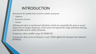
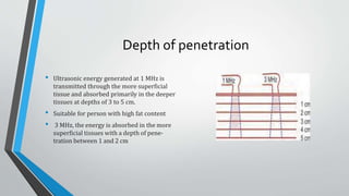
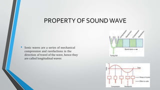
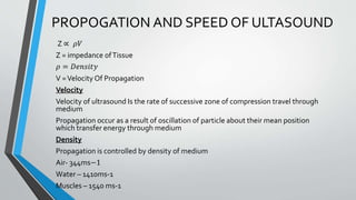
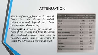
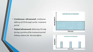
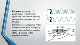
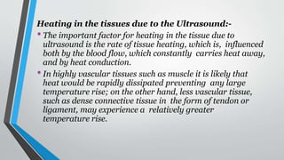
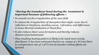
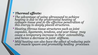
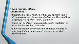
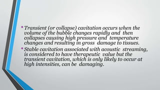
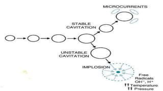
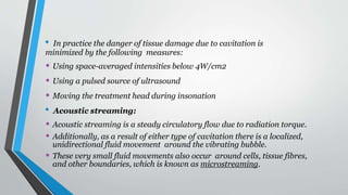
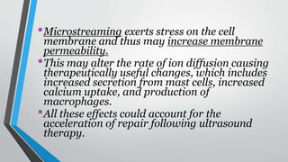
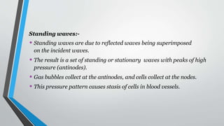
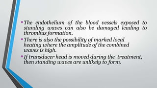
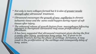
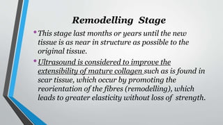
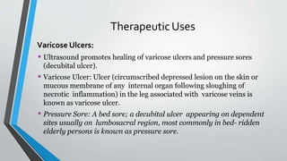
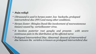
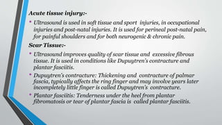
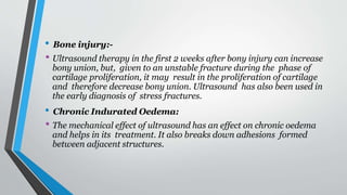
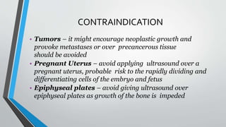
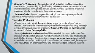
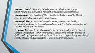
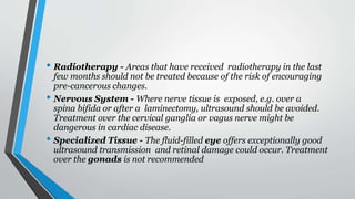
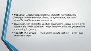
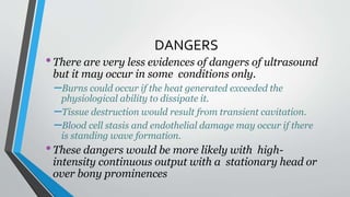
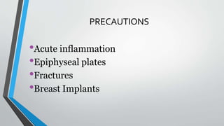
ULTRASOUNDTHERAPY
INTRODUCTION
Ultrasound is the modality that is used for a number of purposes
• diagnosis,
• destruction of tissues
• therapy
Ultrasound refers to mechanical vibrations which are essentially the same as sound
waves but of a higher frequency. Such waves are beyond the range of human hearing
and therefore also be called ultrasonic.
Frequency- above audible range 20-20000 HZ
A generator that can be set between 1 and 3 MHz affords the therapist the treatment
flexibility.
Depth of penetration
• Ultrasonic energy generated at 1 MHz is
transmitted through the more superficial
tissue and absorbed primarily in the deeper
tissues at depths of 3 to 5 cm.
• Suitable for person with high fat content
• 3 MHz, the energy is absorbed in the more
superficial tissues with a depth of pene-
tration between 1 and 2 cm
Medical frequency of Ultrasound
• Physiotherapy Equipment -0.75 MHZ , 1 MHZ , 1 MHZ, 1.5 MHZ and 3.3 MHZ
• Diagnostic – 1 MHZ-10 MHZ
• Surgical Eq1uipment- 1 MHZ-5 MHz
PROPERTY OF SOUND WAVE
• Sonic waves are a series of mechanical
compression and rarefactions in the
direction of travel of the wave, hence they
are called longitudinal waves
PROPOGATION AND SPEED OF ULTASOUND
Z ∝ 𝜌𝑉
Z = impedance ofTissue
𝜌 = 𝐷𝑒𝑛𝑠𝑖𝑡𝑦
V =Velocity Of Propagation
Velocity
Velocity of ultrasound Is the rate of successive zone of compression travel through
medium
Propagation occur as a result of oscillation of particle about their mean position
which transfer energy through medium
Density
Propagation is controlled by density of medium
Air- 344ms−1
Water – 1410ms-1
Muscles – 1540 ms-1
PRODUCTION OF ULTRASOUND
For 1 MHz machine a vibrating source with a frequency of one million cycles per second is needed
. This is achieved by using
• a quartz crystal
• barium titanate crystal
• lead zirconate crystal
• nickel-cobalt ferrite crystal
These crystals deform when subjected to a varying potential difference, this is called piezoelectric
effect
PRODUCTION OF ULTRASOUND
• Power supply on
• Energy to oscillator circuit
• Produces oscillating voltage
• Cystal inside transducer embedded between
the link electrode and metal front plate
respond to oscillating voltage which fed
through coaxial cable
• Crystal expand and contract at same frequency
at which the current changes polarity
• Metal front plate of the treatment head moves
backward and forward generating a stream of
compression waves that form sonic beam
PRODUCTION OF ULTRASOUND
• Intensity controller controls the amplitude of
alternating voltafge thereby controls
amplitude of sonic waves
• Intensity is expressed in watts per square
centimeter (W/cm2 )
Mark space ratio
Mark-space ratio, or mark-to-space ratio, is
another term for the same concept, to
describe the temporal relationship between
two alternating periods of a waveform
Mark space ratio=
Both PW is duration of alternating period
𝑃𝑊𝑂𝑁
𝑃𝑊 𝑂𝐹𝐹
TRANSMISSION OF ULTRASOUND
EFFECT OF BOUNDRIES BERTEEN MEDIUM
Sonic waves involve vibratory motion of molecules so that there is a
characteristic velocity of wave progression for each particular medium.
• Some of the energy is reflected back. The amount of the energy
reflected is proportional to the difference in acoustic impedance
between the two media.
–Water / Glass – 63% of energy is reflected
–Water / Soft tissue – 0.2% of energy is reflected
• Refraction also occurs with sonic waves due to the difference in
acoustic impedance.
• The beam of sonic energy that passes through the second medium
does not continue in a straight line but changes direction at the
boundary because of the different velocities in the two media.
• If the acoustic impedances are closely matched little refraction will
occur.
Absorption of Sonic waves
• Kinetic energy is converted to heat energy as it passes through the
material.
• The energy will decrease exponentially with distance from the source
because a fixed proportion of it is absorbed at each unit distance so
that the remaining amount will become a smaller and smaller
percentage of the initial energy
• The conversion of sonic energy to heat is due to increased molecular
motion
• Half value depth: depth of tissue at which the US intensity is half
its initial intensity
Absorption of sonic energy is greatest in tissues with largest
amounts of structural protein and lowest water content.
• Blood – least protein content and least absorption
• Bone - greatest protein content and greatest absorption
ATTENUATION
• The loss of energy from the ultrasound
beam in the tissues is called
attenuation and depends on both
absorption and scattering
• Absorption accounts for some 60 –
80% of the energy lost from the beam.
The scattered energy may also be
absorbed other than in the region to
which the ultrasound beam is applied.
Scattering is caused by reflections and refractions, which occur at
interfaces throughout the tissues. This is particularly apparent where
there is a large difference in acoustic impedance.
• Spatial average intensity:
Average intensity of the US output over
the area of the transducer
• Spatial peak intensity: Peak intensity
of the ultrasound output over the area of
the transducer. The intensity is usually
great in the centre of the beam and lowest
at the edges of the beam.
• Beam non-uniformity ratio
(BNR): Ratio between peak
intensity and average intensity in
the beam. The lower the BNR the
more uniform the beam
• With BNR 5:1, when the spatial
average intensity is 1W/cm2
,the
spatial peak intensity would be
5W/cm2
• Continuous ultrasound: continuous
delivery of US through out the treatment
period
• Pulsed ultrasound: delivering US only
during a portion of the treatment period.
Pulsing reduces the thermal effects
• Duty cycle: proportion of the total
treatment time that the US is on. This can
be expressed in percentage or a ration
• 20% or 1:5 duty cycle, is on for 20% of
the time and off for the 80% of time.
• Spatial average temporal peak
intensity: spatial average intensity of the
US during the on time
• Clinically US displays SATP intensity and
duty cycle
• Spatial average temporal average
intensity: The spatial average intensity
of the US averaged over both the on time
and the off time
• SATP X duty cycle = SATA
• SATA is frequently used in research and
non clinical literatures
• Frequency: number of
compression- rarefraction
cycles per unit of time, usually
expressed in cycles per second
(Hertz)
• Increasing the frequency of US
causes a decrease in its depth of
penetration and concentration
of the US energy in the
superficial tissues.
• Effective
radiating area
(ERA): The area of
the transducer from
which the US energy
radiates. Since the
crystal doesn’t
vibrate uniformly ,
the ERA is always
smaller than the area
of the treatment
head.
• Some waves cancel out, others reinforce so that the net result is a very
irregular pattern of the sonic waves in the region close to the
transducer face, called the near field or Fresnel zone.
• Beyond this, the far field or Fraunhofer zone, the sonic field
spreads out somewhat and becomes much more regular because of
the differing path lengths from points on the transducer.
• The length of the near field depends directly on the square
of the radius of the transducer face and inversely
proportional to the wavelength of the sonic waves.
• Length of Fresnel zone = r2/ λ
• For practical purposes therapeutic ultrasound utilizes the
near field. The relatively more energy on average is
carried in the central part of the cross-section of the beam.
• The irregularity of the near field can be ‘ironed out’ to some
extent by continuous movement of the treatment head
during the therapy.
•Shear waves can be formed which transmit energy
along the periosteal surface at right angles to the
ultrasound beam.
•Due to the fact that this reflection is quite large
(almost 25%) and that sonic energy is absorbed
almost immediately in bone, there is marked
heating at the bone surface.
•This is considered to account for the periosteal pain
that can arise with excessive doses of therapeutic
ultrasound.
Heating in the tissues due to the Ultrasound:-
•The important factor for heating in the tissue due to
ultrasound is the rate of tissue heating, which is, influenced
both by the blood flow, which constantly carries heat away,
and by heat conduction.
•In highly vascular tissues such as muscle it is likely that
heat would be rapidly dissipated preventing any large
temperature rise; on the other hand, less vascular tissue,
such as dense connective tissue in the form of tendon or
ligament, may experience a relatively greater
temperature rise.
•Moving the transducer head during the treatment is
important because of following effects :-
• To smooth out the irregularities of the near field
• It reduces the irregularities of absorption that might occur due to
reflection at interfaces, standing waves, refraction, and differences
in tissue thermal conduction or blood flow
• It also reduces shear wave formation and thereby reduces
chances of periosteal pain
• Thus resulting heating pattern is likely to be much more evenly
distributed. It has been estimated that for an output of 1 W/cm2 there
is a temperature rise of 0.8°C/min if vascular cooling effects are
ignored
•The effect is not the same because with pulsed treatment
there is time for heat to be dissipated by conduction in the
tissues and in the circulating blood. Therefore, higher
intensities can be safely used in a pulsed treatment because
the average heating is reduced.
•Ultrasound application can increase rates of ion diffusion
across cell membranes; this could be due to increased
particle movement on either side of the membrane and
possibly, increased motion of the phospholipids and
proteins that form the membrane.
• Physical & Physiological effects:
•As oscillation or sonic energy is passed through the body
tissue, it causes transfer of heat energy in the body tissues.
If this energy is not dissipated by normal physiological
response, then there is local rise in temperature, which
accounts for thermal effects.
•If heat dissipation equals heat generation there is no net
rise in temperature and any effects are said to be non-
thermal.
•Using low intensities or pulsing the output achieves
non-thermal effects.
• Thermal effects:
•The advantage of using ultrasound to achieve
heating is due to the preferential heating of
collagen tissue and to the effective penetration of
this energy to deeply placed structures.
•Heating fibrous tissue structures such as joint
capsules, ligaments, tendons, and scar tissue may
cause a temporary increase in their extensibility,
and hence a decrease in joint stiffness.
•Mild heating can also have the effect of reducing pain
and muscle spasm and promoting healing processes.
• Non thermal effects:-
Cavitation:
•Cavitation is the formation of tiny gas bubbles in the
tissues as a result of ultrasound vibration. These bubbles,
generally of a micron (10-6m) diameter.
•These can be of two types, namely stable cavitation or
transient(non-stable) cavitation.
•Stable cavitation occurs when the bubbles oscillate to
and fro within the ultrasound pressure waves but
remain intact.
•Transient (or collapse) cavitation occurs when the
volume of the bubble changes rapidly and then
collapses causing high pressure and temperature
changes and resulting in gross damage to tissues.
•Stable cavitation associated with acoustic streaming,
is considered to have therapeutic value but the
transient cavitation, which is only likely to occur at
high intensities, can be damaging.
• In practice the danger of tissue damage due to cavitation is
minimized by the following measures:
• Using space-averaged intensities below 4W/cm2
• Using a pulsed source of ultrasound
• Moving the treatment head during insonation
• Acoustic streaming:
• Acoustic streaming is a steady circulatory flow due to radiation torque.
• Additionally, as a result of either type of cavitation there is a localized,
unidirectional fluid movement around the vibrating bubble.
• These very small fluid movements also occur around cells, tissue fibres,
and other boundaries, which is known as microstreaming.
•Microstreaming exerts stress on the cell
membrane and thus may increase membrane
permeability.
•This may alter the rate of ion diffusion causing
therapeutically useful changes, which includes
increased secretion from mast cells, increased
calcium uptake, and production of
macrophages.
•All these effects could account for the
acceleration of repair following ultrasound
therapy.
Standing waves:-
• Standing waves are due to reflected waves being superimposed
on the incident waves.
• The result is a set of standing or stationary waves with peaks of high
pressure (antinodes).
• Gas bubbles collect at the antinodes, and cells collect at the nodes.
• This pressure pattern causes stasis of cells in blood vessels.
•The endothelium of the blood vessels exposed to
standing waves can also be damaged leading to
thrombus formation.
•There is also the possibility of marked local
heating where the amplitude of the combined
waves is high.
•If transducer head is moved during the treatment,
then standing waves are unlikely to form.
Micromassage:-
•The micromassage effect of ultrasound occurs at a cellular
level where the cells are alternately compressed and then
pulled further apart.
•The waves of compression and rarefaction may produce a
form of micromassage, which could reduce oedema.
•Ultrasound has been found to be effective at reducing
recent traumatic oedema and chronic indurated oedema.
Acute stage:-
•Stable cavitation and acoustic streaming increases calcium
ion diffusion across the cell membrane, which works as a
cellular ‘secondary messenger’, and thereby increases the
production and release of wound-healing factors.
•These include the release of histamine from mast cells and
growth factors released from macrophages.
•In this way, ultrasound has the potential to accelerate
normal resolution of inflammation providing that the
inflammatory stimulus is removed.
• This acceleration could also be due to the gentle
agitation of the tissue fluid, which may increase the
rate of phagocytosis and movement of particles and
cells.
•Thus, ultrasound has a pro-inflammatory, not an
anti-inflammatory action.
Proliferative (Granulation) stage:-
• This begins approximately 3 days after injury and is the stage at
which the connective tissue framework is laid down by fibroblasts
for the new blood vessels.
• During repair, fibroblasts may be stimulated to produce more
collagen; ultrasound can promote collagen synthesis by increasing
cell membrane permeability, which allows the entry of calcium ions,
which control cellular activity.
• Not only is more collagen formed but it is also of greater tensile
strength after ultrasound treatment.
• Ultrasound encourages the growth of new capillaries in chronic
ischaemic tissue and the same could happen during repair of soft
tissues after injury.
• The enhanced release of growth factors from macrophages following
exposure to therapeutic ultrasound may cause proliferation of
fibroblasts.
• It has been suggested that ultrasound treatment given during the first
2 weeks after injury accelerates bony union, but, if given to an
unstable fracture during the phase of cartilage formation, it may
result in the proliferation of the cartilage and consequently delay of
bony union.
Remodelling Stage
•This stage last months or years until the new
tissue is as near in structure as possible to the
original tissue.
•Ultrasound is considered to improve the
extensibility of mature collagen such as is found in
scar tissue, which occur by promoting the
reorientation of the fibres (remodelling), which
leads to greater elasticity without loss of strength.
TherapeuticUses
Varicose Ulcers:
• Ultrasound promotes healing of varicose ulcers and pressure sores
(decubital ulcer).
• Varicose Ulcer: Ulcer (circumscribed depressed lesion on the skin or
mucous membrane of any internal organ following sloughing of
necrotic inflammation) in the leg associated with varicose veins is
known as varicose ulcer.
• Pressure Sore: A bed sore; a decubital ulcer appearing on dependent
sites usually on lumbosacral region, most commonly in bed- ridden
elderly persons is known as pressure sore.
• Pain relief:
• Ultrasound is used in herpes zoster, low backache, prolapsed
intervertebral disc (PIV) and many other conditions.
• Herpes Zoster: Shingles (band-like involvement of neurocutaneous
tissues) caused by varicellazoster virus.
• It involves posterior root ganglia and presents with severe
continuous pain in the distribution of the affected nerve
• Prolapsed Intervertebral Disc: Abnormal descent of intervertebral
disc between the vertebra is known as prolapsed intervertebral disc
Acute tissue injury:-
• Ultrasound is used in soft tissue and sport injuries, in occupational
injuries and post-natal injuries. It is used for perineal post-natal pain,
for painful shoulders and for both neurogenic & chronic pain.
Scar Tissue:-
• Ultrasound improves quality of scar tissue and excessive fibrous
tissue. It is used in conditions like Dupuytren’s contracture and
plantar fasciitis.
• Dupuytren’s contracture: Thickening and contracture of palmar
fascia, typically affects the ring finger and may involve years later
incompletely little finger is called Dupuytren’s contracture.
• Plantar fasciitis: Tenderness under the heel from plantar
fibromatosis or tear of plantar fascia is called plantar fasciitis.
• Bone injury:-
• Ultrasound therapy in the first 2 weeks after bony injury can increase
bony union, but, given to an unstable fracture during the phase of
cartilage proliferation, it may result in the proliferation of cartilage
and therefore decrease bony union. Ultrasound has also been used in
the early diagnosis of stress fractures.
• Chronic Indurated Oedema:
• The mechanical effect of ultrasound has an effect on chronic oedema
and helps in its treatment. It also breaks down adhesions formed
between adjacent structures.
CONTRAINDICATION
• Tumors – it might encourage neoplastic growth and
provoke metastases or over precancerous tissue
should be avoided
• Pregnant Uterus – avoid applying ultrasound over a
pregnant uterus, probable risk to the rapidly dividing and
differentiating cells of the embryo and fetus
• Epiphyseal plates – avoid giving ultrasound over
epiphyseal plates as growth of the bone is impeded
• Spread of Infection - Bacterial or viral infection could be spread by
ultrasound, presumably by facilitating microorganism movement across
membranes and through the tissues. The low-grade infections of venous
ulcers, or similar, would seem to be safe to treat.
• Tuberculosis - Due to the possible risk of reactivating encapsulated
lesions tuberculous regions should not be treated.
Vascular Problems-
• Circumstances in which hemorrhage might provoke should not be
treated. For example, where bleeding is still occurring or has only recently
been controlled, such as an enlarging haemarthrosis or haematoma
or uncontrollable haemophilia.
• Severely ischaemic tissues should be avoided because of the poor heat
transfer and possible greater risk of arterial thrombosis due to stasis and
endothelial damage. Treatment over recent venous thrombosis might
extend the thrombus or disrupt its attachment to the vein wall forming an
embolus. Areas of atherosclerosis are best avoided for the same reason
• Haemarthrosis: Bleeding into the joint usually from an injury,
which results in a swelling of the joint, is known as haemarthrosis.
• Haematoma: A collection of blood inside the body, caused by bleeding
from an injured vessel is called haematoma.
• Haemophilia: An inherited coagulation defect characterized by a
permanent tendency to hemorrhages due to a defect in the coagulation of
blood is known as haemophilia.
• Atherosclerosis: A condition caused by intramural deposition of Low
Density Lipoprotein (LDL), secondary to exposure of smooth muscles to
lipid, resulting in platelet induced smooth muscle proliferation, formation of
fibrotic plaques and calcification is known as atherosclerosis
• Radiotherapy - Areas that have received radiotherapy in the last
few months should not be treated because of the risk of encouraging
pre-cancerous changes.
• Nervous System - Where nerve tissue is exposed, e.g. over a
spina bifida or after a laminectomy, ultrasound should be avoided.
Treatment over the cervical ganglia or vagus nerve might be
dangerous in cardiac disease.
• Specialized Tissue - The fluid-filled eye offers exceptionally good
ultrasound transmission and retinal damage could occur. Treatment
over the gonads is not recommended
• Implants - Smaller and superficial implants, like metal bone-
fixing pins subcutaneously placed; as a precaution, low doses
should be used in these circumstances.
• Treatment over implanted cardiac pacemakers should not be given
because the sonic vibration may interfere with the pacemaker’s
stimulating frequency
• Anaesthetic areas - High doses should not be given over
anaesthetic areas.
DANGERS
•There are very less evidences of dangers of ultrasound
but it may occur in some conditions only.
–Burns could occur if the heat generated exceeded the
physiological ability to dissipate it.
–Tissue destruction would result from transient cavitation.
–Blood cell stasis and endothelial damage may occur if there
is standing wave formation.
•These dangers would be more likely with high-
intensity continuous output with a stationary head or
over bony prominences
PRECAUTIONS
•Acute inflammation
•Epiphyseal plates
•Fractures
•Breast Implants

ultrsound.pptx

  • 1.
  • 2.
    INTRODUCTION Ultrasound is themodality that is used for a number of purposes • diagnosis, • destruction of tissues • therapy Ultrasound refers to mechanical vibrations which are essentially the same as sound waves but of a higher frequency. Such waves are beyond the range of human hearing and therefore also be called ultrasonic. Frequency- above audible range 20-20000 HZ A generator that can be set between 1 and 3 MHz affords the therapist the treatment flexibility.
  • 3.
    Depth of penetration •Ultrasonic energy generated at 1 MHz is transmitted through the more superficial tissue and absorbed primarily in the deeper tissues at depths of 3 to 5 cm. • Suitable for person with high fat content • 3 MHz, the energy is absorbed in the more superficial tissues with a depth of pene- tration between 1 and 2 cm
  • 4.
    Medical frequency ofUltrasound • Physiotherapy Equipment -0.75 MHZ , 1 MHZ , 1 MHZ, 1.5 MHZ and 3.3 MHZ • Diagnostic – 1 MHZ-10 MHZ • Surgical Eq1uipment- 1 MHZ-5 MHz
  • 5.
    PROPERTY OF SOUNDWAVE • Sonic waves are a series of mechanical compression and rarefactions in the direction of travel of the wave, hence they are called longitudinal waves
  • 6.
    PROPOGATION AND SPEEDOF ULTASOUND Z ∝ 𝜌𝑉 Z = impedance ofTissue 𝜌 = 𝐷𝑒𝑛𝑠𝑖𝑡𝑦 V =Velocity Of Propagation Velocity Velocity of ultrasound Is the rate of successive zone of compression travel through medium Propagation occur as a result of oscillation of particle about their mean position which transfer energy through medium Density Propagation is controlled by density of medium Air- 344ms−1 Water – 1410ms-1 Muscles – 1540 ms-1
  • 7.
    PRODUCTION OF ULTRASOUND For1 MHz machine a vibrating source with a frequency of one million cycles per second is needed . This is achieved by using • a quartz crystal • barium titanate crystal • lead zirconate crystal • nickel-cobalt ferrite crystal These crystals deform when subjected to a varying potential difference, this is called piezoelectric effect
  • 8.
    PRODUCTION OF ULTRASOUND •Power supply on • Energy to oscillator circuit • Produces oscillating voltage • Cystal inside transducer embedded between the link electrode and metal front plate respond to oscillating voltage which fed through coaxial cable • Crystal expand and contract at same frequency at which the current changes polarity • Metal front plate of the treatment head moves backward and forward generating a stream of compression waves that form sonic beam
  • 9.
    PRODUCTION OF ULTRASOUND •Intensity controller controls the amplitude of alternating voltafge thereby controls amplitude of sonic waves • Intensity is expressed in watts per square centimeter (W/cm2 ) Mark space ratio Mark-space ratio, or mark-to-space ratio, is another term for the same concept, to describe the temporal relationship between two alternating periods of a waveform Mark space ratio= Both PW is duration of alternating period 𝑃𝑊𝑂𝑁 𝑃𝑊 𝑂𝐹𝐹
  • 10.
    TRANSMISSION OF ULTRASOUND EFFECTOF BOUNDRIES BERTEEN MEDIUM Sonic waves involve vibratory motion of molecules so that there is a characteristic velocity of wave progression for each particular medium. • Some of the energy is reflected back. The amount of the energy reflected is proportional to the difference in acoustic impedance between the two media. –Water / Glass – 63% of energy is reflected –Water / Soft tissue – 0.2% of energy is reflected
  • 11.
    • Refraction alsooccurs with sonic waves due to the difference in acoustic impedance. • The beam of sonic energy that passes through the second medium does not continue in a straight line but changes direction at the boundary because of the different velocities in the two media. • If the acoustic impedances are closely matched little refraction will occur.
  • 12.
    Absorption of Sonicwaves • Kinetic energy is converted to heat energy as it passes through the material. • The energy will decrease exponentially with distance from the source because a fixed proportion of it is absorbed at each unit distance so that the remaining amount will become a smaller and smaller percentage of the initial energy • The conversion of sonic energy to heat is due to increased molecular motion
  • 13.
    • Half valuedepth: depth of tissue at which the US intensity is half its initial intensity Absorption of sonic energy is greatest in tissues with largest amounts of structural protein and lowest water content. • Blood – least protein content and least absorption • Bone - greatest protein content and greatest absorption
  • 14.
    ATTENUATION • The lossof energy from the ultrasound beam in the tissues is called attenuation and depends on both absorption and scattering • Absorption accounts for some 60 – 80% of the energy lost from the beam. The scattered energy may also be absorbed other than in the region to which the ultrasound beam is applied.
  • 15.
    Scattering is causedby reflections and refractions, which occur at interfaces throughout the tissues. This is particularly apparent where there is a large difference in acoustic impedance.
  • 16.
    • Spatial averageintensity: Average intensity of the US output over the area of the transducer • Spatial peak intensity: Peak intensity of the ultrasound output over the area of the transducer. The intensity is usually great in the centre of the beam and lowest at the edges of the beam.
  • 17.
    • Beam non-uniformityratio (BNR): Ratio between peak intensity and average intensity in the beam. The lower the BNR the more uniform the beam • With BNR 5:1, when the spatial average intensity is 1W/cm2 ,the spatial peak intensity would be 5W/cm2
  • 18.
    • Continuous ultrasound:continuous delivery of US through out the treatment period • Pulsed ultrasound: delivering US only during a portion of the treatment period. Pulsing reduces the thermal effects
  • 19.
    • Duty cycle:proportion of the total treatment time that the US is on. This can be expressed in percentage or a ration • 20% or 1:5 duty cycle, is on for 20% of the time and off for the 80% of time.
  • 20.
    • Spatial averagetemporal peak intensity: spatial average intensity of the US during the on time • Clinically US displays SATP intensity and duty cycle • Spatial average temporal average intensity: The spatial average intensity of the US averaged over both the on time and the off time • SATP X duty cycle = SATA • SATA is frequently used in research and non clinical literatures
  • 21.
    • Frequency: numberof compression- rarefraction cycles per unit of time, usually expressed in cycles per second (Hertz) • Increasing the frequency of US causes a decrease in its depth of penetration and concentration of the US energy in the superficial tissues.
  • 22.
    • Effective radiating area (ERA):The area of the transducer from which the US energy radiates. Since the crystal doesn’t vibrate uniformly , the ERA is always smaller than the area of the treatment head.
  • 23.
    • Some wavescancel out, others reinforce so that the net result is a very irregular pattern of the sonic waves in the region close to the transducer face, called the near field or Fresnel zone. • Beyond this, the far field or Fraunhofer zone, the sonic field spreads out somewhat and becomes much more regular because of the differing path lengths from points on the transducer.
  • 24.
    • The lengthof the near field depends directly on the square of the radius of the transducer face and inversely proportional to the wavelength of the sonic waves. • Length of Fresnel zone = r2/ λ
  • 25.
    • For practicalpurposes therapeutic ultrasound utilizes the near field. The relatively more energy on average is carried in the central part of the cross-section of the beam. • The irregularity of the near field can be ‘ironed out’ to some extent by continuous movement of the treatment head during the therapy.
  • 26.
    •Shear waves canbe formed which transmit energy along the periosteal surface at right angles to the ultrasound beam. •Due to the fact that this reflection is quite large (almost 25%) and that sonic energy is absorbed almost immediately in bone, there is marked heating at the bone surface. •This is considered to account for the periosteal pain that can arise with excessive doses of therapeutic ultrasound.
  • 27.
    Heating in thetissues due to the Ultrasound:- •The important factor for heating in the tissue due to ultrasound is the rate of tissue heating, which is, influenced both by the blood flow, which constantly carries heat away, and by heat conduction. •In highly vascular tissues such as muscle it is likely that heat would be rapidly dissipated preventing any large temperature rise; on the other hand, less vascular tissue, such as dense connective tissue in the form of tendon or ligament, may experience a relatively greater temperature rise.
  • 28.
    •Moving the transducerhead during the treatment is important because of following effects :- • To smooth out the irregularities of the near field • It reduces the irregularities of absorption that might occur due to reflection at interfaces, standing waves, refraction, and differences in tissue thermal conduction or blood flow • It also reduces shear wave formation and thereby reduces chances of periosteal pain • Thus resulting heating pattern is likely to be much more evenly distributed. It has been estimated that for an output of 1 W/cm2 there is a temperature rise of 0.8°C/min if vascular cooling effects are ignored
  • 29.
    •The effect isnot the same because with pulsed treatment there is time for heat to be dissipated by conduction in the tissues and in the circulating blood. Therefore, higher intensities can be safely used in a pulsed treatment because the average heating is reduced. •Ultrasound application can increase rates of ion diffusion across cell membranes; this could be due to increased particle movement on either side of the membrane and possibly, increased motion of the phospholipids and proteins that form the membrane.
  • 30.
    • Physical &Physiological effects: •As oscillation or sonic energy is passed through the body tissue, it causes transfer of heat energy in the body tissues. If this energy is not dissipated by normal physiological response, then there is local rise in temperature, which accounts for thermal effects. •If heat dissipation equals heat generation there is no net rise in temperature and any effects are said to be non- thermal. •Using low intensities or pulsing the output achieves non-thermal effects.
  • 31.
    • Thermal effects: •Theadvantage of using ultrasound to achieve heating is due to the preferential heating of collagen tissue and to the effective penetration of this energy to deeply placed structures. •Heating fibrous tissue structures such as joint capsules, ligaments, tendons, and scar tissue may cause a temporary increase in their extensibility, and hence a decrease in joint stiffness. •Mild heating can also have the effect of reducing pain and muscle spasm and promoting healing processes.
  • 32.
    • Non thermaleffects:- Cavitation: •Cavitation is the formation of tiny gas bubbles in the tissues as a result of ultrasound vibration. These bubbles, generally of a micron (10-6m) diameter. •These can be of two types, namely stable cavitation or transient(non-stable) cavitation. •Stable cavitation occurs when the bubbles oscillate to and fro within the ultrasound pressure waves but remain intact.
  • 33.
    •Transient (or collapse)cavitation occurs when the volume of the bubble changes rapidly and then collapses causing high pressure and temperature changes and resulting in gross damage to tissues. •Stable cavitation associated with acoustic streaming, is considered to have therapeutic value but the transient cavitation, which is only likely to occur at high intensities, can be damaging.
  • 35.
    • In practicethe danger of tissue damage due to cavitation is minimized by the following measures: • Using space-averaged intensities below 4W/cm2 • Using a pulsed source of ultrasound • Moving the treatment head during insonation • Acoustic streaming: • Acoustic streaming is a steady circulatory flow due to radiation torque. • Additionally, as a result of either type of cavitation there is a localized, unidirectional fluid movement around the vibrating bubble. • These very small fluid movements also occur around cells, tissue fibres, and other boundaries, which is known as microstreaming.
  • 36.
    •Microstreaming exerts stresson the cell membrane and thus may increase membrane permeability. •This may alter the rate of ion diffusion causing therapeutically useful changes, which includes increased secretion from mast cells, increased calcium uptake, and production of macrophages. •All these effects could account for the acceleration of repair following ultrasound therapy.
  • 37.
    Standing waves:- • Standingwaves are due to reflected waves being superimposed on the incident waves. • The result is a set of standing or stationary waves with peaks of high pressure (antinodes). • Gas bubbles collect at the antinodes, and cells collect at the nodes. • This pressure pattern causes stasis of cells in blood vessels.
  • 38.
    •The endothelium ofthe blood vessels exposed to standing waves can also be damaged leading to thrombus formation. •There is also the possibility of marked local heating where the amplitude of the combined waves is high. •If transducer head is moved during the treatment, then standing waves are unlikely to form.
  • 39.
    Micromassage:- •The micromassage effectof ultrasound occurs at a cellular level where the cells are alternately compressed and then pulled further apart. •The waves of compression and rarefaction may produce a form of micromassage, which could reduce oedema. •Ultrasound has been found to be effective at reducing recent traumatic oedema and chronic indurated oedema.
  • 40.
    Acute stage:- •Stable cavitationand acoustic streaming increases calcium ion diffusion across the cell membrane, which works as a cellular ‘secondary messenger’, and thereby increases the production and release of wound-healing factors. •These include the release of histamine from mast cells and growth factors released from macrophages. •In this way, ultrasound has the potential to accelerate normal resolution of inflammation providing that the inflammatory stimulus is removed.
  • 41.
    • This accelerationcould also be due to the gentle agitation of the tissue fluid, which may increase the rate of phagocytosis and movement of particles and cells. •Thus, ultrasound has a pro-inflammatory, not an anti-inflammatory action.
  • 42.
    Proliferative (Granulation) stage:- •This begins approximately 3 days after injury and is the stage at which the connective tissue framework is laid down by fibroblasts for the new blood vessels. • During repair, fibroblasts may be stimulated to produce more collagen; ultrasound can promote collagen synthesis by increasing cell membrane permeability, which allows the entry of calcium ions, which control cellular activity.
  • 43.
    • Not onlyis more collagen formed but it is also of greater tensile strength after ultrasound treatment. • Ultrasound encourages the growth of new capillaries in chronic ischaemic tissue and the same could happen during repair of soft tissues after injury. • The enhanced release of growth factors from macrophages following exposure to therapeutic ultrasound may cause proliferation of fibroblasts. • It has been suggested that ultrasound treatment given during the first 2 weeks after injury accelerates bony union, but, if given to an unstable fracture during the phase of cartilage formation, it may result in the proliferation of the cartilage and consequently delay of bony union.
  • 44.
    Remodelling Stage •This stagelast months or years until the new tissue is as near in structure as possible to the original tissue. •Ultrasound is considered to improve the extensibility of mature collagen such as is found in scar tissue, which occur by promoting the reorientation of the fibres (remodelling), which leads to greater elasticity without loss of strength.
  • 45.
    TherapeuticUses Varicose Ulcers: • Ultrasoundpromotes healing of varicose ulcers and pressure sores (decubital ulcer). • Varicose Ulcer: Ulcer (circumscribed depressed lesion on the skin or mucous membrane of any internal organ following sloughing of necrotic inflammation) in the leg associated with varicose veins is known as varicose ulcer. • Pressure Sore: A bed sore; a decubital ulcer appearing on dependent sites usually on lumbosacral region, most commonly in bed- ridden elderly persons is known as pressure sore.
  • 46.
    • Pain relief: •Ultrasound is used in herpes zoster, low backache, prolapsed intervertebral disc (PIV) and many other conditions. • Herpes Zoster: Shingles (band-like involvement of neurocutaneous tissues) caused by varicellazoster virus. • It involves posterior root ganglia and presents with severe continuous pain in the distribution of the affected nerve • Prolapsed Intervertebral Disc: Abnormal descent of intervertebral disc between the vertebra is known as prolapsed intervertebral disc
  • 47.
    Acute tissue injury:- •Ultrasound is used in soft tissue and sport injuries, in occupational injuries and post-natal injuries. It is used for perineal post-natal pain, for painful shoulders and for both neurogenic & chronic pain. Scar Tissue:- • Ultrasound improves quality of scar tissue and excessive fibrous tissue. It is used in conditions like Dupuytren’s contracture and plantar fasciitis. • Dupuytren’s contracture: Thickening and contracture of palmar fascia, typically affects the ring finger and may involve years later incompletely little finger is called Dupuytren’s contracture. • Plantar fasciitis: Tenderness under the heel from plantar fibromatosis or tear of plantar fascia is called plantar fasciitis.
  • 48.
    • Bone injury:- •Ultrasound therapy in the first 2 weeks after bony injury can increase bony union, but, given to an unstable fracture during the phase of cartilage proliferation, it may result in the proliferation of cartilage and therefore decrease bony union. Ultrasound has also been used in the early diagnosis of stress fractures. • Chronic Indurated Oedema: • The mechanical effect of ultrasound has an effect on chronic oedema and helps in its treatment. It also breaks down adhesions formed between adjacent structures.
  • 49.
    CONTRAINDICATION • Tumors –it might encourage neoplastic growth and provoke metastases or over precancerous tissue should be avoided • Pregnant Uterus – avoid applying ultrasound over a pregnant uterus, probable risk to the rapidly dividing and differentiating cells of the embryo and fetus • Epiphyseal plates – avoid giving ultrasound over epiphyseal plates as growth of the bone is impeded
  • 50.
    • Spread ofInfection - Bacterial or viral infection could be spread by ultrasound, presumably by facilitating microorganism movement across membranes and through the tissues. The low-grade infections of venous ulcers, or similar, would seem to be safe to treat. • Tuberculosis - Due to the possible risk of reactivating encapsulated lesions tuberculous regions should not be treated. Vascular Problems- • Circumstances in which hemorrhage might provoke should not be treated. For example, where bleeding is still occurring or has only recently been controlled, such as an enlarging haemarthrosis or haematoma or uncontrollable haemophilia. • Severely ischaemic tissues should be avoided because of the poor heat transfer and possible greater risk of arterial thrombosis due to stasis and endothelial damage. Treatment over recent venous thrombosis might extend the thrombus or disrupt its attachment to the vein wall forming an embolus. Areas of atherosclerosis are best avoided for the same reason
  • 51.
    • Haemarthrosis: Bleedinginto the joint usually from an injury, which results in a swelling of the joint, is known as haemarthrosis. • Haematoma: A collection of blood inside the body, caused by bleeding from an injured vessel is called haematoma. • Haemophilia: An inherited coagulation defect characterized by a permanent tendency to hemorrhages due to a defect in the coagulation of blood is known as haemophilia. • Atherosclerosis: A condition caused by intramural deposition of Low Density Lipoprotein (LDL), secondary to exposure of smooth muscles to lipid, resulting in platelet induced smooth muscle proliferation, formation of fibrotic plaques and calcification is known as atherosclerosis
  • 52.
    • Radiotherapy -Areas that have received radiotherapy in the last few months should not be treated because of the risk of encouraging pre-cancerous changes. • Nervous System - Where nerve tissue is exposed, e.g. over a spina bifida or after a laminectomy, ultrasound should be avoided. Treatment over the cervical ganglia or vagus nerve might be dangerous in cardiac disease. • Specialized Tissue - The fluid-filled eye offers exceptionally good ultrasound transmission and retinal damage could occur. Treatment over the gonads is not recommended
  • 53.
    • Implants -Smaller and superficial implants, like metal bone- fixing pins subcutaneously placed; as a precaution, low doses should be used in these circumstances. • Treatment over implanted cardiac pacemakers should not be given because the sonic vibration may interfere with the pacemaker’s stimulating frequency • Anaesthetic areas - High doses should not be given over anaesthetic areas.
  • 56.
    DANGERS •There are veryless evidences of dangers of ultrasound but it may occur in some conditions only. –Burns could occur if the heat generated exceeded the physiological ability to dissipate it. –Tissue destruction would result from transient cavitation. –Blood cell stasis and endothelial damage may occur if there is standing wave formation. •These dangers would be more likely with high- intensity continuous output with a stationary head or over bony prominences
  • 57.