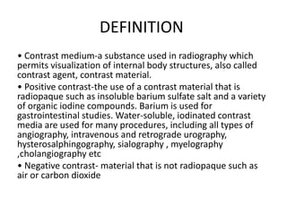
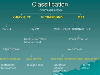
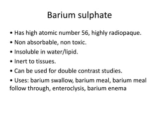
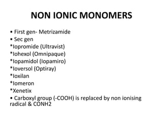
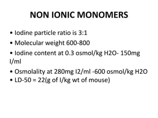
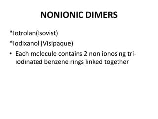
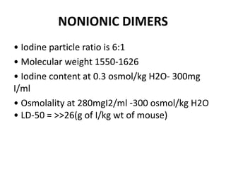
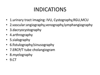
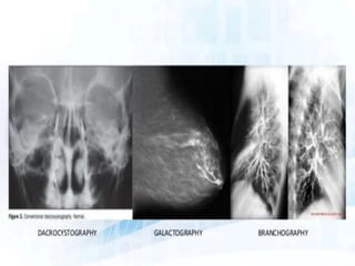
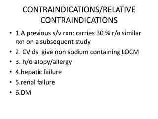
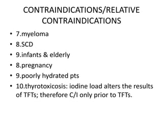
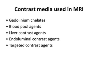
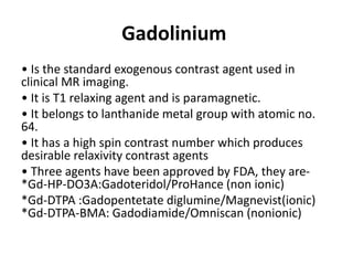
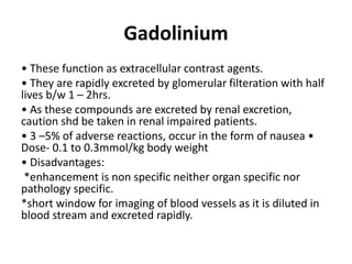
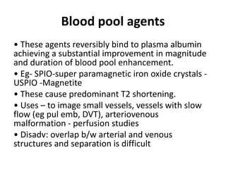
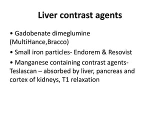
CONTRAST AGENTS
OVERVIEW
• Introduction & Definition
• Classification
• Water soluble iodinated contrast media
• Physiology of contrast media
• Ultrasound contrast media
• MR contrast agents
• Adverse reactions to contrast media
• Contrast induced nephrotoxicity
DEFINITION
• Contrast medium-a substance used in radiography which
permits visualization of internal body structures, also called
contrast agent, contrast material.
• Positive contrast-the use of a contrast material that is
radiopaque such as insoluble barium sulfate salt and a variety
of organic iodine compounds. Barium is used for
gastrointestinal studies. Water-soluble, iodinated contrast
media are used for many procedures, including all types of
angiography, intravenous and retrograde urography,
hysterosalphingography, sialography , myelography
,cholangiography etc
• Negative contrast- material that is not radiopaque such as
air or carbon dioxide
Barium sulphate
• Has high atomic number 56, highly radiopaque.
• Non absorbable, non toxic.
• Insoluble in water/lipid.
• Inert to tissues.
• Can be used for double contrast studies.
• Uses: barium swallow, barium meal, barium meal
follow through, enteroclysis, barium enema
Water soluble iodinated contrast
media
IODINE :
• Atomic number 53 & atomic weight 127
• Radio-opacity is dependent on iodine conc of the solution, so
dependent on number of iodine atoms in each molecule of the
contrast medium.
• Iodine particle ratio: the ratio of number of iodine atoms per
molecule to the number of osmotically active particles per molecule
of solute in solution
• High radio-opacity & low osmolality are required.
• Iodine is preferred because
*High contrast density due to high atomic number
*Allows firm binding to highly variable benzene ring
*Low toxicity
Conventional contrast media / High
osmolar contrast media / Ionic
monomers
• These are salts of sodium or meglumine
cation & triiodinated benzoate anion(C2, C4 &
C6). C3 & C5 are connected to amines which
reduce the toxicity & increase the solubility.
*Diatrizoate (urograffin , angiograffin ,
urovedeo, trazograff)
*Iothalamate(conray , triovideo)
*Ioxithalamate
*Metrizoate
Conventional contrast media / High
osmolar / Ionic monomers
• Iodine particle ratio is 3:2
• Molecular weight 600-800
• Iodine content at 0.3 osmol/kg H2O- 70mg I/ml
• Osmolality at 280mg I2/ml -1500 osmol/kg H2O
• LD-50 = 7(g of I/kg wt of mouse)
• Disadvantage : high osmolality (8 times that of
plasma), responsible for the adverse effects,
because of the non radiopaque cations( Na &
meg)
Low osmolar contrast media
• IONIC DIMERS- Ioxaglate(Hexabrix) Only
compound, mixture of sodium and meglumine
salts
• Two benzene rings (each with 3 iodine atoms)
are linked by a bridge to form a large
compound, carries only one carboxyl group, so
known as monoacid dimers
IONIC DIMERS- Ioxaglate(Hexabrix)
• Iodine particle ratio is 6:2 or 3:1
• Molecular weight is 1269
• Iodine content at 0.3 osmol/kg H2O- 150mg
I/ml
• Osmolality at 280mg I2/ml 560 osmol/kg H2O
• LD-50 = 12(g of I/kg wt of mouse)
NON IONIC MONOMERS
• First gen- Metrizamide
• Sec gen
*Iopromide (Ultravist)
*Iohexol (Omnipaque)
*Iopamidol (Iopamiro)
*Ioversol (Optiray)
*Ioxilan
*Iomeron
*Xenetix
• Carboxyl group (-COOH) is replaced by non ionising
radical & CONH2
NON IONIC MONOMERS
• Iodine particle ratio is 3:1
• Molecular weight 600-800
• Iodine content at 0.3 osmol/kg H2O- 150mg
I/ml
• Osmolality at 280mg I2/ml -600 osmol/kg H2O
• LD-50 = 22(g of I/kg wt of mouse)
NONIONIC DIMERS
*Iotrolan(Isovist)
*Iodixanol (Visipaque)
• Each molecule contains 2 non ionosing tri-
iodinated benzene rings linked together
NONIONIC DIMERS
• Iodine particle ratio is 6:1
• Molecular weight 1550-1626
• Iodine content at 0.3 osmol/kg H2O- 300mg
I/ml
• Osmolality at 280mgI2/ml -300 osmol/kg H2O
• LD-50 = >>26(g of I/kg wt of mouse)
Additives used in contrast media
• Stabilizer – Ca or Na EDTA
• Buffers – stabilizes pH during storage Na acid
phosphates
• Preservatives ( generally not disclosed by the
manufacturers.)
Ideal contrast media should have:
• High water solubility
• Heat & chemical stability(shelf life) ideally- 3 to
5yrs
• Biological inertness( non antigenic)
• Low viscosity
• Low or iso osmolar to plasma
• Selective excretion, like excretion by kidney is
favorable.
• Safety: LD50 (lethal dose) should be high
• Reasonable cost
Physiology
• On intravascular injection , the contrast media
is distributed rapidly by capillary permeability
into extravascular, extra cellular space (except in
CNS).
• They do not enter the interior of blood cells or
tissue cells and they are rapidly excreted, over
90% being eliminated by glomerular filteration
by kidneys within 12 hrs
Imp points to remember
• Contrast media used for myelography are non-ionic CM.
• CM used for cerebral angiography, are CM containing only meglumine
cation.
• CM containing only meglumine cation- conray 280, triovideo 280, trazograff
60% and angiograffin.
• CM which cause max nausea & vomiting are – Ioxaglate (Hexabrix). •
Meglumine salts cause bronchospasm, so CI in bronchial asthma.
• Among newer CM, Iohexol is most hyperosmolar
• Viscosity increases as conc increases & tends to be higher for big sized
molecules (dimers). High viscosity interferes with mixing of contrast media
with plasma & body fluids. Omnipaque240 has least viscosity.
• Meticulous heparinization is required during angiography as incidence of
thrombo embolic phenomenon is high when CM is mixed with blood
INDICATIONS
• 1.urinary tract imaging: IVU, Cystography,RGU,MCU
• 2.vascular:angiography,venography,lymphangiography
• 3.dacryocystography
• 4.arthrography
• 5.sialography
• 6.fistulography/sinusography
• 7.ERCP/T tube cholangiogram
• 8.myelography
• 9.CT
CONTRAINDICATIONS/RELATIVE
CONTRAINDICATIONS
• 1.A previous s/v rxn: carries 30 % r/o similar
rxn on a subsequent study
• 2. CV ds: give non sodium containing LOCM
• 3. h/o atopy/allergy
• 4.hepatic failure
• 5.renal failure
• 6.DM
CONTRAINDICATIONS/RELATIVE
CONTRAINDICATIONS
• 7.myeloma
• 8.SCD
• 9.infants & elderly
• 8.pregnancy
• 9.poorly hydrated pts
• 10.thyrotoxicosis: iodine load alters the results
of TFTs; therefore C/I only prior to TFTs.
Ultrasound contrast agents
• Also called ECHO ENHANCING AGENTS.
• These agents increase the echogenicity of blood, which heightens
the tissue contrast & allows better delineation of body cavities.
• Consist of microscopic gas filled bubbles whose surface reflect sound
waves.
• Their extremely high reflectivity(backscatter) arises from the fact
that microbubbles easily change their size, contracting in compression
part of the ultrasonic cycle & expanding in the rarefaction part.
• Thus they resonate in the ultrasound beam when there is a
mismatch b/w their diameter and ultrasonic wavelength, which occurs
for microbubbles in 2 to 7um at usg freq of 2-10 MHz
Generations of Echo Enhancers
• First gen- unstabilised bubbles in indocyanine
green , cant survive pulmonary passage,
therefore used only for cardiac & large vein
study.
• Second gen- longer lasting bubbles coated
with shells of protein, lipids or synthetic
polymers.
• Third gen- encapsulated emulsions or bubbles,
offer high reflectivity.
Ideal ultrasound contrast agent
• Be injectable by a peripheral vein
• Be non toxic
• Small enough to pass through pulmonary, cardiac
& capillary systems
• Stable enough to undergo the shear forces,
hydrostatic pressure changes & diameter changes
• Half life should be sufficient to allow complete
examination
• Should require little preparation
Mechanism of action
• Pri mechanism of signal enhancement is microbubble
backscatter, which relates to differences in microbubble
versus blood compressibility.
• Increased echogenicity may be seen as an increased signal in
color or spectral doppler signal strength or gray scale image
intensity.
• The half life or persistence of microbubble depends on –
*size(<7um passes through pul cirltn)
*surface tension & gas diffusion across the bubble shell.
*transducer frequency & power
• Mechanical index (MI) –peak pressure of usg beam
calculated from frequency & power of usg beam. Higher the
MI, more likely the bubble will break
Doppler rescue:
• Application of UCA results in enhancement of
colour, power & spectral doppler waveform &
this improves doppler imaging & is termed as
“doppler rescue “
Applications
• Evaluating normal, increased or decreased
vascularity.
• Detecting vascular stenosis & occlusions
• Improving neoplasm detection
• Analysing & characterizing tumour neovascularity
• Differentiating normal variants such as renal
column of bertin from neoplasm.
• Echocardiography – cardiac cavities, valves,
coronary artery & myocardial viability
Artifacts
• Colour blooming – grey scale pixels are
displayed as colour pixel in areas that lack flow,
occurs when high conc of UCA is delivered by
bolus inj.
• Bubble noise – audible sound accompanied on
visible spectral doppler tracing blips
• An increase (17 to 45 %) in maximum doppler
shift frequency
Contrast media used in MRI
• Gadolinium chelates
• Blood pool agents
• Liver contrast agents
• Endoluminal contrast agents
• Targeted contrast agents
Gadolinium
• Is the standard exogenous contrast agent used in
clinical MR imaging.
• It is T1 relaxing agent and is paramagnetic.
• It belongs to lanthanide metal group with atomic no.
64.
• It has a high spin contrast number which produces
desirable relaxivity contrast agents
• Three agents have been approved by FDA, they are-
*Gd-HP-DO3A:Gadoteridol/ProHance (non ionic)
*Gd-DTPA :Gadopentetate diglumine/Magnevist(ionic)
*Gd-DTPA-BMA: Gadodiamide/Omniscan (nonionic)
Gadolinium
• These function as extracellular contrast agents.
• They are rapidly excreted by glomerular filteration with half
lives b/w 1 – 2hrs.
• As these compounds are excreted by renal excretion,
caution shd be taken in renal impaired patients.
• 3 –5% of adverse reactions, occur in the form of nausea •
Dose- 0.1 to 0.3mmol/kg body weight
• Disadvantages:
*enhancement is non specific neither organ specific nor
pathology specific.
*short window for imaging of blood vessels as it is diluted in
blood stream and excreted rapidly.
Blood pool agents
• These agents reversibly bind to plasma albumin
achieving a substantial improvement in magnitude
and duration of blood pool enhancement.
• Eg- SPIO-super paramagnetic iron oxide crystals -
USPIO -Magnetite
• These cause predominant T2 shortening.
• Uses – to image small vessels, vessels with slow
flow (eg pul emb, DVT), arteriovenous
malformation - perfusion studies
• Disadv: overlap b/w arterial and venous
structures and separation is difficult
Liver contrast agents
• Gadobenate dimeglumine
(MultiHance,Bracco)
• Small iron particles- Endorem & Resovist
• Manganese containing contrast agents-
Teslascan – absorbed by liver, pancreas and
cortex of kidneys, T1 relaxation
Endoluminal contrast agents
• Negative contrast agents, based on iron
particles(Abdoscan, Nycomed-Amersham) for use in
MR enteroclysis & MR imaging of rectal cancer.
• Combination of methyl cellulose solution for
bowel distention & iv gadopentate dimeglumine for
bowel wall enhancement.
• Natural contrast- blueberry juice acts as a
negative contrast in upper abdominal MR imaging,
eg MRCP
Targeted contrast agents
• Blood pool agents
• Liver specific agents
• Necrosis specific agents (bis-gadolinium-
mesoporphyrin)
• Lymphographic contrast agents
• Agents targeted at inflammation detection.
References
• Grainger & Allison –Diagnostic radiology-5th
edition.
• Radiological procedures- Dr.Bhushan N
Lakhkar.

CONTRAST AGENTS PPT.pptx

  • 1.
  • 2.
    OVERVIEW • Introduction &Definition • Classification • Water soluble iodinated contrast media • Physiology of contrast media • Ultrasound contrast media • MR contrast agents • Adverse reactions to contrast media • Contrast induced nephrotoxicity
  • 3.
    DEFINITION • Contrast medium-asubstance used in radiography which permits visualization of internal body structures, also called contrast agent, contrast material. • Positive contrast-the use of a contrast material that is radiopaque such as insoluble barium sulfate salt and a variety of organic iodine compounds. Barium is used for gastrointestinal studies. Water-soluble, iodinated contrast media are used for many procedures, including all types of angiography, intravenous and retrograde urography, hysterosalphingography, sialography , myelography ,cholangiography etc • Negative contrast- material that is not radiopaque such as air or carbon dioxide
  • 5.
    Barium sulphate • Hashigh atomic number 56, highly radiopaque. • Non absorbable, non toxic. • Insoluble in water/lipid. • Inert to tissues. • Can be used for double contrast studies. • Uses: barium swallow, barium meal, barium meal follow through, enteroclysis, barium enema
  • 10.
    Water soluble iodinatedcontrast media IODINE : • Atomic number 53 & atomic weight 127 • Radio-opacity is dependent on iodine conc of the solution, so dependent on number of iodine atoms in each molecule of the contrast medium. • Iodine particle ratio: the ratio of number of iodine atoms per molecule to the number of osmotically active particles per molecule of solute in solution • High radio-opacity & low osmolality are required. • Iodine is preferred because *High contrast density due to high atomic number *Allows firm binding to highly variable benzene ring *Low toxicity
  • 11.
    Conventional contrast media/ High osmolar contrast media / Ionic monomers • These are salts of sodium or meglumine cation & triiodinated benzoate anion(C2, C4 & C6). C3 & C5 are connected to amines which reduce the toxicity & increase the solubility. *Diatrizoate (urograffin , angiograffin , urovedeo, trazograff) *Iothalamate(conray , triovideo) *Ioxithalamate *Metrizoate
  • 12.
    Conventional contrast media/ High osmolar / Ionic monomers • Iodine particle ratio is 3:2 • Molecular weight 600-800 • Iodine content at 0.3 osmol/kg H2O- 70mg I/ml • Osmolality at 280mg I2/ml -1500 osmol/kg H2O • LD-50 = 7(g of I/kg wt of mouse) • Disadvantage : high osmolality (8 times that of plasma), responsible for the adverse effects, because of the non radiopaque cations( Na & meg)
  • 14.
    Low osmolar contrastmedia • IONIC DIMERS- Ioxaglate(Hexabrix) Only compound, mixture of sodium and meglumine salts • Two benzene rings (each with 3 iodine atoms) are linked by a bridge to form a large compound, carries only one carboxyl group, so known as monoacid dimers
  • 15.
    IONIC DIMERS- Ioxaglate(Hexabrix) •Iodine particle ratio is 6:2 or 3:1 • Molecular weight is 1269 • Iodine content at 0.3 osmol/kg H2O- 150mg I/ml • Osmolality at 280mg I2/ml 560 osmol/kg H2O • LD-50 = 12(g of I/kg wt of mouse)
  • 16.
    NON IONIC MONOMERS •First gen- Metrizamide • Sec gen *Iopromide (Ultravist) *Iohexol (Omnipaque) *Iopamidol (Iopamiro) *Ioversol (Optiray) *Ioxilan *Iomeron *Xenetix • Carboxyl group (-COOH) is replaced by non ionising radical & CONH2
  • 17.
    NON IONIC MONOMERS •Iodine particle ratio is 3:1 • Molecular weight 600-800 • Iodine content at 0.3 osmol/kg H2O- 150mg I/ml • Osmolality at 280mg I2/ml -600 osmol/kg H2O • LD-50 = 22(g of I/kg wt of mouse)
  • 18.
    NONIONIC DIMERS *Iotrolan(Isovist) *Iodixanol (Visipaque) •Each molecule contains 2 non ionosing tri- iodinated benzene rings linked together
  • 19.
    NONIONIC DIMERS • Iodineparticle ratio is 6:1 • Molecular weight 1550-1626 • Iodine content at 0.3 osmol/kg H2O- 300mg I/ml • Osmolality at 280mgI2/ml -300 osmol/kg H2O • LD-50 = >>26(g of I/kg wt of mouse)
  • 20.
    Additives used incontrast media • Stabilizer – Ca or Na EDTA • Buffers – stabilizes pH during storage Na acid phosphates • Preservatives ( generally not disclosed by the manufacturers.)
  • 21.
    Ideal contrast mediashould have: • High water solubility • Heat & chemical stability(shelf life) ideally- 3 to 5yrs • Biological inertness( non antigenic) • Low viscosity • Low or iso osmolar to plasma • Selective excretion, like excretion by kidney is favorable. • Safety: LD50 (lethal dose) should be high • Reasonable cost
  • 22.
    Physiology • On intravascularinjection , the contrast media is distributed rapidly by capillary permeability into extravascular, extra cellular space (except in CNS). • They do not enter the interior of blood cells or tissue cells and they are rapidly excreted, over 90% being eliminated by glomerular filteration by kidneys within 12 hrs
  • 23.
    Imp points toremember • Contrast media used for myelography are non-ionic CM. • CM used for cerebral angiography, are CM containing only meglumine cation. • CM containing only meglumine cation- conray 280, triovideo 280, trazograff 60% and angiograffin. • CM which cause max nausea & vomiting are – Ioxaglate (Hexabrix). • Meglumine salts cause bronchospasm, so CI in bronchial asthma. • Among newer CM, Iohexol is most hyperosmolar • Viscosity increases as conc increases & tends to be higher for big sized molecules (dimers). High viscosity interferes with mixing of contrast media with plasma & body fluids. Omnipaque240 has least viscosity. • Meticulous heparinization is required during angiography as incidence of thrombo embolic phenomenon is high when CM is mixed with blood
  • 24.
    INDICATIONS • 1.urinary tractimaging: IVU, Cystography,RGU,MCU • 2.vascular:angiography,venography,lymphangiography • 3.dacryocystography • 4.arthrography • 5.sialography • 6.fistulography/sinusography • 7.ERCP/T tube cholangiogram • 8.myelography • 9.CT
  • 27.
    CONTRAINDICATIONS/RELATIVE CONTRAINDICATIONS • 1.A previouss/v rxn: carries 30 % r/o similar rxn on a subsequent study • 2. CV ds: give non sodium containing LOCM • 3. h/o atopy/allergy • 4.hepatic failure • 5.renal failure • 6.DM
  • 28.
    CONTRAINDICATIONS/RELATIVE CONTRAINDICATIONS • 7.myeloma • 8.SCD •9.infants & elderly • 8.pregnancy • 9.poorly hydrated pts • 10.thyrotoxicosis: iodine load alters the results of TFTs; therefore C/I only prior to TFTs.
  • 29.
    Ultrasound contrast agents •Also called ECHO ENHANCING AGENTS. • These agents increase the echogenicity of blood, which heightens the tissue contrast & allows better delineation of body cavities. • Consist of microscopic gas filled bubbles whose surface reflect sound waves. • Their extremely high reflectivity(backscatter) arises from the fact that microbubbles easily change their size, contracting in compression part of the ultrasonic cycle & expanding in the rarefaction part. • Thus they resonate in the ultrasound beam when there is a mismatch b/w their diameter and ultrasonic wavelength, which occurs for microbubbles in 2 to 7um at usg freq of 2-10 MHz
  • 30.
    Generations of EchoEnhancers • First gen- unstabilised bubbles in indocyanine green , cant survive pulmonary passage, therefore used only for cardiac & large vein study. • Second gen- longer lasting bubbles coated with shells of protein, lipids or synthetic polymers. • Third gen- encapsulated emulsions or bubbles, offer high reflectivity.
  • 32.
    Ideal ultrasound contrastagent • Be injectable by a peripheral vein • Be non toxic • Small enough to pass through pulmonary, cardiac & capillary systems • Stable enough to undergo the shear forces, hydrostatic pressure changes & diameter changes • Half life should be sufficient to allow complete examination • Should require little preparation
  • 33.
    Mechanism of action •Pri mechanism of signal enhancement is microbubble backscatter, which relates to differences in microbubble versus blood compressibility. • Increased echogenicity may be seen as an increased signal in color or spectral doppler signal strength or gray scale image intensity. • The half life or persistence of microbubble depends on – *size(<7um passes through pul cirltn) *surface tension & gas diffusion across the bubble shell. *transducer frequency & power • Mechanical index (MI) –peak pressure of usg beam calculated from frequency & power of usg beam. Higher the MI, more likely the bubble will break
  • 34.
    Doppler rescue: • Applicationof UCA results in enhancement of colour, power & spectral doppler waveform & this improves doppler imaging & is termed as “doppler rescue “
  • 35.
    Applications • Evaluating normal,increased or decreased vascularity. • Detecting vascular stenosis & occlusions • Improving neoplasm detection • Analysing & characterizing tumour neovascularity • Differentiating normal variants such as renal column of bertin from neoplasm. • Echocardiography – cardiac cavities, valves, coronary artery & myocardial viability
  • 39.
    Artifacts • Colour blooming– grey scale pixels are displayed as colour pixel in areas that lack flow, occurs when high conc of UCA is delivered by bolus inj. • Bubble noise – audible sound accompanied on visible spectral doppler tracing blips • An increase (17 to 45 %) in maximum doppler shift frequency
  • 40.
    Contrast media usedin MRI • Gadolinium chelates • Blood pool agents • Liver contrast agents • Endoluminal contrast agents • Targeted contrast agents
  • 41.
    Gadolinium • Is thestandard exogenous contrast agent used in clinical MR imaging. • It is T1 relaxing agent and is paramagnetic. • It belongs to lanthanide metal group with atomic no. 64. • It has a high spin contrast number which produces desirable relaxivity contrast agents • Three agents have been approved by FDA, they are- *Gd-HP-DO3A:Gadoteridol/ProHance (non ionic) *Gd-DTPA :Gadopentetate diglumine/Magnevist(ionic) *Gd-DTPA-BMA: Gadodiamide/Omniscan (nonionic)
  • 42.
    Gadolinium • These functionas extracellular contrast agents. • They are rapidly excreted by glomerular filteration with half lives b/w 1 – 2hrs. • As these compounds are excreted by renal excretion, caution shd be taken in renal impaired patients. • 3 –5% of adverse reactions, occur in the form of nausea • Dose- 0.1 to 0.3mmol/kg body weight • Disadvantages: *enhancement is non specific neither organ specific nor pathology specific. *short window for imaging of blood vessels as it is diluted in blood stream and excreted rapidly.
  • 43.
    Blood pool agents •These agents reversibly bind to plasma albumin achieving a substantial improvement in magnitude and duration of blood pool enhancement. • Eg- SPIO-super paramagnetic iron oxide crystals - USPIO -Magnetite • These cause predominant T2 shortening. • Uses – to image small vessels, vessels with slow flow (eg pul emb, DVT), arteriovenous malformation - perfusion studies • Disadv: overlap b/w arterial and venous structures and separation is difficult
  • 44.
    Liver contrast agents •Gadobenate dimeglumine (MultiHance,Bracco) • Small iron particles- Endorem & Resovist • Manganese containing contrast agents- Teslascan – absorbed by liver, pancreas and cortex of kidneys, T1 relaxation
  • 45.
    Endoluminal contrast agents •Negative contrast agents, based on iron particles(Abdoscan, Nycomed-Amersham) for use in MR enteroclysis & MR imaging of rectal cancer. • Combination of methyl cellulose solution for bowel distention & iv gadopentate dimeglumine for bowel wall enhancement. • Natural contrast- blueberry juice acts as a negative contrast in upper abdominal MR imaging, eg MRCP
  • 46.
    Targeted contrast agents •Blood pool agents • Liver specific agents • Necrosis specific agents (bis-gadolinium- mesoporphyrin) • Lymphographic contrast agents • Agents targeted at inflammation detection.
  • 47.
    References • Grainger &Allison –Diagnostic radiology-5th edition. • Radiological procedures- Dr.Bhushan N Lakhkar.