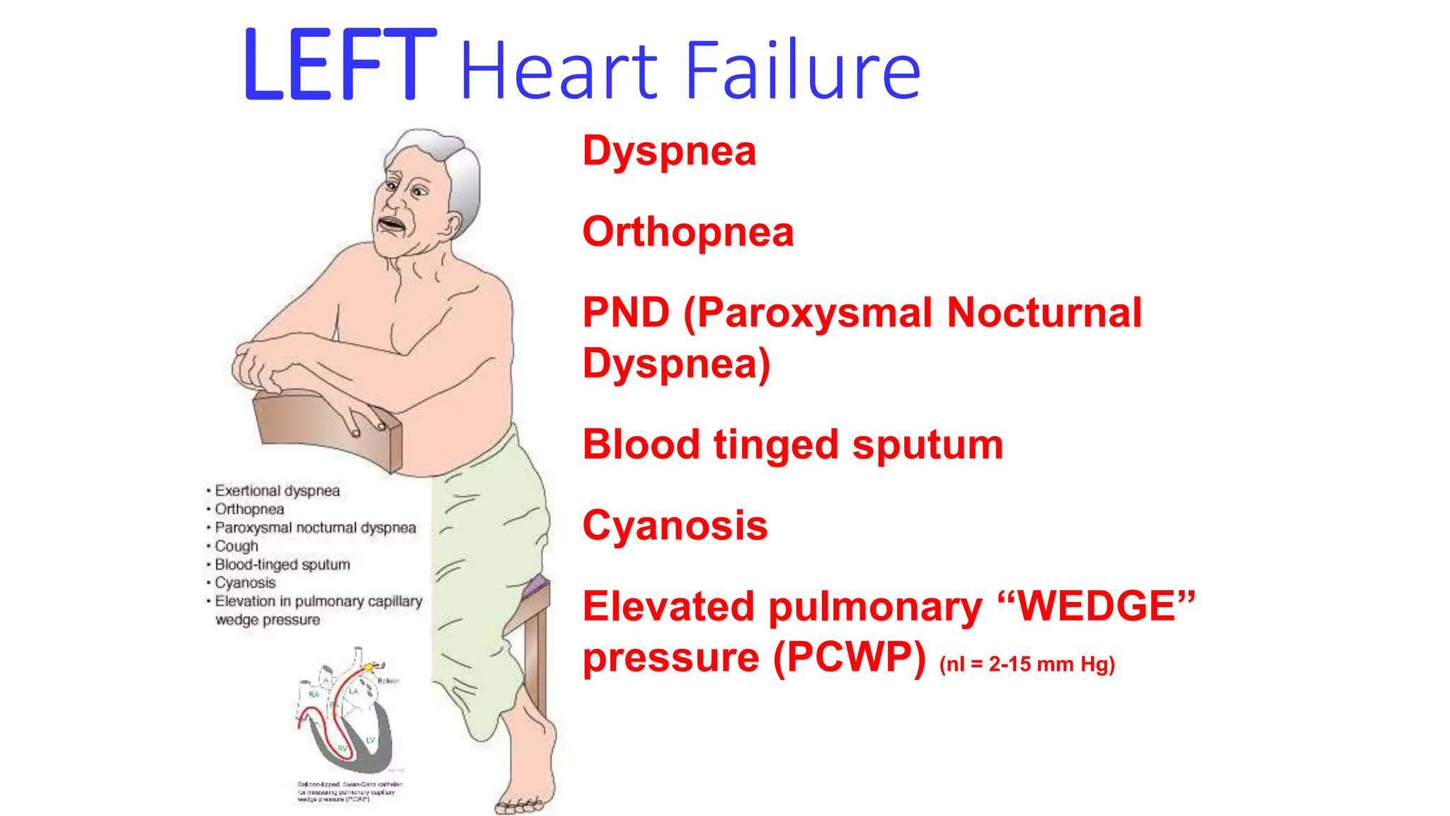
Heart failure
Dr kamalesh lenka
• Heart failure, often referred to as congestive heart failure
• (CHF), is the common end point for many forms of cardiac
• disease and typically is a progressive condition with a
• poor prognosis.
• CHF occurs when the heart cannot generate sufficient output to
meet the metabolic demands of the tissues, or can only do so at
higher-than-normal filling pressures
• greatly increased tissue demands, as in hyperthyroidism, or
decreased oxygen carrying capacity, as in anemia (highoutput failure).
• Heart failure may result from systolic or diastolic dysfunction
• Systolic dysfunction results from inadequate myocardial contractile
function, usually as a consequence of ischemic heart disease or
hypertension
• Diastolic dysfunction refers to an inability of the heart to adequately
relax and fill, which may be a consequence of massive left ventricular
hypertrophy, myocardial fibrosis, amyloid deposition, or constrictive
pericarditis.
• When, the failing heart can no longer efficiently pump blood, there is
an increase in end-diastolic ventricular volumes, increased end-
diastolic pressures, and elevated venous pressures. Thus, inadequate
cardiac output— called forward failure—
• is almost always accompanied by increased congestion of the
venous circulation—that is, backward failure.
• The cardiovascular system attempts to compensate for reduced
myocardial contractility or increased hemodynamic burden through
several homeostatic mechanisms:
• Frank-Starling mechanism.-
• Increased end-diastolic filling volumes dilate the heart and cause
increased cardiac myofiber stretching; these lengthened fibers
contract more forcibly, thereby increasing cardiac output. If the
dilated ventricle is able to maintain cardiac output by this means, the
patient is said to be in compensated heart failure.
• However, ventricular dilation comes at the expense of increased wall
tension and magnifies the oxygen requirements of an already-
compromised myocardium. With time, the failing muscle is no longer
able to propel sufficient blood to meet the needs of the body, and the
patient develops decompensated heart failure
• Activation of neurohumoral systems:-
• Release of the neurotransmitter norepinephrine by the autonomic
nervous system increases heart rate and augments myocardial
contractility and vascular resistance.
• Activation of the renin-angiotensin-aldosterone system spurs water
and salt retention (augmenting circulatory volume) and increases
vascular tone.
• Release of atrial natriuretic peptide acts to balance the renin-
angiotensin-aldosterone system through diuresis and vascular smooth
muscle relaxation
• Myocardial structural changes, including augmented muscle mass
• Cardiac myocytes adapt to increased workload by assembling new
sarcomeres, a change that is accompanied by myocyte enlargement
• In pressure overload states-e.g., hypertension or valvular stenosis),
new sarcomeres tend to be added parallel to the long axis of the
myocytes, adjacent to existing sarcomeres. The growing muscle fiber
diameter thus results in concentric hypertrophy—the ventricular wall
thickness increases without an increase in the size of the chamber.
• In volume overload states (e.g., valvular regurgitation or shunts), the
new sarcomeres are added in series with existing sarcomeres, so that
the muscle fiber length increases. Consequently, the ventricle tends
to dilate, and the resulting wall thickness can be increased, normal, or
decreased; thus, heart weight—rather than wall thickness—is the
best measure of hypertrophy in volume-overloaded hearts.
Left-Sided Heart Failure
• Heart failure can affect predominantly the left or the right side of the
heart or may involve both sides
• The most common causes of left-sided cardiac failure are
• ischemic heart disease (IHD),
• systemic hypertension,
• mitral or aortic valve disease, and
• primary diseases of the myocardium (e.g., amyloidosis).
MORPHOLOGY
• Heart.-
• the left ventricle usually is hypertrophied
• The microscopic changes in heart failure are nonspecific, consisting
primarily of myocyte hypertrophy with interstitial fibrosis of variable
severity.
• Lungs
• congestion and edema as well as pleural effusion due to an increase
in hydrostatic pressure in the venules of the visceral pleura
• The lungs are heavy and boggy, and microscopically show perivascular
and interstitial transudates, alveolar septal edema, and accumulation
of edema fluid in the alveolar spaces
LEFT Heart Failure
Dyspnea
Orthopnea
PND (Paroxysmal Nocturnal
Dyspnea)
Blood tinged sputum
Cyanosis
Elevated pulmonary “WEDGE”
pressure (PCWP) (nl = 2-15 mm Hg)
Right-Sided Heart Failure
• Right-sided heart failure is usually the consequence of left-sided
heart failure, since any pressure increase in the pulmonary
circulation inevitably produces an increased burden on the right side
of the heart
• causes of right-sided heart failure include all of those that induce left-
sided heart failure.
• Isolated right-sided heart failure is infrequent and typically occurs in
patients with one of a variety of disorders affecting the lungs; hence it
is often referred to as cor pulmonale.
• The common feature of these disorders is pulmonary hypertension
(discussed later), which results in hypertrophy and dilation of the
right side of the heart.
• In cor pulmonale,myocardial hypertrophy and dilation generally are
confined to the right ventricle and atrium, although bulging of the
ventricular septum to the left can reduce cardiac output by causing
outflow tract obstruction.
MORPHOLOGY
• Liver and Portal System
• The liver usually is increased in size and weight (congestive
hepatomegaly). A cut section displays prominent passive congestion, a
pattern referred to as nutmeg liver
• When left-sided heart failure is also present, severe central hypoxia
produces centrilobular necrosis
• Right-sided heart failure can also lead to elevated pressure in the portal
vein and its tributaries (portal hypertension), with vascular congestion
producing a tense, enlarged spleen (congestive splenomegaly).
• Pleural, Pericardial, and Peritoneal Spaces
• venous congestion due to right-sided heart failure can lead to
• transudates (effusions) in the pleural and pericardial spaces,
• Subcutaneous Tissues. Edema of dependent portions of
• the body, especially the feet and lower legs, is a hallmark of rightsided
chf
RIGHT Heart Failure
FATIGUE
“Dependent” edema
JVD
Hepatomegaly (congestion)
ASCITES, PLEURAL EFFUSION
GI
Cyanosis
Increased peripheral venous pressure
(CVP) (nl = 2-6 mm Hg)

Heart failure

  • 1.
  • 2.
    • Heart failure,often referred to as congestive heart failure • (CHF), is the common end point for many forms of cardiac • disease and typically is a progressive condition with a • poor prognosis.
  • 3.
    • CHF occurswhen the heart cannot generate sufficient output to meet the metabolic demands of the tissues, or can only do so at higher-than-normal filling pressures • greatly increased tissue demands, as in hyperthyroidism, or decreased oxygen carrying capacity, as in anemia (highoutput failure).
  • 4.
    • Heart failuremay result from systolic or diastolic dysfunction • Systolic dysfunction results from inadequate myocardial contractile function, usually as a consequence of ischemic heart disease or hypertension • Diastolic dysfunction refers to an inability of the heart to adequately relax and fill, which may be a consequence of massive left ventricular hypertrophy, myocardial fibrosis, amyloid deposition, or constrictive pericarditis.
  • 5.
    • When, thefailing heart can no longer efficiently pump blood, there is an increase in end-diastolic ventricular volumes, increased end- diastolic pressures, and elevated venous pressures. Thus, inadequate cardiac output— called forward failure— • is almost always accompanied by increased congestion of the venous circulation—that is, backward failure.
  • 6.
    • The cardiovascularsystem attempts to compensate for reduced myocardial contractility or increased hemodynamic burden through several homeostatic mechanisms: • Frank-Starling mechanism.- • Increased end-diastolic filling volumes dilate the heart and cause increased cardiac myofiber stretching; these lengthened fibers contract more forcibly, thereby increasing cardiac output. If the dilated ventricle is able to maintain cardiac output by this means, the patient is said to be in compensated heart failure.
  • 7.
    • However, ventriculardilation comes at the expense of increased wall tension and magnifies the oxygen requirements of an already- compromised myocardium. With time, the failing muscle is no longer able to propel sufficient blood to meet the needs of the body, and the patient develops decompensated heart failure
  • 8.
    • Activation ofneurohumoral systems:- • Release of the neurotransmitter norepinephrine by the autonomic nervous system increases heart rate and augments myocardial contractility and vascular resistance. • Activation of the renin-angiotensin-aldosterone system spurs water and salt retention (augmenting circulatory volume) and increases vascular tone. • Release of atrial natriuretic peptide acts to balance the renin- angiotensin-aldosterone system through diuresis and vascular smooth muscle relaxation
  • 9.
    • Myocardial structuralchanges, including augmented muscle mass • Cardiac myocytes adapt to increased workload by assembling new sarcomeres, a change that is accompanied by myocyte enlargement • In pressure overload states-e.g., hypertension or valvular stenosis), new sarcomeres tend to be added parallel to the long axis of the myocytes, adjacent to existing sarcomeres. The growing muscle fiber diameter thus results in concentric hypertrophy—the ventricular wall thickness increases without an increase in the size of the chamber.
  • 10.
    • In volumeoverload states (e.g., valvular regurgitation or shunts), the new sarcomeres are added in series with existing sarcomeres, so that the muscle fiber length increases. Consequently, the ventricle tends to dilate, and the resulting wall thickness can be increased, normal, or decreased; thus, heart weight—rather than wall thickness—is the best measure of hypertrophy in volume-overloaded hearts.
  • 12.
    Left-Sided Heart Failure •Heart failure can affect predominantly the left or the right side of the heart or may involve both sides • The most common causes of left-sided cardiac failure are • ischemic heart disease (IHD), • systemic hypertension, • mitral or aortic valve disease, and • primary diseases of the myocardium (e.g., amyloidosis).
  • 13.
    MORPHOLOGY • Heart.- • theleft ventricle usually is hypertrophied • The microscopic changes in heart failure are nonspecific, consisting primarily of myocyte hypertrophy with interstitial fibrosis of variable severity. • Lungs • congestion and edema as well as pleural effusion due to an increase in hydrostatic pressure in the venules of the visceral pleura • The lungs are heavy and boggy, and microscopically show perivascular and interstitial transudates, alveolar septal edema, and accumulation of edema fluid in the alveolar spaces
  • 14.
    LEFT Heart Failure Dyspnea Orthopnea PND(Paroxysmal Nocturnal Dyspnea) Blood tinged sputum Cyanosis Elevated pulmonary “WEDGE” pressure (PCWP) (nl = 2-15 mm Hg)
  • 15.
    Right-Sided Heart Failure •Right-sided heart failure is usually the consequence of left-sided heart failure, since any pressure increase in the pulmonary circulation inevitably produces an increased burden on the right side of the heart • causes of right-sided heart failure include all of those that induce left- sided heart failure. • Isolated right-sided heart failure is infrequent and typically occurs in patients with one of a variety of disorders affecting the lungs; hence it is often referred to as cor pulmonale.
  • 16.
    • The commonfeature of these disorders is pulmonary hypertension (discussed later), which results in hypertrophy and dilation of the right side of the heart. • In cor pulmonale,myocardial hypertrophy and dilation generally are confined to the right ventricle and atrium, although bulging of the ventricular septum to the left can reduce cardiac output by causing outflow tract obstruction.
  • 17.
    MORPHOLOGY • Liver andPortal System • The liver usually is increased in size and weight (congestive hepatomegaly). A cut section displays prominent passive congestion, a pattern referred to as nutmeg liver • When left-sided heart failure is also present, severe central hypoxia produces centrilobular necrosis • Right-sided heart failure can also lead to elevated pressure in the portal vein and its tributaries (portal hypertension), with vascular congestion producing a tense, enlarged spleen (congestive splenomegaly).
  • 18.
    • Pleural, Pericardial,and Peritoneal Spaces • venous congestion due to right-sided heart failure can lead to • transudates (effusions) in the pleural and pericardial spaces, • Subcutaneous Tissues. Edema of dependent portions of • the body, especially the feet and lower legs, is a hallmark of rightsided chf
  • 20.
    RIGHT Heart Failure FATIGUE “Dependent”edema JVD Hepatomegaly (congestion) ASCITES, PLEURAL EFFUSION GI Cyanosis Increased peripheral venous pressure (CVP) (nl = 2-6 mm Hg)