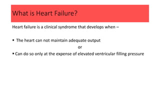
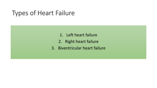
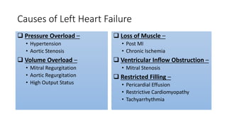
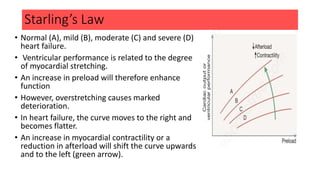
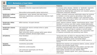
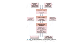
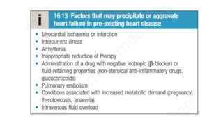
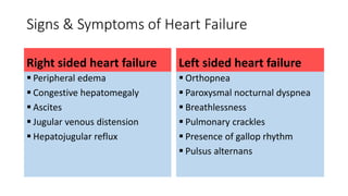
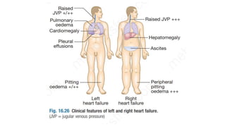
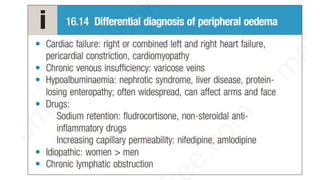
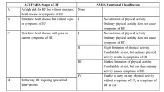
Heart Failure
Dr. Sayem Bin Latif
MD (Cardiology) Phase A Resident
Chittagong Medical College & Hospital
07 Aug 2018
sayembinlatif@gmail.com
What is Heart Failure?
Heart failure is a clinical syndrome that develops when –
 The heart can not maintain adequate output
or
 Can do so only at the expense of elevated ventricular filling pressure
Types of Heart Failure
1. Left heart failure
2. Right heart failure
3. Biventricular heart failure
Left Heart Failure
Left heart failure is characterized by –
 Reduction in left ventricular output
and
 Increase in left atrial and pulmonary venous pressure
Causes of Left Heart Failure
 Pressure Overload –
• Hypertension
• Aortic Stenosis
 Volume Overload –
• Mitral Regurgitation
• Aortic Regurgitation
• High Output Status
 Loss of Muscle –
• Post MI
• Chronic Ischemia
 Ventricular Inflow Obstruction –
• Mitral Stenosis
 Restricted Filling –
• Pericardial Effusion
• Restrictive Cardiomyopathy
• Tachyarrhythmia
Acute Left Heart Failure
If left heart failure occurs suddenly (ex. after acute MI)
Rapid increase in left atrial pressure
Causes pulmonary edema
If the rise in left atrial pressure is more gradual (ex. In MS)
There is reflex pulmonary vasoconstriction
It protects against pulmonary edema but causes pulmonary
hypertension
Which in turn impairs right ventricular function.
Chronic Left Heart Failure
Right Heart Failure
Right heart failure is characterized by –
 Reduction in right ventricular output
and
 Increase in right atrial and systemic venous pressure
Causes of Right Heart Failure
• Secondary to left heart failure
• Chronic lung disease
• Pulmonary embolism
• Pulmonary valvular stenosis
Cor Pulmonale
Cor pulmonale is used to describe –
Right heart failure that is secondary to chronic lung disease
Biventricular Heart Failure
In biventricular failure, both sides of heart are affected
Causes of Biventricular Heart Failure
• Dilated cardiomyopathy
• Ischemic heart disease
Epidemiology
 Heart failure predominantly affects the elderly
 Prevalence –
 1% at age 50–59 years
 10% in those aged 80–89 years
 Approximately 50% of patients with severe heart failure die within 2
years because of either pump failure or malignant ventricular
arrhythmias
Pathogenesis
• Heart failure occurs when cardiac output fails to meet the demands
of the circulation.
• Cardiac output is determined by –
I. Preload (the volume and pressure of blood in the ventricles at the
end of diastole)
II. Afterload (the volume and pressure of blood in the ventricles
during systole) and
III. Myocardial contractility
Starling’s Law
• Normal (A), mild (B), moderate (C) and severe (D)
heart failure.
• Ventricular performance is related to the degree
of myocardial stretching.
• An increase in preload will therefore enhance
function
• However, overstretching causes marked
deterioration.
• In heart failure, the curve moves to the right and
becomes flatter.
• An increase in myocardial contractility or a
reduction in afterload will shift the curve upwards
and to the left (green arrow).
Signs & Symptoms of Heart Failure
Right sided heart failure
 Peripheral edema
 Congestive hepatomegaly
 Ascites
 Jugular venous distension
 Hepatojugular reflux
Left sided heart failure
 Orthopnea
 Paroxysmal nocturnal dyspnea
 Breathlessness
 Pulmonary crackles
 Presence of gallop rhythm
 Pulsus alternans
Complications of Heart Failure
Renal Failure
• Renal failure is caused by poor renal perfusion due to low cardiac
output and
• May be exacerbated by diuretic therapy, ACE inhibitors and
angiotensin receptor blockers (ARBs)
Hypokalaemia
Hypokalaemia may be the result of –
• Treatment with potassium-losing diuretics or
• Hyperaldosteronism caused by activation of the renin–angiotensin system
and
• Impaired aldosterone metabolism due to hepatic congestion.
Most of the body’s potassium is intracellular and there may be
substantial depletion of potassium stores, even when the plasma
concentration is in the reference range
Hyperkalaemia
• Hyperkalaemia may be due to the effects of drugs that promote renal
resorption of potassium, in particular the combination of ACEi, ARBs
and mineralocorticoid receptor antagonists.
• These effects are amplified if there is renal dysfunction due to low
cardiac output or atherosclerotic renal vascular disease
Hyponatraemia
• Hyponatraemia is a feature of severe heart failure
• Is a poor prognostic sign
It may be caused by –
Diuretic therapy
Inappropriate water retention due to high vasopressin secretion, or
Failure of the cell membrane ion pump
Impaired Liver Function
• Impaired liver function is caused by -
 Hepatic venous congestion and
 Poor arterial perfusion
• It frequently causes -
 Mild jaundice and
 Abnormal liver function tests
• Reduced synthesis of clotting factors can make anticoagulant control
difficult
Thromboembolism
• Deep vein thrombosis and pulmonary embolism may occur due to the
effects of a low cardiac output and enforced immobility.
• Systemic emboli occur in patients with atrial fibrillation or flutter, or
with intracardiac thrombus complicating conditions such as mitral
stenosis, MI or left ventricular aneurysm
Arrhythmia
• Atrial and ventricular arrhythmias are very common
• May be related to –
Electrolyte changes such as hypokalaemia and hypomagnesaemia,
The underlying cardiac disease
Pro-arrhythmic effects of sympathetic activation.
• Atrial fibrillation occurs in approximately 20% of patients with heart
failure and causes further impairment of cardiac function.
• Ventricular ectopic beats and runs of non-sustained ventricular
tachycardia are common findings in patients with heart failure and
are associated with an adverse prognosis.
Sudden Death
• Sudden death occurs in up to 50% of patients with heart failure and is
most probably due to ventricular fibrillation
Investigations
Chest X-ray
A chest X-ray should be performed in all cases
This may show -
• abnormal distension of the upper lobe pulmonary veins
• Vascularity of the lung fields becomes more prominent and
• The right and left pulmonary arteries dilate.
• Interstitial oedema causes thickened interlobular septa and dilated
lymphatics. These are evident as horizontal lines in the costophrenic
angles (septal or ‘Kerley B’ lines)
Echocardiography
Echocardiography should be considered in all patients with
heart failure in order to –
• Determine the etiology
• Detect unsuspected valvular heart disease, such as occult mitral
stenosis
• Identify patients who will benefit from long-term drug therapy.
• Serum urea, creatinine and electrolytes, haemoglobin and thyroid
function may help to establish the nature and severity of the
underlying heart disease and detect any complications.
• BNP is elevated in heart failure and is a prognostic marker, as well as
being useful in differentiating heart failure from other causes of
breathlessness or peripheral oedema
Management
Management of Acute Heart Failure
• Acute heart failure with pulmonary oedema is a medical emergency
that should be treated urgently.
• The patient should initially be kept rested, with continuous
monitoring of cardiac rhythm, BP and p ulse oximetry.
• Intravenous opiates can be of value in distressed patients but must be
used sparingly, as they may cause respiratory depression and
exacerbation of hypoxaemia and hypercapnia.
• If these measures prove ineffective, inotropic agents such as
dobutamine (2.5–10 µg/kg/min) may be required to augment cardiac
output, particularly in hypotensive patients.
• Insertion of an intra-aortic balloon pump may be beneficial in patients
with acute cardiogenic pulmonary oedema and shock.
• Following management of the acute episode, additional measures
must be instituted to control heart failure in the longer term.
Management of Chronic Heart Failure
Diuretics:
• Diuretics promote urinary sodium and water excretion
• Leading to a reduction in blood plasma volume, which in turn reduces
preload and improves pulmonary and systemic venous congestion.
• They may also reduce afterload and ventricular volume, leading to a
fall in ventricular wall tension and increased cardiac efficiency.
Angiotensin-converting enzyme inhibitors:
• ACE inhibitors play a central role in the management of heart failure
• They interrupt the vicious circle of neurohumoral activation that is
characteristic of the disease by preventing the conversion of
angiotensin I to angiotensin II.
• Effect of ACEi –
 Reduces peripheral vasoconstriction
 Decreases activation of the sympathetic nervous system
 Reduces salt and water retention due to aldosterone release
 Prevents the activation of the renin–angiotensin system caused by diuretic
therapy
• ACE inhibitors can produce a substantial improvement in effort
tolerance and in mortality.
• They can also improve outcome and prevent the onset of overt heart
failure in patients with poor residual left ventricular function
following MI.
• Adverse effects of ACE inhibitors include symptomatic hypotension
and impairment of renal function, hyperkalaemia especially in
patients with bilateral renal artery stenosis or those with pre-existing
renal disease.
Angiotensin receptor blockers:
• ARBs act by blocking the action of angiotensin II on the heart,
peripheral vasculature and kidney.
• In heart failure, they produce beneficial haemodynamic changes that
are similar to the effects of ACE inhibitors but are generally better
tolerated.
• They have comparable effects on mortality and are a useful
alternative for patients who cannot tolerate ACE inhibitors.
Neprilysin inhibitors:
• The only drug currently in this class is sacubitril, a small-molecule
inhibitor of neprilysin, which is responsible for the breakdown of the
endogenous diuretics ANP and BNP.
• Used in combination with the ARB valsartan (sacubitril–valsartan), it
has been shown to produce additional symptomatic and mortality
benefit over ACE inhibition
• It is now recommended in the management of resistant heart failure
Vasodilators:
• These drugs are valuable in chronic heart failure, when ACE inhibitors
or ARBs are contraindicated.
• Venodilators, such as nitrates, reduce preload
• Arterial dilators, such as hydralazine, reduce afterload
• Their use is limited by pharmacological tolerance and hypotension
Beta-adrenoceptor blockers:
• Beta-blockade helps to counteract the deleterious effects of
enhanced sympathetic stimulation and reduces the risk of
arrhythmias and sudden death.
• When initiated in standard doses β-blockers may precipitate acute-
on-chronic heart failure, but when given in small incremental doses
they can increase ejection fraction, improve symptoms, reduce the
frequency of hospitalisation and reduce mortality in patients with
chronic heart failure.
• A typical regimen is bisoprolol, starting at a dose of 1.25 mg daily and
increased gradually over a 12-week period to a target maintenance
dose of 10 mg daily.
• Beta-blockers are more effective at reducing mortality than ACE
inhibitors, with a relative risk reduction of 33% versus 20%,
respectively
Ivabradine :
• Ivabradine acts on the If inward current in the SA node, resulting in
reduction of heart rate.
• It reduces hospital admission and mortality rates in patients with
heart failure due to moderate or severe left ventricular systolic
impairment.
• Its effects were most marked in patients with a relatively high heart
rate (over 77/min)
• Ivabradine is best suited to patients who cannot take β-blockers or
whose heart rate remains high despite β-blockade. It is ineffective in
patients with atrial fibrillation
Digoxin:
• Can be used to provide rate control in patients with heart failure and
atrial fibrillation.
• In patients with severe heart failure (NYHA class III–IV), digoxin
reduces the likelihood of hospitalisation for heart failure, although it
has no effect on long-term survival
Non-pharmacological treatments
• Implantable cardiac defibrillators
• Resynchronisation devices
• Coronary revascularization
• Cardiac transplantation
• Ventricular assist devices
Thank You
sayembinlatif@gmail.com

Heart failure

  • 1.
    Heart Failure Dr. SayemBin Latif MD (Cardiology) Phase A Resident Chittagong Medical College & Hospital 07 Aug 2018 sayembinlatif@gmail.com
  • 2.
    What is HeartFailure? Heart failure is a clinical syndrome that develops when –  The heart can not maintain adequate output or  Can do so only at the expense of elevated ventricular filling pressure
  • 3.
    Types of HeartFailure 1. Left heart failure 2. Right heart failure 3. Biventricular heart failure
  • 4.
    Left Heart Failure Leftheart failure is characterized by –  Reduction in left ventricular output and  Increase in left atrial and pulmonary venous pressure
  • 5.
    Causes of LeftHeart Failure  Pressure Overload – • Hypertension • Aortic Stenosis  Volume Overload – • Mitral Regurgitation • Aortic Regurgitation • High Output Status  Loss of Muscle – • Post MI • Chronic Ischemia  Ventricular Inflow Obstruction – • Mitral Stenosis  Restricted Filling – • Pericardial Effusion • Restrictive Cardiomyopathy • Tachyarrhythmia
  • 6.
    Acute Left HeartFailure If left heart failure occurs suddenly (ex. after acute MI) Rapid increase in left atrial pressure Causes pulmonary edema
  • 7.
    If the risein left atrial pressure is more gradual (ex. In MS) There is reflex pulmonary vasoconstriction It protects against pulmonary edema but causes pulmonary hypertension Which in turn impairs right ventricular function. Chronic Left Heart Failure
  • 8.
    Right Heart Failure Rightheart failure is characterized by –  Reduction in right ventricular output and  Increase in right atrial and systemic venous pressure
  • 9.
    Causes of RightHeart Failure • Secondary to left heart failure • Chronic lung disease • Pulmonary embolism • Pulmonary valvular stenosis
  • 10.
    Cor Pulmonale Cor pulmonaleis used to describe – Right heart failure that is secondary to chronic lung disease
  • 11.
    Biventricular Heart Failure Inbiventricular failure, both sides of heart are affected Causes of Biventricular Heart Failure • Dilated cardiomyopathy • Ischemic heart disease
  • 12.
    Epidemiology  Heart failurepredominantly affects the elderly  Prevalence –  1% at age 50–59 years  10% in those aged 80–89 years  Approximately 50% of patients with severe heart failure die within 2 years because of either pump failure or malignant ventricular arrhythmias
  • 13.
    Pathogenesis • Heart failureoccurs when cardiac output fails to meet the demands of the circulation. • Cardiac output is determined by – I. Preload (the volume and pressure of blood in the ventricles at the end of diastole) II. Afterload (the volume and pressure of blood in the ventricles during systole) and III. Myocardial contractility
  • 14.
    Starling’s Law • Normal(A), mild (B), moderate (C) and severe (D) heart failure. • Ventricular performance is related to the degree of myocardial stretching. • An increase in preload will therefore enhance function • However, overstretching causes marked deterioration. • In heart failure, the curve moves to the right and becomes flatter. • An increase in myocardial contractility or a reduction in afterload will shift the curve upwards and to the left (green arrow).
  • 18.
    Signs & Symptomsof Heart Failure Right sided heart failure  Peripheral edema  Congestive hepatomegaly  Ascites  Jugular venous distension  Hepatojugular reflux Left sided heart failure  Orthopnea  Paroxysmal nocturnal dyspnea  Breathlessness  Pulmonary crackles  Presence of gallop rhythm  Pulsus alternans
  • 22.
  • 23.
    Renal Failure • Renalfailure is caused by poor renal perfusion due to low cardiac output and • May be exacerbated by diuretic therapy, ACE inhibitors and angiotensin receptor blockers (ARBs)
  • 24.
    Hypokalaemia Hypokalaemia may bethe result of – • Treatment with potassium-losing diuretics or • Hyperaldosteronism caused by activation of the renin–angiotensin system and • Impaired aldosterone metabolism due to hepatic congestion. Most of the body’s potassium is intracellular and there may be substantial depletion of potassium stores, even when the plasma concentration is in the reference range
  • 25.
    Hyperkalaemia • Hyperkalaemia maybe due to the effects of drugs that promote renal resorption of potassium, in particular the combination of ACEi, ARBs and mineralocorticoid receptor antagonists. • These effects are amplified if there is renal dysfunction due to low cardiac output or atherosclerotic renal vascular disease
  • 26.
    Hyponatraemia • Hyponatraemia isa feature of severe heart failure • Is a poor prognostic sign It may be caused by – Diuretic therapy Inappropriate water retention due to high vasopressin secretion, or Failure of the cell membrane ion pump
  • 27.
    Impaired Liver Function •Impaired liver function is caused by -  Hepatic venous congestion and  Poor arterial perfusion • It frequently causes -  Mild jaundice and  Abnormal liver function tests • Reduced synthesis of clotting factors can make anticoagulant control difficult
  • 28.
    Thromboembolism • Deep veinthrombosis and pulmonary embolism may occur due to the effects of a low cardiac output and enforced immobility. • Systemic emboli occur in patients with atrial fibrillation or flutter, or with intracardiac thrombus complicating conditions such as mitral stenosis, MI or left ventricular aneurysm
  • 29.
    Arrhythmia • Atrial andventricular arrhythmias are very common • May be related to – Electrolyte changes such as hypokalaemia and hypomagnesaemia, The underlying cardiac disease Pro-arrhythmic effects of sympathetic activation. • Atrial fibrillation occurs in approximately 20% of patients with heart failure and causes further impairment of cardiac function. • Ventricular ectopic beats and runs of non-sustained ventricular tachycardia are common findings in patients with heart failure and are associated with an adverse prognosis.
  • 30.
    Sudden Death • Suddendeath occurs in up to 50% of patients with heart failure and is most probably due to ventricular fibrillation
  • 31.
  • 32.
    Chest X-ray A chestX-ray should be performed in all cases This may show - • abnormal distension of the upper lobe pulmonary veins • Vascularity of the lung fields becomes more prominent and • The right and left pulmonary arteries dilate. • Interstitial oedema causes thickened interlobular septa and dilated lymphatics. These are evident as horizontal lines in the costophrenic angles (septal or ‘Kerley B’ lines)
  • 34.
    Echocardiography Echocardiography should beconsidered in all patients with heart failure in order to – • Determine the etiology • Detect unsuspected valvular heart disease, such as occult mitral stenosis • Identify patients who will benefit from long-term drug therapy.
  • 35.
    • Serum urea,creatinine and electrolytes, haemoglobin and thyroid function may help to establish the nature and severity of the underlying heart disease and detect any complications. • BNP is elevated in heart failure and is a prognostic marker, as well as being useful in differentiating heart failure from other causes of breathlessness or peripheral oedema
  • 36.
  • 38.
    Management of AcuteHeart Failure • Acute heart failure with pulmonary oedema is a medical emergency that should be treated urgently. • The patient should initially be kept rested, with continuous monitoring of cardiac rhythm, BP and p ulse oximetry. • Intravenous opiates can be of value in distressed patients but must be used sparingly, as they may cause respiratory depression and exacerbation of hypoxaemia and hypercapnia.
  • 39.
    • If thesemeasures prove ineffective, inotropic agents such as dobutamine (2.5–10 µg/kg/min) may be required to augment cardiac output, particularly in hypotensive patients. • Insertion of an intra-aortic balloon pump may be beneficial in patients with acute cardiogenic pulmonary oedema and shock. • Following management of the acute episode, additional measures must be instituted to control heart failure in the longer term.
  • 41.
    Management of ChronicHeart Failure Diuretics: • Diuretics promote urinary sodium and water excretion • Leading to a reduction in blood plasma volume, which in turn reduces preload and improves pulmonary and systemic venous congestion. • They may also reduce afterload and ventricular volume, leading to a fall in ventricular wall tension and increased cardiac efficiency.
  • 42.
    Angiotensin-converting enzyme inhibitors: •ACE inhibitors play a central role in the management of heart failure • They interrupt the vicious circle of neurohumoral activation that is characteristic of the disease by preventing the conversion of angiotensin I to angiotensin II. • Effect of ACEi –  Reduces peripheral vasoconstriction  Decreases activation of the sympathetic nervous system  Reduces salt and water retention due to aldosterone release  Prevents the activation of the renin–angiotensin system caused by diuretic therapy
  • 43.
    • ACE inhibitorscan produce a substantial improvement in effort tolerance and in mortality. • They can also improve outcome and prevent the onset of overt heart failure in patients with poor residual left ventricular function following MI. • Adverse effects of ACE inhibitors include symptomatic hypotension and impairment of renal function, hyperkalaemia especially in patients with bilateral renal artery stenosis or those with pre-existing renal disease.
  • 44.
    Angiotensin receptor blockers: •ARBs act by blocking the action of angiotensin II on the heart, peripheral vasculature and kidney. • In heart failure, they produce beneficial haemodynamic changes that are similar to the effects of ACE inhibitors but are generally better tolerated. • They have comparable effects on mortality and are a useful alternative for patients who cannot tolerate ACE inhibitors.
  • 45.
    Neprilysin inhibitors: • Theonly drug currently in this class is sacubitril, a small-molecule inhibitor of neprilysin, which is responsible for the breakdown of the endogenous diuretics ANP and BNP. • Used in combination with the ARB valsartan (sacubitril–valsartan), it has been shown to produce additional symptomatic and mortality benefit over ACE inhibition • It is now recommended in the management of resistant heart failure
  • 46.
    Vasodilators: • These drugsare valuable in chronic heart failure, when ACE inhibitors or ARBs are contraindicated. • Venodilators, such as nitrates, reduce preload • Arterial dilators, such as hydralazine, reduce afterload • Their use is limited by pharmacological tolerance and hypotension
  • 47.
    Beta-adrenoceptor blockers: • Beta-blockadehelps to counteract the deleterious effects of enhanced sympathetic stimulation and reduces the risk of arrhythmias and sudden death. • When initiated in standard doses β-blockers may precipitate acute- on-chronic heart failure, but when given in small incremental doses they can increase ejection fraction, improve symptoms, reduce the frequency of hospitalisation and reduce mortality in patients with chronic heart failure.
  • 48.
    • A typicalregimen is bisoprolol, starting at a dose of 1.25 mg daily and increased gradually over a 12-week period to a target maintenance dose of 10 mg daily. • Beta-blockers are more effective at reducing mortality than ACE inhibitors, with a relative risk reduction of 33% versus 20%, respectively
  • 49.
    Ivabradine : • Ivabradineacts on the If inward current in the SA node, resulting in reduction of heart rate. • It reduces hospital admission and mortality rates in patients with heart failure due to moderate or severe left ventricular systolic impairment. • Its effects were most marked in patients with a relatively high heart rate (over 77/min) • Ivabradine is best suited to patients who cannot take β-blockers or whose heart rate remains high despite β-blockade. It is ineffective in patients with atrial fibrillation
  • 50.
    Digoxin: • Can beused to provide rate control in patients with heart failure and atrial fibrillation. • In patients with severe heart failure (NYHA class III–IV), digoxin reduces the likelihood of hospitalisation for heart failure, although it has no effect on long-term survival
  • 51.
    Non-pharmacological treatments • Implantablecardiac defibrillators • Resynchronisation devices • Coronary revascularization • Cardiac transplantation • Ventricular assist devices
  • 52.