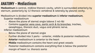
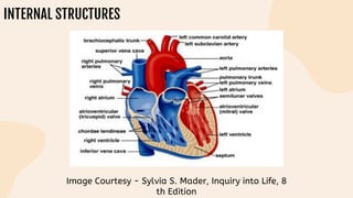
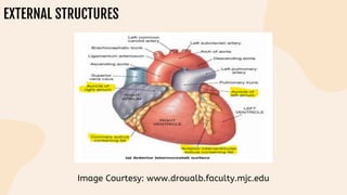
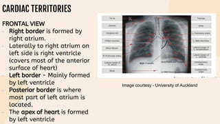
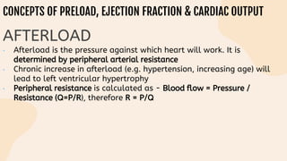
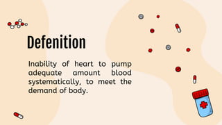
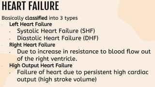
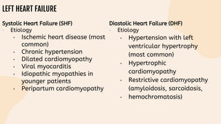
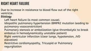
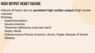
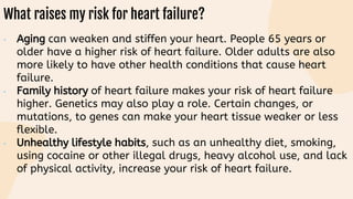
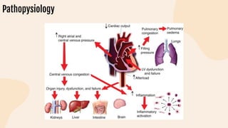
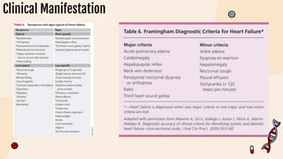
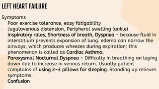
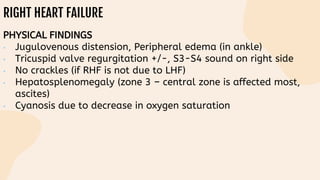
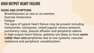
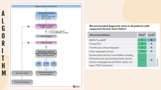
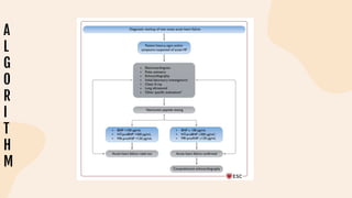
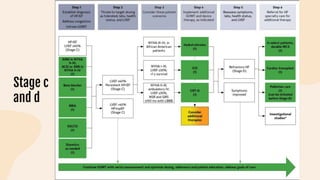
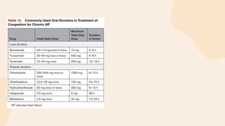
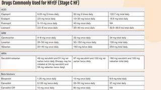
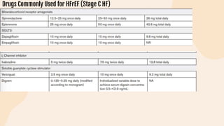
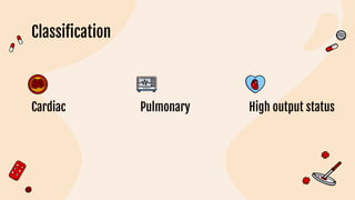
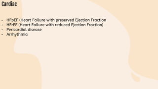
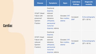
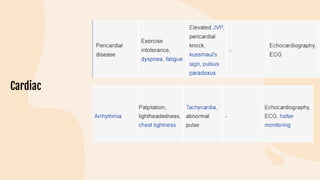
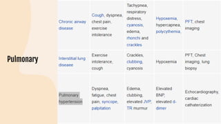
HEART FAILURE
Table of content
Physiology of heart
and defenition of HF
03
Classification of HF
Etiology, risk factor,
patohysiology of HF
Diagnosis and Clinical
Manifestation
Table of content
Treatment of HF
07
Diffrential diagnosis
of HF
Defenition, etiology,
pathophysiology RHD
Diagnosis for RHD
Table of content
Treatment of CHD
11
Complication,
prognosis of RHD
Education
Physiology of heart and
defenition of HF
01
ANATOMY - Mediastinum
Mediastinum is central, midline thoracic cavity, which is surrounded anteriorly by
sternum, posteriorly by 12 thoracic vertebrae & laterally by pleural cavity.
Mediastinum is divided into superior mediastinum & inferior mediastinum.
• Superior mediastinum
• Above the plane of sternal angle (above 2 nd rib)
• Contains superior vena cava, aortic arch & its branches, trachea,
oesophagus, thoracic duct, vagus and phrenic nerve.
• Inferior mediastinum
• Below the plane of sternal angle
• Further divided into 3 parts - anterior, middle & posterior mediastinum.
• Anterior mediastinum is anterior to the heart
• Middle mediastinum contains the heart and great vessels
• Posterior mediastinum contains everything that is below the posterior
margin of heart i.e. thoracic aorta
INTERNAL STRUCTURES
Image Courtesy - Sylvia S. Mader, Inquiry into Life, 8
th Edition
EXTERNAL STRUCTURES
Image Courtesy: www.droualb.faculty.mjc.edu
CARDIAC TERRITORIES
FRONTAL VIEW
• Right border is formed by
right atrium.
• Laterally to right atrium on
left side is right ventricle
(covers most of the anterior
surface of heart)
• Left border - Mainly formed
by left ventricle
• Posterior border is where
most part of left atrium is
located.
• The apex of heart is formed
by left ventricle
Image courtesy - University of Auckland
CONDUCTION PATHWAY
• Speed of conduction -
Purkinje > Atria >
Ventricles > AV node
• Pacemakers rhythm
generation - SA (60-
100/min) > AV (40-
60/min) > bundle of
His/Purkinje (20-40/min).
• Pacemaker - Uses calcium
to generate action
potential. Atrial and
ventricular muscle
depolarization is sodium
dependent
Image courtesy : Hole’s human anatomy and physiology , 7 th
edition by Shier
CORONARY ARTERIES
• The left coronary artery is
further divided into left
circumflex artery and left
anterior descending artery
aka anterior
interventricular branch
• The right coronary artery
is further divided into
marginal arteries, nodal
arteries & posterior
interventricular branch.
CONCEPTS OF PRELOAD, EJECTION FRACTION & CARDIAC OUTPUT
PRELOAD
• Preload is the load on ventricular
muscles at the end of diastole. It
is determined mainly by left
ventricular end diastolic volume &
left ventricular end diastolic
pressure, in other words - by
venous return.
• Increase in preload results in
increase in contractility that in
turn increases stroke volume &
thus increase in ejection fraction
• Chronic increase in preload is
responsible for dilated
cardiomyopathy
CONCEPTS OF PRELOAD, EJECTION FRACTION & CARDIAC OUTPUT
STROKE VOLUME
• Stroke volume is the amount of blood that heart pump out with each beat. It is
affected by contractility, afterload & preload
• SV = EDV (End Diastolic Volume) – ESV (End Systolic Volume)
EJECTION FRACTION
• Is the fraction of blood that heart pump out during 1 contraction which
is usually 60- 70% in healthy normal adult
• Ejection Fraction = Stroke volume/End diastolic volume; therefore, EF =
EDV – ESV/EDV
CARDIAC OUTPUT
• Is the amount of blood that heart pump out during 1 minute
• Cardiac output is calculated as: CO = Heart rate * Stroke volume.
CONCEPTS OF PRELOAD, EJECTION FRACTION & CARDIAC OUTPUT
AFTERLOAD
• Afterload is the pressure against which heart will work. It is
determined by peripheral arterial resistance
• Chronic increase in afterload (e.g. hypertension, increasing age) will
lead to left ventricular hypertrophy
• Peripheral resistance is calculated as - Blood flow = Pressure /
Resistance (Q=P/R), therefore R = P/Q
CONCEPTS OF VELOCITY, AREA,
RESISTANCE AND FLOW
HEART
FAILURE
Defenition
Classification of HF
02
HEART FAILURE
Basically classified into 3 types
• Left Heart Failure
• Systolic Heart Failure (SHF)
• Diastolic Heart Failure (DHF)
• Right Heart Failure
• Due to increase in resistance to blood flow out
of the right ventricle.
• High Output Heart Failure
• Failure of heart due to persistent high cardiac
output (high stroke volume)
Etiology, risk factor,
patohysiology of HF
03
Etiology
LEFT HEART FAILURE
Systolic Heart Failure (SHF)
• Etiology
• Ischemic heart disease (most
common)
• Chronic hypertension
• Dilated cardiomyopathy
• Viral myocarditis
• Idiopathic myopathies in
younger patients
• Peripartum cardiomyopathy
Diastolic Heart Failure (DHF)
• Etiology
• Hypertension with left
ventricular hypertrophy
(most common)
• Hypertrophic
cardiomyopathy
• Restrictive cardiomyopathy
(amyloidosis, sarcoidosis,
• hemochromatosis)
RIGHT HEART FAILURE
Due to increase in resistance to blood flow out of the right
ventricle.
Etiology
• Left heart failure (is most common cause).
• Idiopathic pulmonary hypertension (BMPR2 mutation leading to
pulmonary vasoconstriction)
• Pulmonary stenosis or embolization (give thrombolytic to break
embolus in hemodynamically unstable patient)
• Right ventricular infarction (clear lungs, hypotension, JVD
elevation)
• Restrictive cardiomyopathy, Tricuspid or Pulmonary
regurgitation
HIGH OUTPUT HEART FAILURE
Failure of heart due to persistent high cardiac output (high stroke
volume)
Etiology
• Hyperthyroidism
• Severe anemia
• Thiamine deficiency (wet beri beri)
• Septic shock
• Arteriovenous fistula (trauma, shunt, Paget disease of bone)
• Obesity
Risk Factor
What raises my risk for heart failure?
• Aging can weaken and stiffen your heart. People 65 years or
older have a higher risk of heart failure. Older adults are also
more likely to have other health conditions that cause heart
failure.
• Family history of heart failure makes your risk of heart failure
higher. Genetics may also play a role. Certain changes, or
mutations, to genes can make your heart tissue weaker or less
flexible.
• Unhealthy lifestyle habits, such as an unhealthy diet, smoking,
using cocaine or other illegal drugs, heavy alcohol use, and lack
of physical activity, increase your risk of heart failure.
What raises my risk for heart failure?
• Heart or blood vessel conditions, serious lung disease, or
infections such as HIV or SARS-CoV-2 raise your risk. This is also
true for long-term health conditions such as obesity, high blood
pressure, diabetes, sleep apnea, chronic kidney disease, anemia,
thyroid disease, or iron overload.
• Black and African American people are more likely to have heart
failure than people of other races, often have more serious
cases of heart failure and experience heart failure at a younger
age
pathopysiology
Pathopysiology
Clinical Manifestation and
Diagnosis
04
Clinical Manifestation
Diagnosis HF
LEFT HEART FAILURE
Symptoms
• Poor exercise tolerance, easy fatigability
• Jugulovenous distension, Peripheral swelling (ankle)
• Inspiratory rales, Shortness of breath, Dyspnea - because fluid in
interstitium prevents expansion of lung. edema can narrow the
airways, which produces wheezes during expiration; this
phenomenon is called as Cardiac Asthma.
• Paroxysmal Nocturnal Dyspnea – Difficulty in breathing on laying
down due to increase in venous return. Usually patient
complains of using 2-3 pillows for sleeping. Standing up relieves
symptoms.
• Confusion
LEFT HEART FAILURE
Findings
• Cardiomegaly, Jugular venous distension, S3 in SHF (rapid filling
of ventricles), S4 in DHF (atrial contract against stiffened
ventricles).
• Congested lungs, Pulmonary edema (transudate fluid due to
increase in pulmonary capillary hydrostatic pressure).
• If pulmonary capillary rupture then heart failure cells in alveoli
(alveolar macrophage containing hemosiderin)
New York Heart Association functional classification based on severity of symptoms and physical activity
INVESTIGATIONS
• BNP level – Use in emergent situation when you are not clear about
CHF.
• BNP level – Use in emergent situation when you are not
clear about CHF.
• High level cannot differentiate SHF versus DHF and so
do transthoracic echocardiography
• If the BNP levels remain high after treatment – sign of bad prognosis.
• If BNP is < 100 pg/mL – Heart failure is highly unlikely.
• If BNP is 100-500 pg/mL - Results are uncertain but
suspicious
• If BNP is > 500 pg/ml - Heart failure is highly likely.
INVESTIGATIONS
• On X-Ray: Cardiomegaly, Kerley B lines (septal edema), pulmonary
vasculature congestion, air bronchogram
• On ECG - Left ventricular hypertrophy (S wave in V1 + R in V5 or V6 >
35 mm, > 7 large squares).
• ECG might show ischemic heart disease, arrhythmias and ventricular
hypertrophy.
RIGHT HEART FAILURE
PHYSICAL FINDINGS
• Jugulovenous distension, Peripheral edema (in ankle)
• Tricuspid valve regurgitation +/-, S3-S4 sound on right side
• No crackles (if RHF is not due to LHF)
• Hepatosplenomegaly (zone 3 – central zone is affected most,
ascites)
• Cyanosis due to decrease in oxygen saturation
HIGH OUTPUT HEART FAILURE
SIGNS AND SYMPTOMS
• Breathlessness at rest or on exertion
• Exercise intolerance
• Fatigue
• The signs of typical heart failure may be present including
tachycardia, tachypnea, raised jugular venous pressure,
pulmonary rales, pleural effusion and peripheral edema
• In high output heart failure, patients are likely to have warm
rather than cold peripheries due to low systemic vascular
resistance and peripheral vasodilatation.
A
L
G
O
R
I
T
H
M
A
L
G
O
R
I
T
H
M
Diagnosis RHD
Clinical
manifestat
ion
MAJOR
MANIFESTA
TION
Arthritis
MAJOR
MANIFESTA
TION
Chorea
MAJOR
MANIFESTA
TION
Subcutaneous nodules
MAJOR
MANIFESTA
TION
Erythema marginatum
LAB. FINDINGS
• Positive throat culture for group a beta hemolitic streptococci
• Elevated acute pahse reactants
• Erythrocyte sedimentation rate
• C- reactive protein
• Leukocytosis
• Prolong P-R interval
IMAGING
Laki-laki usia 61 tahun datang ke poli dengan Riwayat DM
lama. Minum glibenclamid fan metformin. Tidak ada obat
lain. Sudah seminggu ini sesak napas saat aktifitas sehari-
hari. Tidur harus setengah duduk 3 hari ini. Kaki pernah
bengkak. Diberi furosemide dan berkurang. Ronki minimal di
basal. Td 100/60
TREATMENT FOR HF
05
Classification
• Positive throat culture for group a beta hemolitic streptococci
• Elevated acute pahse reactants
• Erythrocyte sedimentation rate
• C- reactive protein
• Leukocytosis
• Prolong P-R interval
C
l
a
s
si
fi
c
a
ti
o
n
Diagnostic
Algorithm
Recommen
dations
(Class 1
and 2a) for
Patients at
Risk of HF
Stage c
and d
Drugs Commonly Used for HFrEF (Stage C HF)
Recommendations for
Patients With Mildly
Reduced LVEF (41%–
49%)
Recommendations
for Patients With
Preserved LVEF
(≥50%).
RHD
Progression of
rheumatic heart
disease (RHD)
Recommended
antibiotic regimens
for secondary
prophylaxis
ANTIBIOTIC TREATMENT
• Low-grade fever does not
require specific treatment
• Fever alone, or fever with
mild arthralgia or arthritis,
may not require NSAIDs but
can instead be treated with
paracetamol
• Salicylates (aspirin) have
traditionally been
recommended as first-line
treatment
Laki-laki usia 61 tahun datang ke poli dengan Riwayat DM
lama. Minum glibenclamid fan metformin. Tidak ada obat
lain. Sudah seminggu ini sesak napas saat aktifitas sehari-
hari. Tidur harus setengah duduk 3 hari ini. Kaki pernah
bengkak. Diberi furosemide dan berkurang. Ronki minimal di
basal. Td 100/60
RAWAT JALAN
• Furosemide 1x 20mg
• Spironolakton 1 x 25mg
• Ramipril 1x 1,25 mg
• Ivabradine 2x 2.5mg
• Digoxin 1x1/2 tab
• Glimepiride 1x 2mg
• Atrovastatin
DIFFERENTIAL DIAGNOSIS
06
Heart failure should be
distinguished from other
conditions that cause
dyspnea, fatigue and
edema
Classification
Cardiac Pulmonary High output status
• HFpEF (Heart Failure with preserved Ejection Fraction
• HFrEF (Heart Failure with reduced Ejection Fraction)
• Pericardial disease
• Arrhythmia
• Chronic airway disease
• Pulmonary hypertension
• Anemia
• Thyrotoxicosis
THANK YOU!
References
References

HEART FAILURE.pptx

  • 1.
  • 2.
    Table of content Physiologyof heart and defenition of HF 03 Classification of HF Etiology, risk factor, patohysiology of HF Diagnosis and Clinical Manifestation
  • 3.
    Table of content Treatmentof HF 07 Diffrential diagnosis of HF Defenition, etiology, pathophysiology RHD Diagnosis for RHD
  • 4.
    Table of content Treatmentof CHD 11 Complication, prognosis of RHD Education
  • 5.
    Physiology of heartand defenition of HF 01
  • 6.
    ANATOMY - Mediastinum Mediastinumis central, midline thoracic cavity, which is surrounded anteriorly by sternum, posteriorly by 12 thoracic vertebrae & laterally by pleural cavity. Mediastinum is divided into superior mediastinum & inferior mediastinum. • Superior mediastinum • Above the plane of sternal angle (above 2 nd rib) • Contains superior vena cava, aortic arch & its branches, trachea, oesophagus, thoracic duct, vagus and phrenic nerve. • Inferior mediastinum • Below the plane of sternal angle • Further divided into 3 parts - anterior, middle & posterior mediastinum. • Anterior mediastinum is anterior to the heart • Middle mediastinum contains the heart and great vessels • Posterior mediastinum contains everything that is below the posterior margin of heart i.e. thoracic aorta
  • 7.
    INTERNAL STRUCTURES Image Courtesy- Sylvia S. Mader, Inquiry into Life, 8 th Edition
  • 8.
    EXTERNAL STRUCTURES Image Courtesy:www.droualb.faculty.mjc.edu
  • 9.
    CARDIAC TERRITORIES FRONTAL VIEW •Right border is formed by right atrium. • Laterally to right atrium on left side is right ventricle (covers most of the anterior surface of heart) • Left border - Mainly formed by left ventricle • Posterior border is where most part of left atrium is located. • The apex of heart is formed by left ventricle Image courtesy - University of Auckland
  • 10.
    CONDUCTION PATHWAY • Speedof conduction - Purkinje > Atria > Ventricles > AV node • Pacemakers rhythm generation - SA (60- 100/min) > AV (40- 60/min) > bundle of His/Purkinje (20-40/min). • Pacemaker - Uses calcium to generate action potential. Atrial and ventricular muscle depolarization is sodium dependent Image courtesy : Hole’s human anatomy and physiology , 7 th edition by Shier
  • 11.
    CORONARY ARTERIES • Theleft coronary artery is further divided into left circumflex artery and left anterior descending artery aka anterior interventricular branch • The right coronary artery is further divided into marginal arteries, nodal arteries & posterior interventricular branch.
  • 12.
    CONCEPTS OF PRELOAD,EJECTION FRACTION & CARDIAC OUTPUT PRELOAD • Preload is the load on ventricular muscles at the end of diastole. It is determined mainly by left ventricular end diastolic volume & left ventricular end diastolic pressure, in other words - by venous return. • Increase in preload results in increase in contractility that in turn increases stroke volume & thus increase in ejection fraction • Chronic increase in preload is responsible for dilated cardiomyopathy
  • 13.
    CONCEPTS OF PRELOAD,EJECTION FRACTION & CARDIAC OUTPUT STROKE VOLUME • Stroke volume is the amount of blood that heart pump out with each beat. It is affected by contractility, afterload & preload • SV = EDV (End Diastolic Volume) – ESV (End Systolic Volume) EJECTION FRACTION • Is the fraction of blood that heart pump out during 1 contraction which is usually 60- 70% in healthy normal adult • Ejection Fraction = Stroke volume/End diastolic volume; therefore, EF = EDV – ESV/EDV CARDIAC OUTPUT • Is the amount of blood that heart pump out during 1 minute • Cardiac output is calculated as: CO = Heart rate * Stroke volume.
  • 14.
    CONCEPTS OF PRELOAD,EJECTION FRACTION & CARDIAC OUTPUT AFTERLOAD • Afterload is the pressure against which heart will work. It is determined by peripheral arterial resistance • Chronic increase in afterload (e.g. hypertension, increasing age) will lead to left ventricular hypertrophy • Peripheral resistance is calculated as - Blood flow = Pressure / Resistance (Q=P/R), therefore R = P/Q
  • 15.
    CONCEPTS OF VELOCITY,AREA, RESISTANCE AND FLOW
  • 16.
  • 17.
  • 18.
  • 19.
    HEART FAILURE Basically classifiedinto 3 types • Left Heart Failure • Systolic Heart Failure (SHF) • Diastolic Heart Failure (DHF) • Right Heart Failure • Due to increase in resistance to blood flow out of the right ventricle. • High Output Heart Failure • Failure of heart due to persistent high cardiac output (high stroke volume)
  • 20.
  • 21.
  • 22.
    LEFT HEART FAILURE SystolicHeart Failure (SHF) • Etiology • Ischemic heart disease (most common) • Chronic hypertension • Dilated cardiomyopathy • Viral myocarditis • Idiopathic myopathies in younger patients • Peripartum cardiomyopathy Diastolic Heart Failure (DHF) • Etiology • Hypertension with left ventricular hypertrophy (most common) • Hypertrophic cardiomyopathy • Restrictive cardiomyopathy (amyloidosis, sarcoidosis, • hemochromatosis)
  • 23.
    RIGHT HEART FAILURE Dueto increase in resistance to blood flow out of the right ventricle. Etiology • Left heart failure (is most common cause). • Idiopathic pulmonary hypertension (BMPR2 mutation leading to pulmonary vasoconstriction) • Pulmonary stenosis or embolization (give thrombolytic to break embolus in hemodynamically unstable patient) • Right ventricular infarction (clear lungs, hypotension, JVD elevation) • Restrictive cardiomyopathy, Tricuspid or Pulmonary regurgitation
  • 24.
    HIGH OUTPUT HEARTFAILURE Failure of heart due to persistent high cardiac output (high stroke volume) Etiology • Hyperthyroidism • Severe anemia • Thiamine deficiency (wet beri beri) • Septic shock • Arteriovenous fistula (trauma, shunt, Paget disease of bone) • Obesity
  • 25.
  • 26.
    What raises myrisk for heart failure? • Aging can weaken and stiffen your heart. People 65 years or older have a higher risk of heart failure. Older adults are also more likely to have other health conditions that cause heart failure. • Family history of heart failure makes your risk of heart failure higher. Genetics may also play a role. Certain changes, or mutations, to genes can make your heart tissue weaker or less flexible. • Unhealthy lifestyle habits, such as an unhealthy diet, smoking, using cocaine or other illegal drugs, heavy alcohol use, and lack of physical activity, increase your risk of heart failure.
  • 27.
    What raises myrisk for heart failure? • Heart or blood vessel conditions, serious lung disease, or infections such as HIV or SARS-CoV-2 raise your risk. This is also true for long-term health conditions such as obesity, high blood pressure, diabetes, sleep apnea, chronic kidney disease, anemia, thyroid disease, or iron overload. • Black and African American people are more likely to have heart failure than people of other races, often have more serious cases of heart failure and experience heart failure at a younger age
  • 28.
  • 29.
  • 30.
  • 31.
  • 32.
  • 33.
    LEFT HEART FAILURE Symptoms •Poor exercise tolerance, easy fatigability • Jugulovenous distension, Peripheral swelling (ankle) • Inspiratory rales, Shortness of breath, Dyspnea - because fluid in interstitium prevents expansion of lung. edema can narrow the airways, which produces wheezes during expiration; this phenomenon is called as Cardiac Asthma. • Paroxysmal Nocturnal Dyspnea – Difficulty in breathing on laying down due to increase in venous return. Usually patient complains of using 2-3 pillows for sleeping. Standing up relieves symptoms. • Confusion
  • 34.
    LEFT HEART FAILURE Findings •Cardiomegaly, Jugular venous distension, S3 in SHF (rapid filling of ventricles), S4 in DHF (atrial contract against stiffened ventricles). • Congested lungs, Pulmonary edema (transudate fluid due to increase in pulmonary capillary hydrostatic pressure). • If pulmonary capillary rupture then heart failure cells in alveoli (alveolar macrophage containing hemosiderin)
  • 35.
    New York HeartAssociation functional classification based on severity of symptoms and physical activity
  • 36.
    INVESTIGATIONS • BNP level– Use in emergent situation when you are not clear about CHF. • BNP level – Use in emergent situation when you are not clear about CHF. • High level cannot differentiate SHF versus DHF and so do transthoracic echocardiography • If the BNP levels remain high after treatment – sign of bad prognosis. • If BNP is < 100 pg/mL – Heart failure is highly unlikely. • If BNP is 100-500 pg/mL - Results are uncertain but suspicious • If BNP is > 500 pg/ml - Heart failure is highly likely.
  • 37.
    INVESTIGATIONS • On X-Ray:Cardiomegaly, Kerley B lines (septal edema), pulmonary vasculature congestion, air bronchogram • On ECG - Left ventricular hypertrophy (S wave in V1 + R in V5 or V6 > 35 mm, > 7 large squares). • ECG might show ischemic heart disease, arrhythmias and ventricular hypertrophy.
  • 38.
    RIGHT HEART FAILURE PHYSICALFINDINGS • Jugulovenous distension, Peripheral edema (in ankle) • Tricuspid valve regurgitation +/-, S3-S4 sound on right side • No crackles (if RHF is not due to LHF) • Hepatosplenomegaly (zone 3 – central zone is affected most, ascites) • Cyanosis due to decrease in oxygen saturation
  • 39.
    HIGH OUTPUT HEARTFAILURE SIGNS AND SYMPTOMS • Breathlessness at rest or on exertion • Exercise intolerance • Fatigue • The signs of typical heart failure may be present including tachycardia, tachypnea, raised jugular venous pressure, pulmonary rales, pleural effusion and peripheral edema • In high output heart failure, patients are likely to have warm rather than cold peripheries due to low systemic vascular resistance and peripheral vasodilatation.
  • 40.
  • 41.
  • 42.
  • 43.
  • 44.
  • 45.
  • 46.
  • 47.
  • 48.
    LAB. FINDINGS • Positivethroat culture for group a beta hemolitic streptococci • Elevated acute pahse reactants • Erythrocyte sedimentation rate • C- reactive protein • Leukocytosis • Prolong P-R interval
  • 49.
  • 50.
    Laki-laki usia 61tahun datang ke poli dengan Riwayat DM lama. Minum glibenclamid fan metformin. Tidak ada obat lain. Sudah seminggu ini sesak napas saat aktifitas sehari- hari. Tidur harus setengah duduk 3 hari ini. Kaki pernah bengkak. Diberi furosemide dan berkurang. Ronki minimal di basal. Td 100/60
  • 53.
  • 54.
    Classification • Positive throatculture for group a beta hemolitic streptococci • Elevated acute pahse reactants • Erythrocyte sedimentation rate • C- reactive protein • Leukocytosis • Prolong P-R interval
  • 55.
  • 56.
  • 57.
    Recommen dations (Class 1 and 2a)for Patients at Risk of HF
  • 58.
  • 60.
    Drugs Commonly Usedfor HFrEF (Stage C HF)
  • 61.
    Drugs Commonly Usedfor HFrEF (Stage C HF)
  • 62.
    Recommendations for Patients WithMildly Reduced LVEF (41%– 49%)
  • 63.
  • 64.
  • 65.
  • 66.
  • 67.
    • Low-grade feverdoes not require specific treatment • Fever alone, or fever with mild arthralgia or arthritis, may not require NSAIDs but can instead be treated with paracetamol
  • 68.
    • Salicylates (aspirin)have traditionally been recommended as first-line treatment
  • 69.
    Laki-laki usia 61tahun datang ke poli dengan Riwayat DM lama. Minum glibenclamid fan metformin. Tidak ada obat lain. Sudah seminggu ini sesak napas saat aktifitas sehari- hari. Tidur harus setengah duduk 3 hari ini. Kaki pernah bengkak. Diberi furosemide dan berkurang. Ronki minimal di basal. Td 100/60
  • 70.
    RAWAT JALAN • Furosemide1x 20mg • Spironolakton 1 x 25mg • Ramipril 1x 1,25 mg • Ivabradine 2x 2.5mg • Digoxin 1x1/2 tab • Glimepiride 1x 2mg • Atrovastatin
  • 71.
  • 72.
    Heart failure shouldbe distinguished from other conditions that cause dyspnea, fatigue and edema
  • 73.
  • 74.
    • HFpEF (HeartFailure with preserved Ejection Fraction • HFrEF (Heart Failure with reduced Ejection Fraction) • Pericardial disease • Arrhythmia
  • 77.
    • Chronic airwaydisease • Pulmonary hypertension
  • 79.
  • 81.
  • 82.
  • 83.