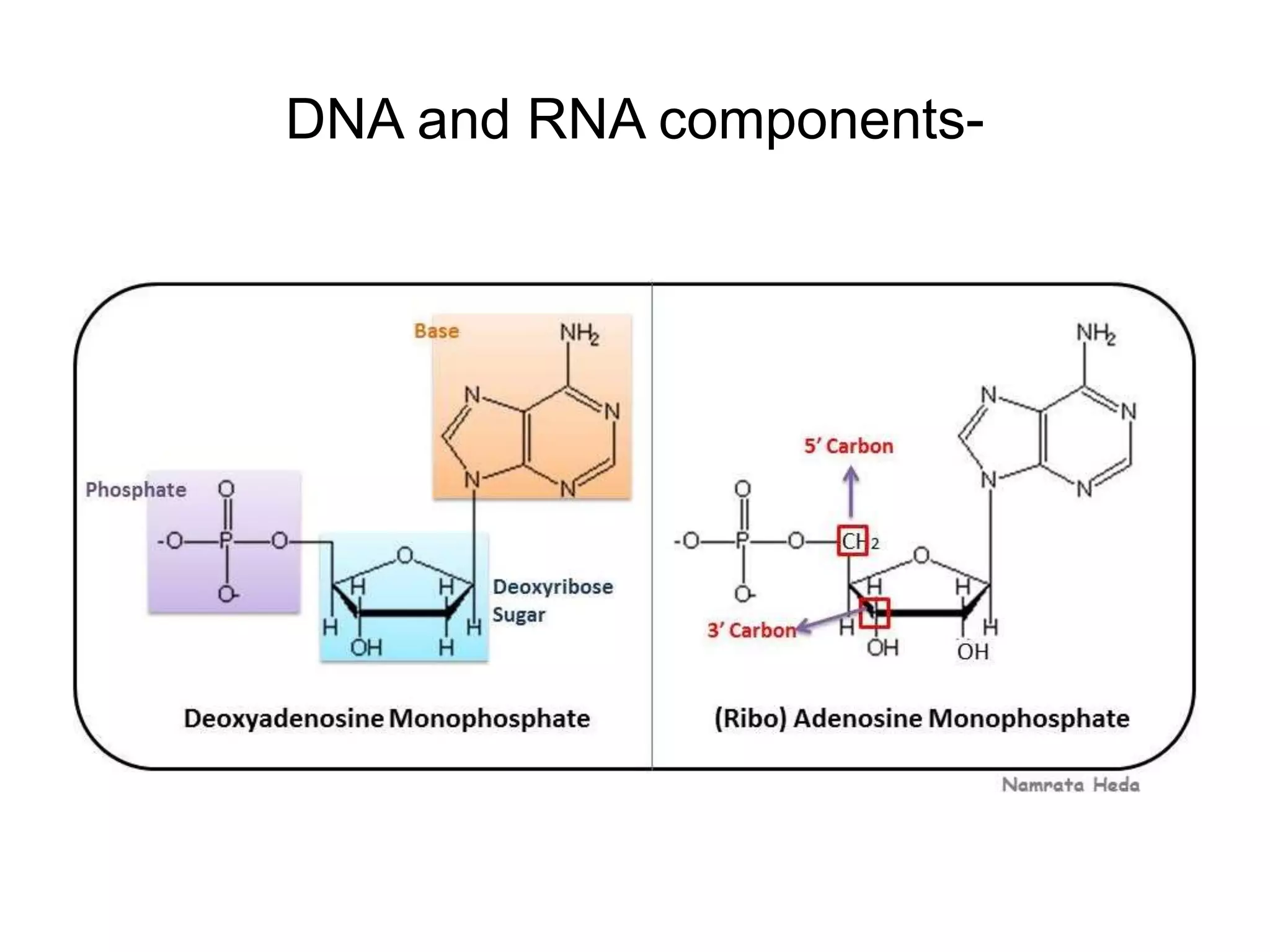
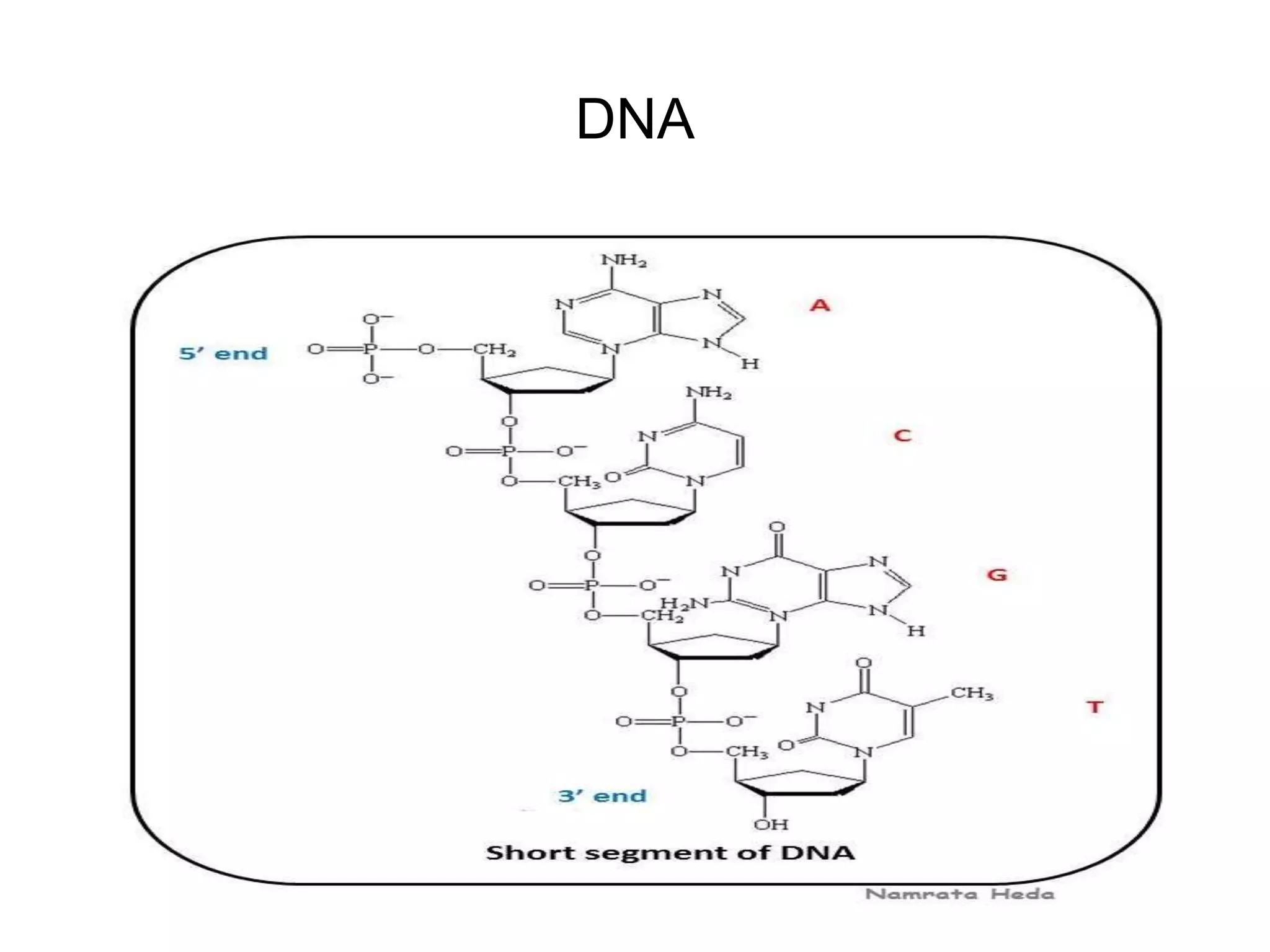
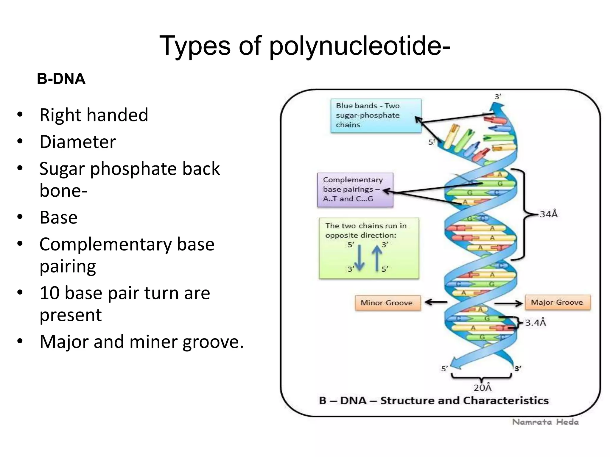
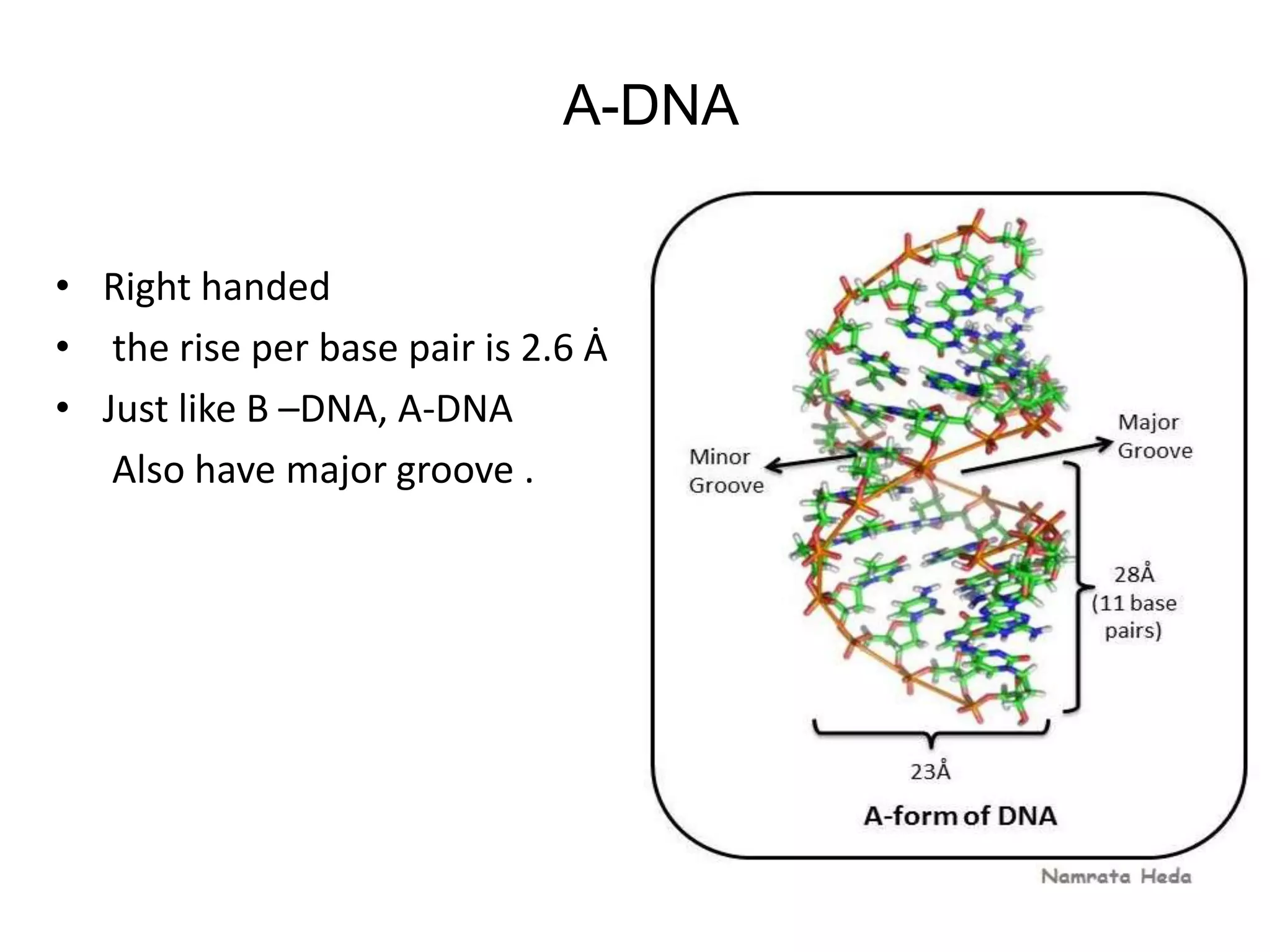
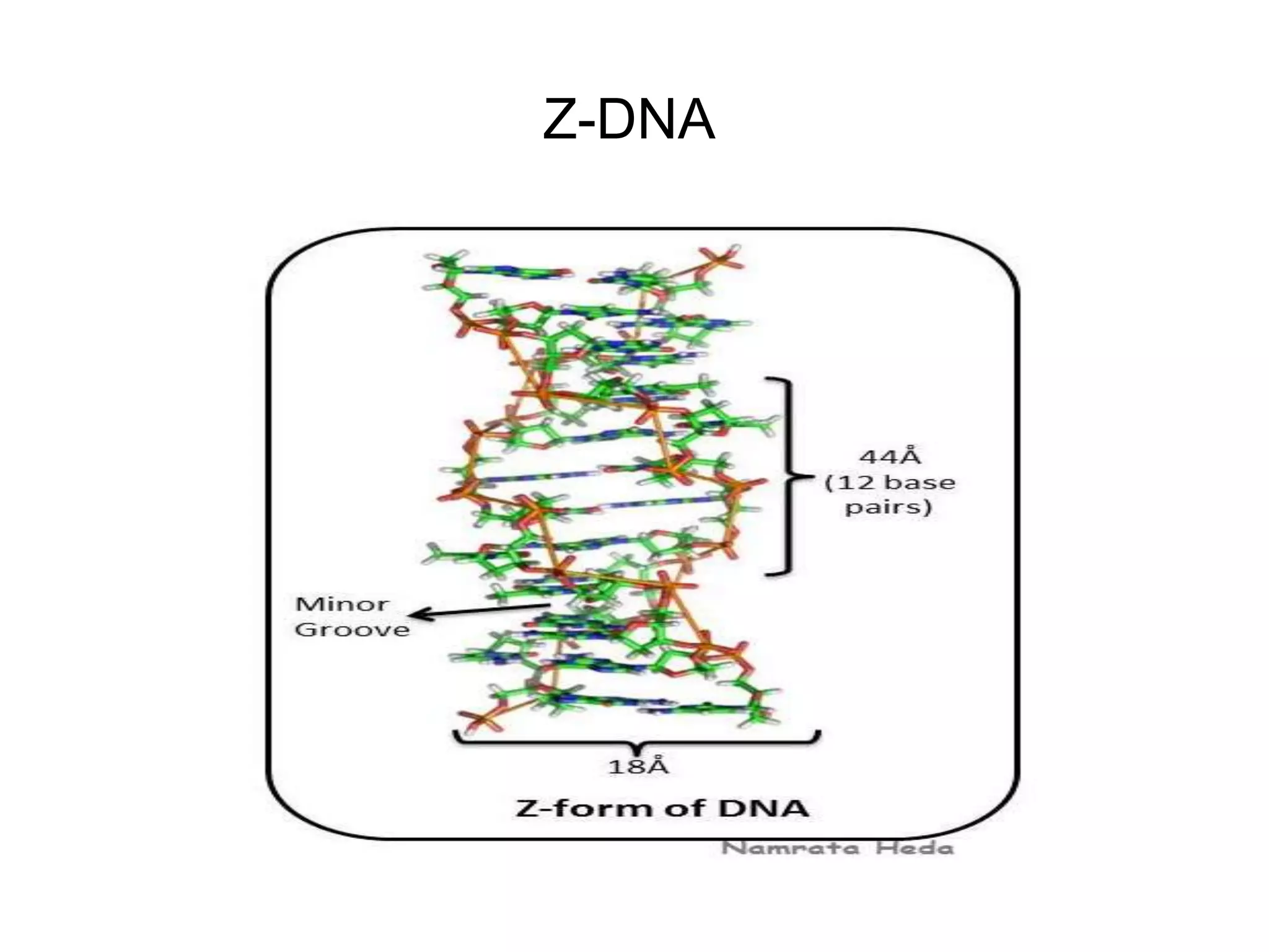
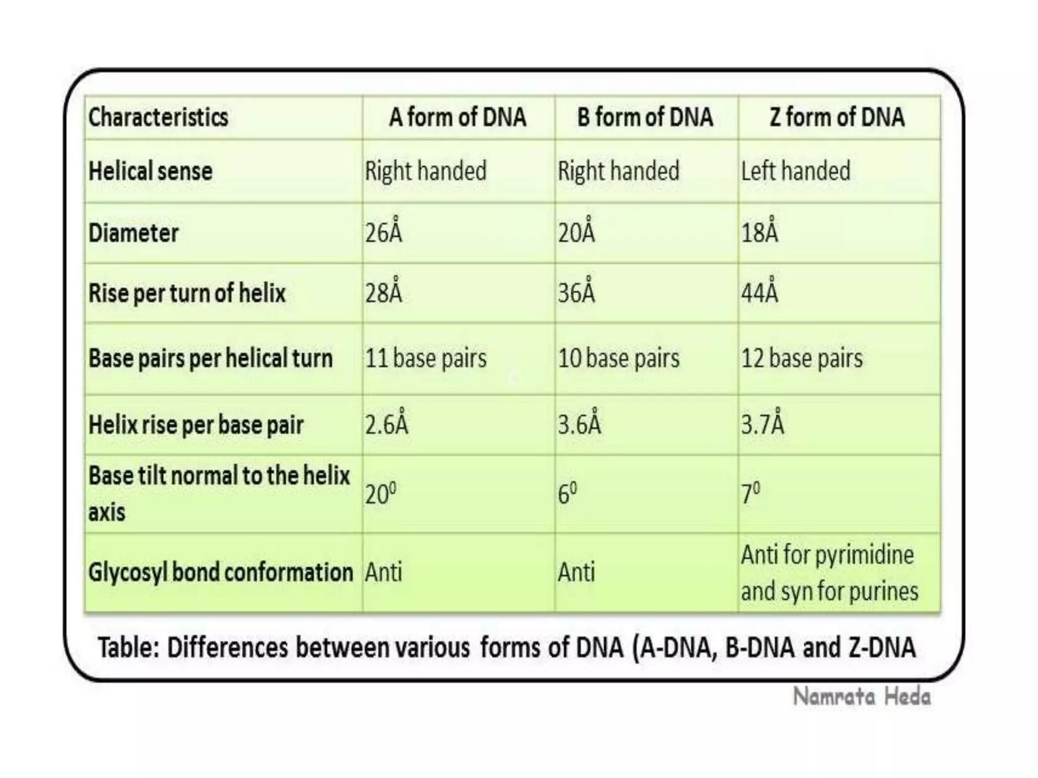
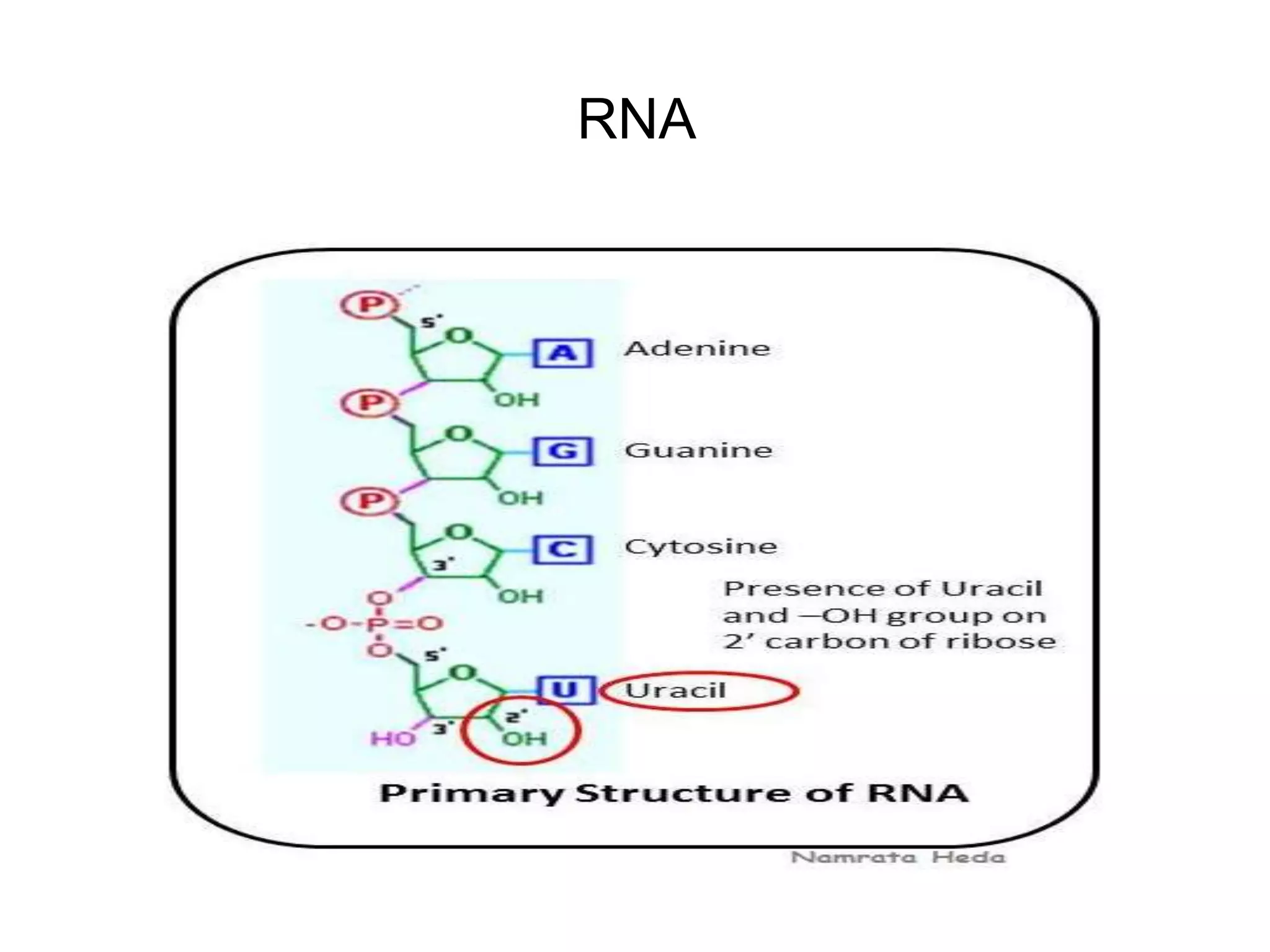
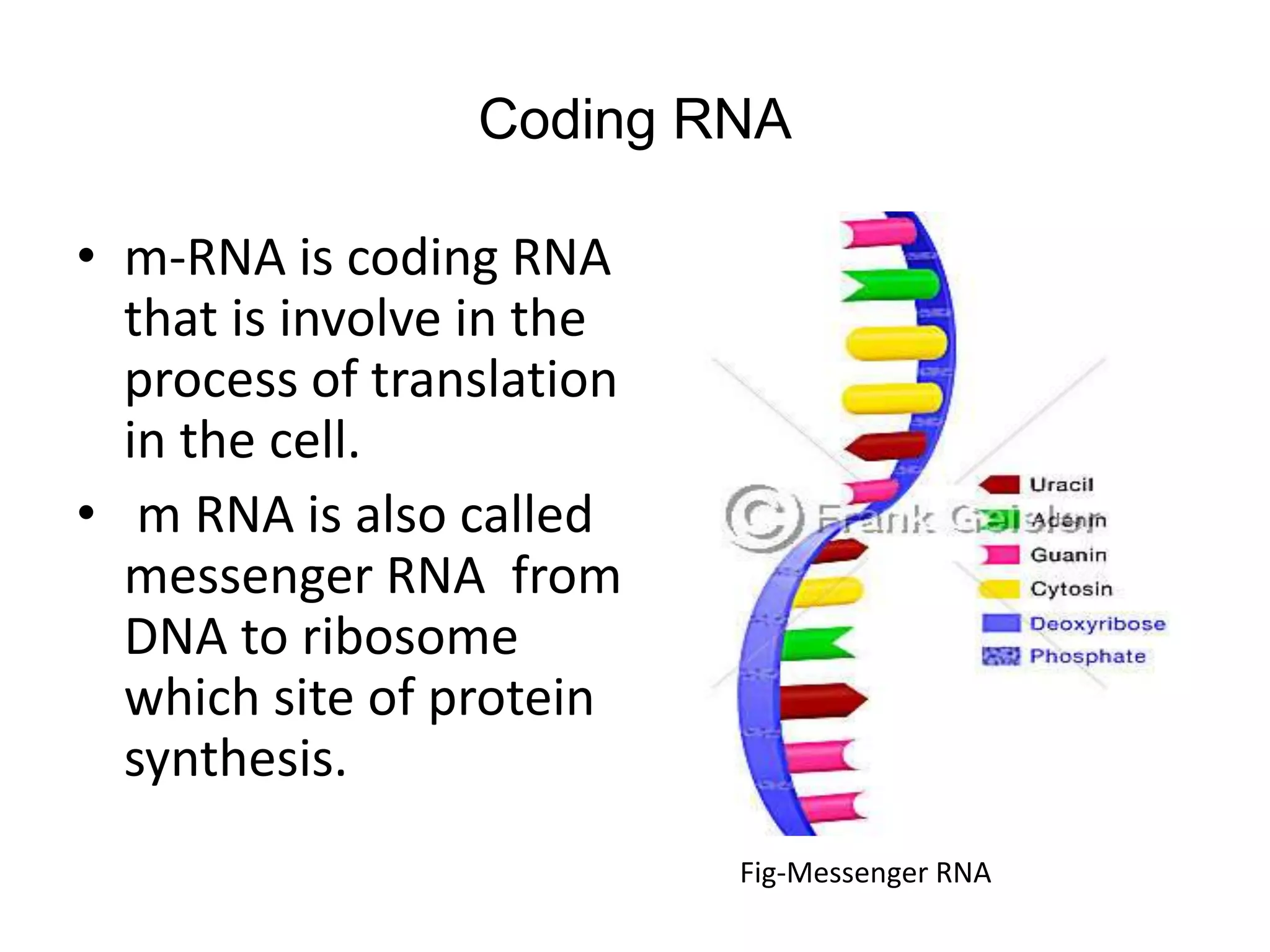
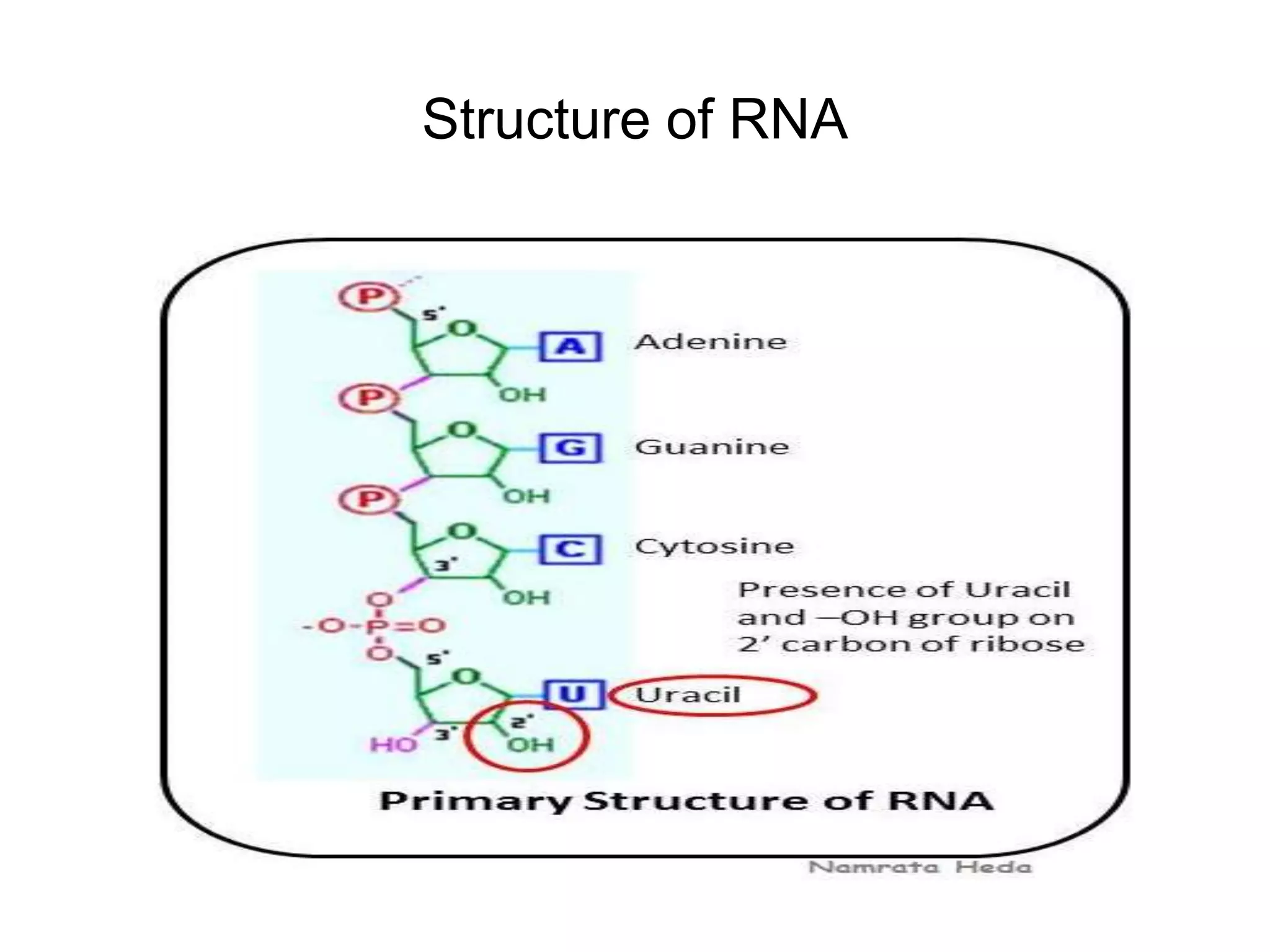
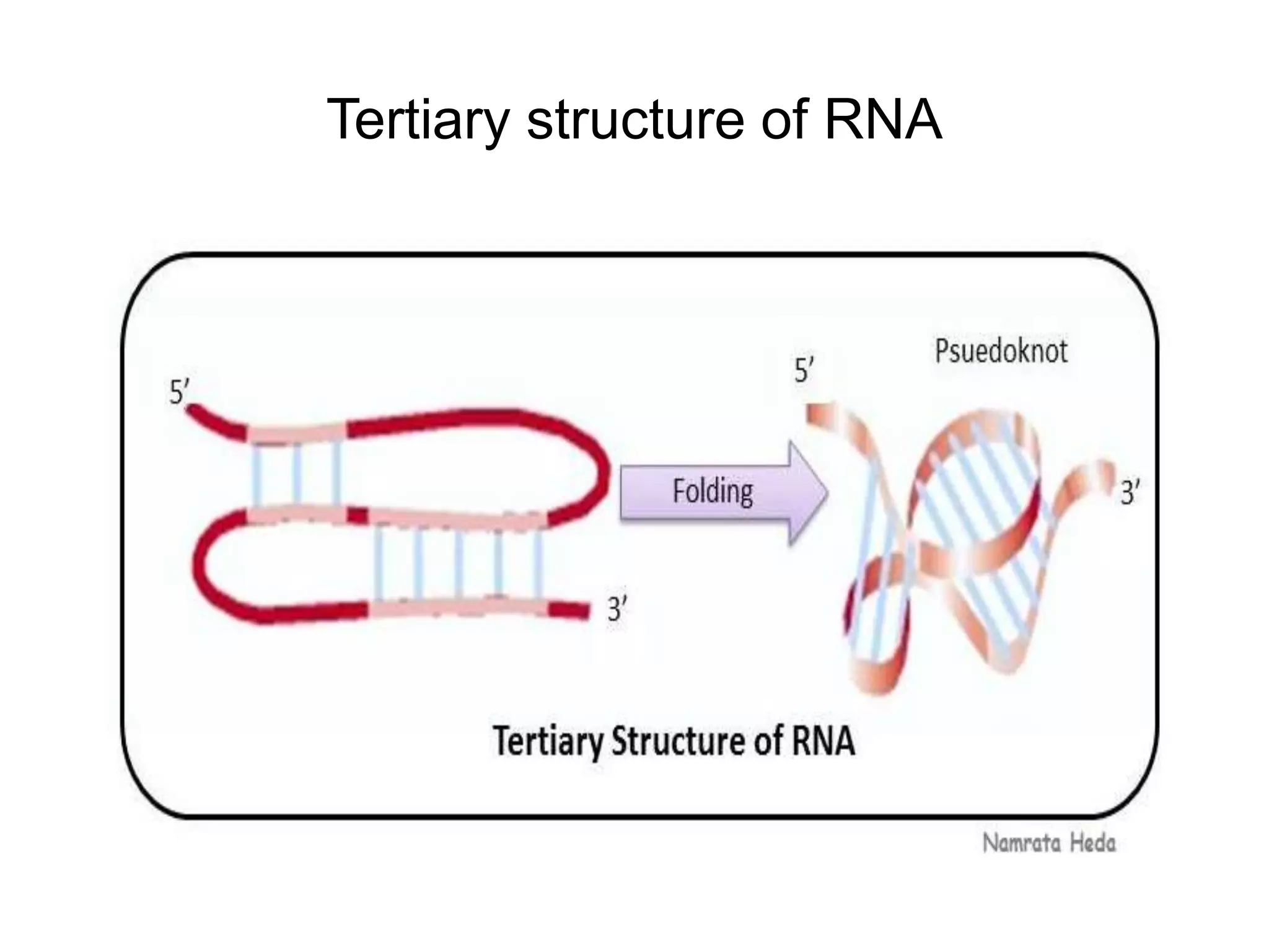
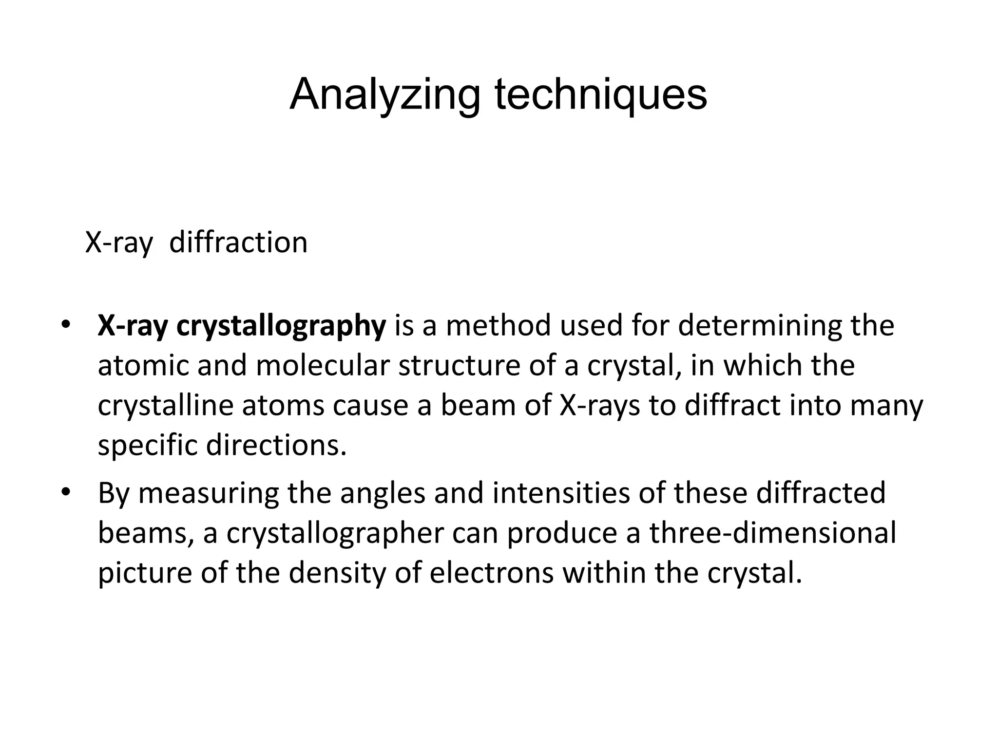
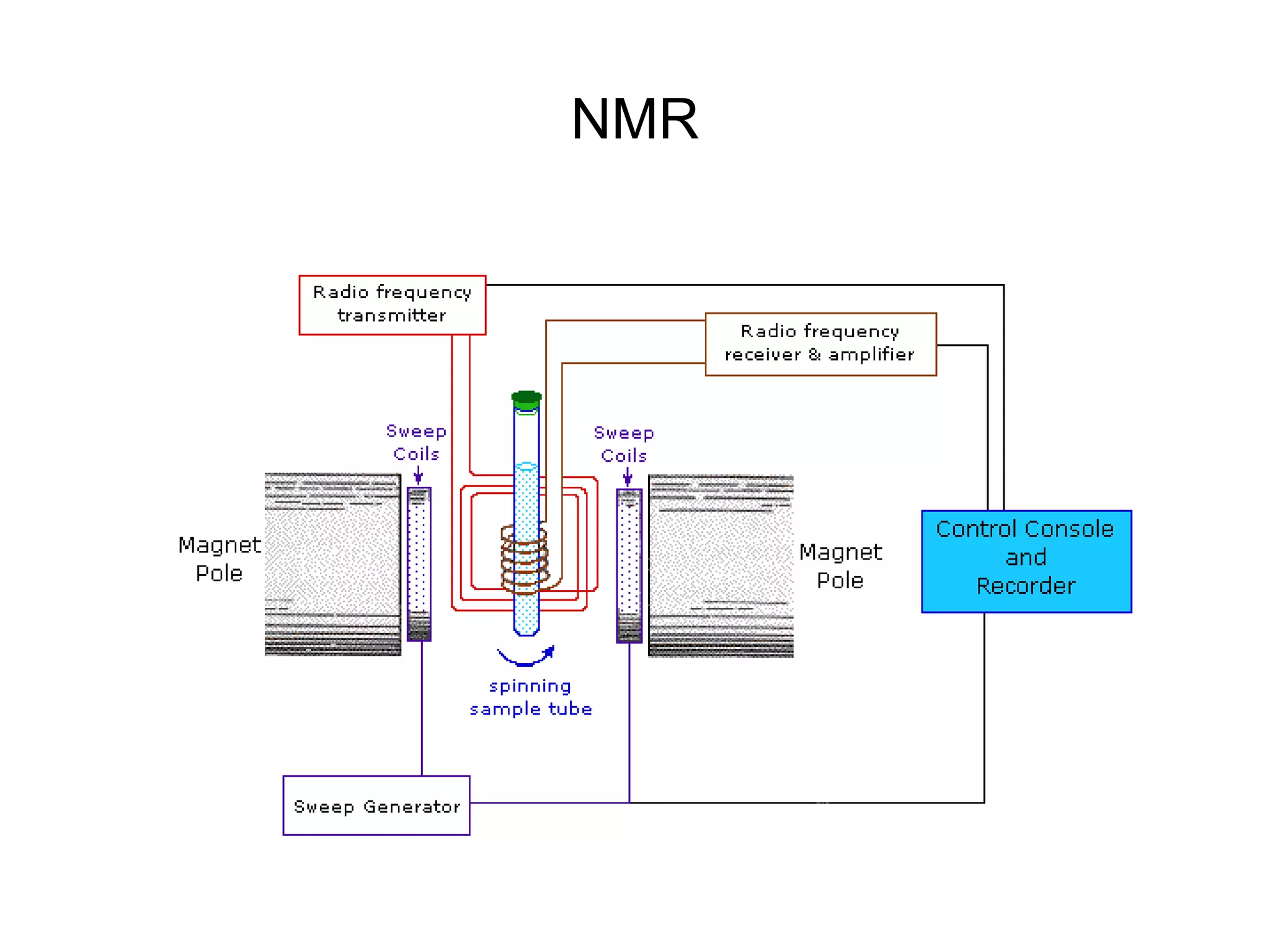
Polynucleotide
Experimental study of conformational
properties
By
KAUSHAL KUMAR SAHU
Assistant Professor (Ad Hoc)
Department of Biotechnology
Govt. Digvijay Autonomous P. G. College
Raj-Nandgaon ( C. G. )
synopsis
• Introduction
• History
• The conformation of nucleic acid
• Types of polynucleotide
DNA- types and conformation of DNA
B- DNA
A-DNA
Z-DNA
RNA – types and conformation of RNA
Coding DNA
Non coding DNA
structure of RNA
Primary structure of RNA
Secondary structure of RNA
Tertiary structure of RNA
• Analyzing techniques
• Conclusion
• References
Introduction
• The polynucleotide are polymeric compound consisting of 15
or more nucleotide monomer covalently bound to gather in a
chain .
• in polynucleotide pentose sugar carbon 5’ and carbon 3’(-
OH) bind with phosphate These bond is also called
phosphodiaster bound
• The chain contributes more and more nucleotide and it is
become polypeptide
History
• The DNA was first discover I 1968 by Friedrich Miesgher he
isolated a compound from the nuclei of White blood cell.
• In 1953 Watson and Crick was discovered double stranded
model of DNA.
• Erwin Chargaff was done chemical analysis of base
composition of DNA
The components of nucleic acid
DNA and RNA components-
DNA
Types of polynucleotide-
• Right handed
• Diameter
• Sugar phosphate back
bone-
• Base
• Complementary base
pairing
• 10 base pair turn are
present
• Major and miner groove.
B-DNA
A-DNA
• Right handed
• the rise per base pair is 2.6 Ȧ
• Just like B –DNA, A-DNA
Also have major groove .
Z-DNA
RNA
Coding RNA
• m-RNA is coding RNA
that is involve in the
process of translation
in the cell.
• m RNA is also called
messenger RNA from
DNA to ribosome
which site of protein
synthesis.
Fig-Messenger RNA
Non coding RNA
• It have no coding RNAs
• some RNA involve in gene
regulation and RNA
Processing
• some RNA act as a catalytic
enzyme is called the
Ribozyme.
Structure of RNA
Secondary structure of RNA
Tertiary structure of RNA
Analyzing techniques
• X-ray crystallography is a method used for determining the
atomic and molecular structure of a crystal, in which the
crystalline atoms cause a beam of X-rays to diffract into many
specific directions.
• By measuring the angles and intensities of these diffracted
beams, a crystallographer can produce a three-dimensional
picture of the density of electrons within the crystal.
X-ray diffraction
NMR
Spectrophotometric method
• The DNA and RNA are absorb light in the ultraviolet region at
260 nm.
• heating is use full method of absorbing of light .
• EtBr- DNA is often in amount too small to be detected by
direct spectroscopy In this case the florescent dye EtBr can be
used to amplify the absorption
References
Books
• principle of biochemistry-Nelson and Cox 5th edition
Net source-
• www/blc.Arizona.edu/molecular- trap
• http/en.m.wiki pedia.org/wiki/x-ray crystal.
• http/ namrata had.blogspot.in

Conformational study of polynucleotide

  • 1.
    Polynucleotide Experimental study ofconformational properties By KAUSHAL KUMAR SAHU Assistant Professor (Ad Hoc) Department of Biotechnology Govt. Digvijay Autonomous P. G. College Raj-Nandgaon ( C. G. )
  • 2.
    synopsis • Introduction • History •The conformation of nucleic acid • Types of polynucleotide DNA- types and conformation of DNA B- DNA A-DNA Z-DNA RNA – types and conformation of RNA Coding DNA Non coding DNA structure of RNA Primary structure of RNA Secondary structure of RNA Tertiary structure of RNA • Analyzing techniques • Conclusion • References
  • 3.
    Introduction • The polynucleotideare polymeric compound consisting of 15 or more nucleotide monomer covalently bound to gather in a chain . • in polynucleotide pentose sugar carbon 5’ and carbon 3’(- OH) bind with phosphate These bond is also called phosphodiaster bound • The chain contributes more and more nucleotide and it is become polypeptide
  • 4.
    History • The DNAwas first discover I 1968 by Friedrich Miesgher he isolated a compound from the nuclei of White blood cell. • In 1953 Watson and Crick was discovered double stranded model of DNA. • Erwin Chargaff was done chemical analysis of base composition of DNA
  • 5.
    The components ofnucleic acid
  • 6.
    DNA and RNAcomponents-
  • 7.
  • 8.
    Types of polynucleotide- •Right handed • Diameter • Sugar phosphate back bone- • Base • Complementary base pairing • 10 base pair turn are present • Major and miner groove. B-DNA
  • 9.
    A-DNA • Right handed •the rise per base pair is 2.6 Ȧ • Just like B –DNA, A-DNA Also have major groove .
  • 10.
  • 12.
  • 13.
    Coding RNA • m-RNAis coding RNA that is involve in the process of translation in the cell. • m RNA is also called messenger RNA from DNA to ribosome which site of protein synthesis. Fig-Messenger RNA
  • 14.
    Non coding RNA •It have no coding RNAs • some RNA involve in gene regulation and RNA Processing • some RNA act as a catalytic enzyme is called the Ribozyme.
  • 15.
  • 16.
  • 17.
  • 18.
    Analyzing techniques • X-raycrystallography is a method used for determining the atomic and molecular structure of a crystal, in which the crystalline atoms cause a beam of X-rays to diffract into many specific directions. • By measuring the angles and intensities of these diffracted beams, a crystallographer can produce a three-dimensional picture of the density of electrons within the crystal. X-ray diffraction
  • 20.
  • 21.
    Spectrophotometric method • TheDNA and RNA are absorb light in the ultraviolet region at 260 nm. • heating is use full method of absorbing of light . • EtBr- DNA is often in amount too small to be detected by direct spectroscopy In this case the florescent dye EtBr can be used to amplify the absorption
  • 22.
    References Books • principle ofbiochemistry-Nelson and Cox 5th edition Net source- • www/blc.Arizona.edu/molecular- trap • http/en.m.wiki pedia.org/wiki/x-ray crystal. • http/ namrata had.blogspot.in