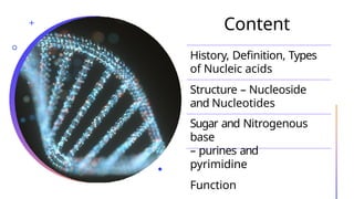
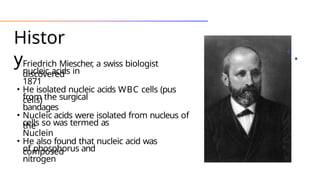
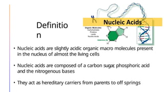
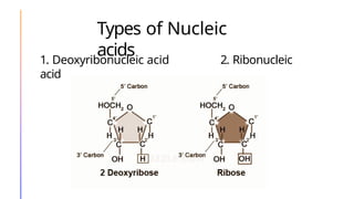
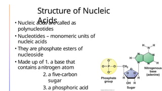
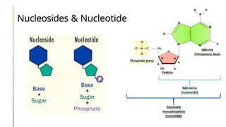
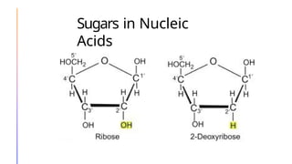
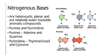
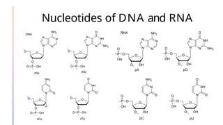
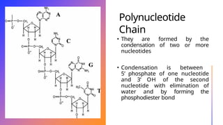
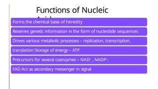
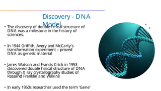
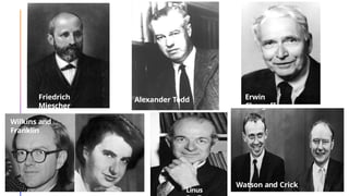
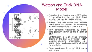
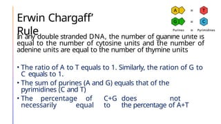
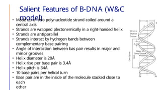
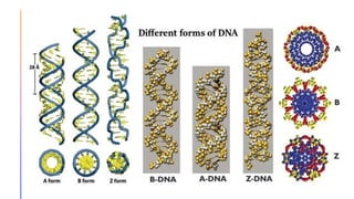
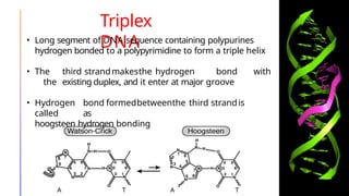
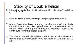
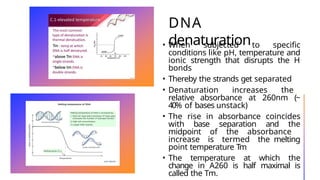
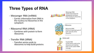
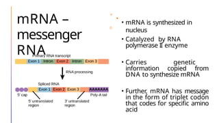
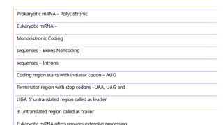
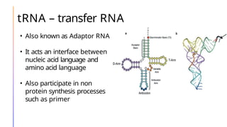
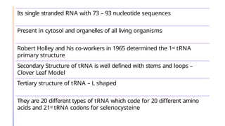
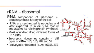
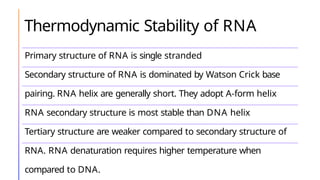
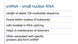
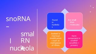
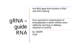
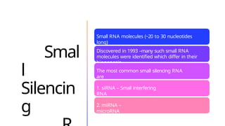
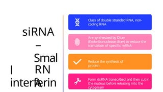
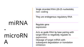
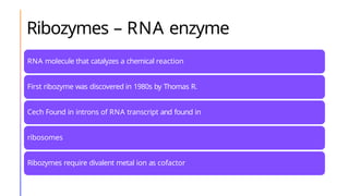
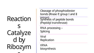
NUCLEIC ACIDS
Dr. Rashmi
Nagesh
Content
History, Definition, Types
of Nucleic acids
Structure – Nucleoside
and Nucleotides
Sugar and Nitrogenous
base
– purines and
pyrimidine
Function
Histor
y
• Friedrich Miescher
, a swiss biologist
discovered
nucleic acids in
1871
• He isolated nucleic acids WBC cells (pus
cells)
from the surgical
bandages
• Nucleic acids were isolated from nucleus of
the
cells so was termed as
Nuclein
• He also found that nucleic acid was
composed
of phosphorus and
nitrogen
Definitio
n
• Nucleic acids are slightly acidic organic macro molecules present
in the nucleus of almost the living cells
• Nucleic acids are composed of a carbon sugar
, phosphoric acid
and the nitrogenous bases
• They act as hereditary carriers from parents to off springs
Types of Nucleic
acids
1. Deoxyribonucleic acid 2. Ribonucleic
acid
Structure of Nucleic
Acids
• Nucleic acids are called as
polynucleotides
• Nucleotides – monomeric units of
nucleic acids
• They are phosphate esters of
nucleoside
• Made up of 1. a base that
contains anitrogen atom
2. a five-carbon
sugar
3. a phosphoric acid
Nucleosides & Nucleotides
Sugars in Nucleic
Acids
Nitrogenous Bases
• Are heterocyclic, planar and
are relatively water insoluble
aromatic compounds
• Purines and Pyrimidine
• Purines – Adenine and
Guanine
• Pyrimidine – Thymine/Uracil
and Cytosine
Nucleotides of DNA and RNA
Polynucleotide
Chain
• They are formed by the
condensation of two or more
nucleotides
• Condensation is between
5’ phosphate of one nucleotide
and 3’ OH of the second
nucleotide with elimination of
water and by forming the
phosphodiester bond
Functions of Nucleic
Acids
Forms the chemical basis of heredity
Reserves genetic information in the form of nucleotide sequences
Drives various metabolic processes – replication, transcription,
translation Storage of energy – ATP
Precursors for several coenzymes – NAD+ , NADP+,
FAD Act as secondary messenger in signal
transduction –cAMP
Discovery - DNA
Model
• The discovery of double helical structure of
DNA was a milestone in the history of
sciences.
• In 1944 Griffith, Avery and McCarty's
transformation experiment – proved
DNA as genetic material
• James Watson and Francis Crick in 1953
discovered double helical structure of DNA
through X ray crystallography studies of
Rosalind Franklin and Wilkins
• In early 1950s researcher used the term ‘Gene’
Alexander
Todd
Friedrich
Miescher
Alexander Todd Erwin
Chargaff
Wilkins and
Franklin
Linus
Watson and Crick
Watson and Crick DNA
Model
• They described the structure in 1953 using
X ray diffraction data of DNA fibers
obtained by R. Franklin and M. Wilkins.
• Watson, Crick and Wilkins were awarded
Nobel prize for Medicine for discovering
the molecular structure of DNA in 1962.
• DNA structure discovered by them
were popularly known as the B form of
DNA.
• Conformation of DNA would primarily
depend on the level of hydration, DNA
sequence, chemical modification of
bases, type and concentration of metal
ion in solution.
• Other well-known forms of DNA are A
and Z form.
Erwin Chargaff’
Rule
In any double stranded DNA, the number of guanine unite is
equal to the number of cytosine units and the number of
adenine units are equal to the number of thymine units
• The ratio of A to T equals to 1. Similarly, the ration of G to
C equals to 1.
• The sum of purines (A and G) equals that of the
pyrimidines (C and T)
• The percentage of C+G does not
necessarily equal to the percentage of A+T
Salient Features of B-DNA (W&C
model)
• Made up of two polynucleotide strand coiled around a
central axis
• Strands are wrapped plectonemically in a right-handed helix
• Strands are antiparallel
• Strands interact by hydrogen bands between
complementary base pairing
• Angle of interaction between bas pair results in major and
minor grooves
• Helix diameter is 20Å
• Helix rise per base pair is 3.4Å
• Helix pitch is 34Å
• 10 base pairs per helical turn
• Base pair are in the inside of the molecule stacked close to
each
other
Differences between different forms of
DNA
Triplex
DNA
• Long segment of DNA sequence containing polypurines
hydrogen bonded to a polypyrimidine to form a triple helix
• The third strandmakesthe hydrogen bond with
the existing duplex, and it enter at major groove
• Hydrogen bond formedbetweenthe third strandis
called as
hoogsteen hydrogen bonding
Stability of Double helical
DNA
1. Internal H bonds that stabilizes the double helix. A to T and G to
C
2. External H bond between sugar and phosphate backbone
3. Apart from the base stacking in the core of the helix
various interactions like hydrophobic and a combination of
Vander Waals and dipole interactions between base pairs
contribute from the overall stability.
4. The –vely charged phosphate situated external surface are
free to interact electrostatically with cations in solution such as
Mg+
DNA
denaturation
• When subjected to specific
conditions like pH, temperature and
ionic strength that disrupts the H
bonds
• Thereby the strands get separated
• Denaturation increases the
relative absorbance at 260nm (~
40% of bases unstack)
• The rise in absorbance coincides
with base separation and the
midpoint of the absorbance
increase is termed the melting
point temperature Tm
• The temperature at which the
change in A260 is half maximal is
called the Tm.
• Ionic strength : Lower the ionic strength lower the
melting temperature
• pH: Basic pH (10) extensive protonation of bases
occurs destroying their H bonds and denature the
duplex
Similarly, below pH 3 protonation of bases
occurs and denaturation of helix
Ribonuclei
c
Acid
Contains the coded message essential for
protein synthesis
Regulates the level of gene expression
Some RNA may be genetic or non
genetic
Some maybe catalytic or non-
catalytic
Act as enzyme –
Ribozymes
Some are coding (mRNA) and non-coding (tRNA
and rRNA
As genetic material – RNA viruses and viroid's
mRNA –
messenger
RNA
• mRNA is synthesized in
nucleus
• Catalyzed by RNA
polymerase II enzyme
• Carries genetic
information copied from
DNA to synthesize mRNA
• Further, mRNA has message
in the form of triplet codon
that codes for specific amino
acid
Prokaryotic mRNA – Polycistronic
Eukaryotic mRNA –
Monocistronic Coding
sequences – Exons Noncoding
sequences – Introns
Coding region starts with initiator codon – AUG
Terminator region with stop codons –UAA, UAG and
UGA 5’ untranslated region called as leader
3’ untranslated region called as trailer
tRNA – transfer RNA
• Also known as Adaptor RNA
• It acts an interface between
nucleic acid language and
amino acid language
• Also participate in non
protein synthesis processes
such as primer
Its single stranded RNA with 73 – 93 nucleotide sequences
Present in cytosol and organelles of all living organisms
Robert Holley and his co-workers in 1965 determined the 1st tRNA
primary structure
Secondary Structure of tRNA is well defined with stems and loops –
Clover Leaf Model
Tertiary structure of tRNA – L shaped
They are 20 different types of tRNA which code for 20 different amino
acids and 21st tRNA codons for selenocysteine
rRNA – ribosomal
RNA
• Is a component of ribosome –
protein synthesis factory of the cell
• rRNA are synthesized is nucleolus and
later exported to nucleus to mature
and assume its role in protein synthesis.
• Most abundant along different forms of
RNA (80%)
• Eukaryotic ribosomes contain 4 diff
types of rRNA: 18S, 5.8S, 28S and 5S
• Prokaryotic ribosomal RNAs: 16S,5S, 23S
Thermodynamic Stability of RNA
Primary structure of RNA is single stranded
Secondary structure of RNA is dominated by Watson Crick base
pairing. RNA helix are generally short. They adopt A-form helix
RNA secondary structure is most stable than DNA helix
Tertiary structure are weaker compared to secondary structure of
RNA. RNA denaturation requires higher temperature when
compared to DNA.
snRNA – small nuclear RNA
Length of above 150 nucleotide sequence
Found within nucleus of eukaryotic
cells Involved in RNA splicing
Helps in maintenance of telomers
Often associated with specific
proteins and form snRNP
snoRNA
–
smal
l
nucleola
RN
A
Found
in
nucleolu
s
Are small
RNA
molecules
Involved in
chemical
modification
s such as
methylation
of rRNAs
Form
component in
snoRNP
(snoRNA +
protein)
gRNA –
guide
RNA
Are RNA gene that function in RNA
and DNA editing
First reported in mitochondria of
kinetoplastids in which mRNAs were
edited by inserting or deleting
stretches of uridine.
Ex: CRISPR
Cas9
Smal
l
Silencin
g
Small RNA molecules (~20 to 30 nucleotides
long)
Discovered in 1993 –many such small RNA
molecules were identified which differ in their
biogenesis.
The most common small silencing RNA
are
1. siRNA – Small interfering
RNA
2. miRNA –
microRNA
siRNA
–
Smal
l
interferin
RN
A
Class of double stranded RNA, non-
coding RNA
Are synthesized by Dicer
(Endoribonuclease dicer) to reduce the
translation of specific mRNA
Reduce the synthesis of
protein
Form dsRNA transcribed and then cut in
the nucleus before releasing into the
cytoplasm
miRNA
-
microRN
A
Single stranded RNA (20-25 nucleotide),
non- coding
They are endogenous regulatory RNA
Regulate gene
expression
Acts as guide RNA by base pairing with
target RNA to negatively regulate its
expression
Cleavage of target mRNA with
subsequent degradation or translation
inhibition
Ribozymes – RNA enzyme
RNA molecule that catalyzes a chemical reaction
First ribozyme was discovered in 1980s by Thomas R.
Cech Found in introns of RNA transcript and found in
ribosomes
Ribozymes require divalent metal ion as cofactor
Ex: RNAse P
, Group I and II introns, hairpin ribozymes, hammerhead ribozyme, hepatitis
delta virus ribozyme and tetrahymena ribozyme
Reaction
s
Catalyze
d by
Ribozym
Cleavage of phosphodiester
bonds (Rnase P
, group I and II
intron)
Synthesis of peptide bonds
(Peptidyl transferase)
RNA processing –
Splicing
Viral
Replication
tRNA
biosynthesis
Biological Macromolecule - Nucleic Acids.pptx

Biological Macromolecule - Nucleic Acids.pptx

  • 1.
  • 2.
    Content History, Definition, Types ofNucleic acids Structure – Nucleoside and Nucleotides Sugar and Nitrogenous base – purines and pyrimidine Function
  • 3.
    Histor y • Friedrich Miescher ,a swiss biologist discovered nucleic acids in 1871 • He isolated nucleic acids WBC cells (pus cells) from the surgical bandages • Nucleic acids were isolated from nucleus of the cells so was termed as Nuclein • He also found that nucleic acid was composed of phosphorus and nitrogen
  • 4.
    Definitio n • Nucleic acidsare slightly acidic organic macro molecules present in the nucleus of almost the living cells • Nucleic acids are composed of a carbon sugar , phosphoric acid and the nitrogenous bases • They act as hereditary carriers from parents to off springs
  • 5.
    Types of Nucleic acids 1.Deoxyribonucleic acid 2. Ribonucleic acid
  • 6.
    Structure of Nucleic Acids •Nucleic acids are called as polynucleotides • Nucleotides – monomeric units of nucleic acids • They are phosphate esters of nucleoside • Made up of 1. a base that contains anitrogen atom 2. a five-carbon sugar 3. a phosphoric acid
  • 7.
  • 8.
  • 9.
    Nitrogenous Bases • Areheterocyclic, planar and are relatively water insoluble aromatic compounds • Purines and Pyrimidine • Purines – Adenine and Guanine • Pyrimidine – Thymine/Uracil and Cytosine
  • 10.
  • 11.
    Polynucleotide Chain • They areformed by the condensation of two or more nucleotides • Condensation is between 5’ phosphate of one nucleotide and 3’ OH of the second nucleotide with elimination of water and by forming the phosphodiester bond
  • 12.
    Functions of Nucleic Acids Formsthe chemical basis of heredity Reserves genetic information in the form of nucleotide sequences Drives various metabolic processes – replication, transcription, translation Storage of energy – ATP Precursors for several coenzymes – NAD+ , NADP+, FAD Act as secondary messenger in signal transduction –cAMP
  • 13.
    Discovery - DNA Model •The discovery of double helical structure of DNA was a milestone in the history of sciences. • In 1944 Griffith, Avery and McCarty's transformation experiment – proved DNA as genetic material • James Watson and Francis Crick in 1953 discovered double helical structure of DNA through X ray crystallography studies of Rosalind Franklin and Wilkins • In early 1950s researcher used the term ‘Gene’
  • 14.
  • 15.
    Watson and CrickDNA Model • They described the structure in 1953 using X ray diffraction data of DNA fibers obtained by R. Franklin and M. Wilkins. • Watson, Crick and Wilkins were awarded Nobel prize for Medicine for discovering the molecular structure of DNA in 1962. • DNA structure discovered by them were popularly known as the B form of DNA. • Conformation of DNA would primarily depend on the level of hydration, DNA sequence, chemical modification of bases, type and concentration of metal ion in solution. • Other well-known forms of DNA are A and Z form.
  • 16.
    Erwin Chargaff’ Rule In anydouble stranded DNA, the number of guanine unite is equal to the number of cytosine units and the number of adenine units are equal to the number of thymine units • The ratio of A to T equals to 1. Similarly, the ration of G to C equals to 1. • The sum of purines (A and G) equals that of the pyrimidines (C and T) • The percentage of C+G does not necessarily equal to the percentage of A+T
  • 17.
    Salient Features ofB-DNA (W&C model) • Made up of two polynucleotide strand coiled around a central axis • Strands are wrapped plectonemically in a right-handed helix • Strands are antiparallel • Strands interact by hydrogen bands between complementary base pairing • Angle of interaction between bas pair results in major and minor grooves • Helix diameter is 20Å • Helix rise per base pair is 3.4Å • Helix pitch is 34Å • 10 base pairs per helical turn • Base pair are in the inside of the molecule stacked close to each other
  • 19.
  • 20.
    Triplex DNA • Long segmentof DNA sequence containing polypurines hydrogen bonded to a polypyrimidine to form a triple helix • The third strandmakesthe hydrogen bond with the existing duplex, and it enter at major groove • Hydrogen bond formedbetweenthe third strandis called as hoogsteen hydrogen bonding
  • 21.
    Stability of Doublehelical DNA 1. Internal H bonds that stabilizes the double helix. A to T and G to C 2. External H bond between sugar and phosphate backbone 3. Apart from the base stacking in the core of the helix various interactions like hydrophobic and a combination of Vander Waals and dipole interactions between base pairs contribute from the overall stability. 4. The –vely charged phosphate situated external surface are free to interact electrostatically with cations in solution such as Mg+
  • 22.
    DNA denaturation • When subjectedto specific conditions like pH, temperature and ionic strength that disrupts the H bonds • Thereby the strands get separated • Denaturation increases the relative absorbance at 260nm (~ 40% of bases unstack) • The rise in absorbance coincides with base separation and the midpoint of the absorbance increase is termed the melting point temperature Tm • The temperature at which the change in A260 is half maximal is called the Tm.
  • 23.
    • Ionic strength: Lower the ionic strength lower the melting temperature • pH: Basic pH (10) extensive protonation of bases occurs destroying their H bonds and denature the duplex Similarly, below pH 3 protonation of bases occurs and denaturation of helix
  • 24.
    Ribonuclei c Acid Contains the codedmessage essential for protein synthesis Regulates the level of gene expression Some RNA may be genetic or non genetic Some maybe catalytic or non- catalytic Act as enzyme – Ribozymes Some are coding (mRNA) and non-coding (tRNA and rRNA As genetic material – RNA viruses and viroid's
  • 26.
    mRNA – messenger RNA • mRNAis synthesized in nucleus • Catalyzed by RNA polymerase II enzyme • Carries genetic information copied from DNA to synthesize mRNA • Further, mRNA has message in the form of triplet codon that codes for specific amino acid
  • 27.
    Prokaryotic mRNA –Polycistronic Eukaryotic mRNA – Monocistronic Coding sequences – Exons Noncoding sequences – Introns Coding region starts with initiator codon – AUG Terminator region with stop codons –UAA, UAG and UGA 5’ untranslated region called as leader 3’ untranslated region called as trailer
  • 28.
    tRNA – transferRNA • Also known as Adaptor RNA • It acts an interface between nucleic acid language and amino acid language • Also participate in non protein synthesis processes such as primer
  • 29.
    Its single strandedRNA with 73 – 93 nucleotide sequences Present in cytosol and organelles of all living organisms Robert Holley and his co-workers in 1965 determined the 1st tRNA primary structure Secondary Structure of tRNA is well defined with stems and loops – Clover Leaf Model Tertiary structure of tRNA – L shaped They are 20 different types of tRNA which code for 20 different amino acids and 21st tRNA codons for selenocysteine
  • 30.
    rRNA – ribosomal RNA •Is a component of ribosome – protein synthesis factory of the cell • rRNA are synthesized is nucleolus and later exported to nucleus to mature and assume its role in protein synthesis. • Most abundant along different forms of RNA (80%) • Eukaryotic ribosomes contain 4 diff types of rRNA: 18S, 5.8S, 28S and 5S • Prokaryotic ribosomal RNAs: 16S,5S, 23S
  • 31.
    Thermodynamic Stability ofRNA Primary structure of RNA is single stranded Secondary structure of RNA is dominated by Watson Crick base pairing. RNA helix are generally short. They adopt A-form helix RNA secondary structure is most stable than DNA helix Tertiary structure are weaker compared to secondary structure of RNA. RNA denaturation requires higher temperature when compared to DNA.
  • 32.
    snRNA – smallnuclear RNA Length of above 150 nucleotide sequence Found within nucleus of eukaryotic cells Involved in RNA splicing Helps in maintenance of telomers Often associated with specific proteins and form snRNP
  • 33.
  • 34.
    gRNA – guide RNA Are RNAgene that function in RNA and DNA editing First reported in mitochondria of kinetoplastids in which mRNAs were edited by inserting or deleting stretches of uridine. Ex: CRISPR Cas9
  • 35.
    Smal l Silencin g Small RNA molecules(~20 to 30 nucleotides long) Discovered in 1993 –many such small RNA molecules were identified which differ in their biogenesis. The most common small silencing RNA are 1. siRNA – Small interfering RNA 2. miRNA – microRNA
  • 36.
    siRNA – Smal l interferin RN A Class of doublestranded RNA, non- coding RNA Are synthesized by Dicer (Endoribonuclease dicer) to reduce the translation of specific mRNA Reduce the synthesis of protein Form dsRNA transcribed and then cut in the nucleus before releasing into the cytoplasm
  • 37.
    miRNA - microRN A Single stranded RNA(20-25 nucleotide), non- coding They are endogenous regulatory RNA Regulate gene expression Acts as guide RNA by base pairing with target RNA to negatively regulate its expression Cleavage of target mRNA with subsequent degradation or translation inhibition
  • 38.
    Ribozymes – RNAenzyme RNA molecule that catalyzes a chemical reaction First ribozyme was discovered in 1980s by Thomas R. Cech Found in introns of RNA transcript and found in ribosomes Ribozymes require divalent metal ion as cofactor Ex: RNAse P , Group I and II introns, hairpin ribozymes, hammerhead ribozyme, hepatitis delta virus ribozyme and tetrahymena ribozyme
  • 39.
    Reaction s Catalyze d by Ribozym Cleavage ofphosphodiester bonds (Rnase P , group I and II intron) Synthesis of peptide bonds (Peptidyl transferase) RNA processing – Splicing Viral Replication tRNA biosynthesis

Editor's Notes

  • #14 Alexander todd – Chemical synthesis of nucleotides led to the discovery for the structural elucidation of DNA Linus Pauling – Primary structural elucidation of Protein insulin