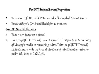
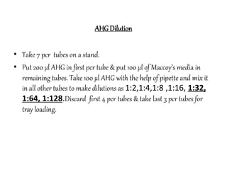
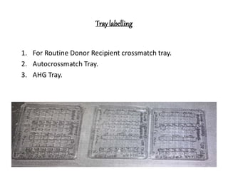
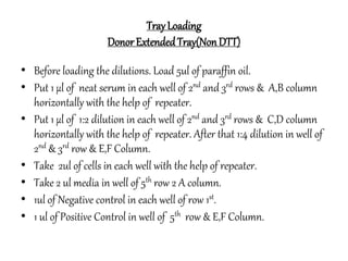
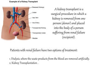
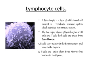
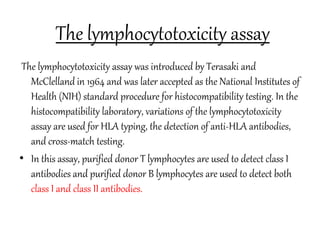
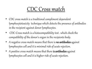
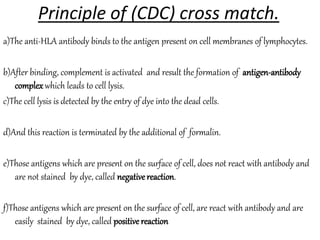
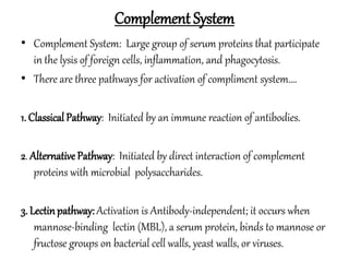
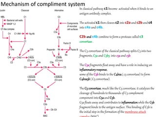
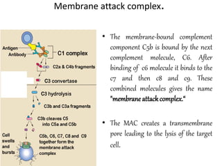
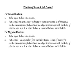
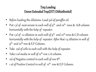
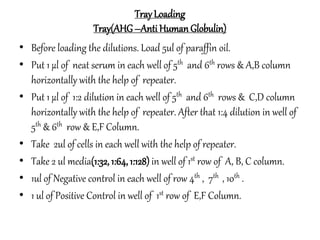
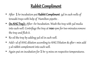
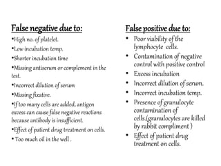
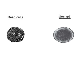
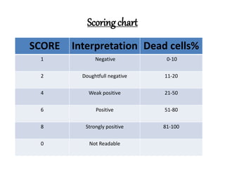
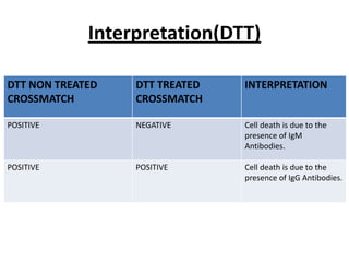
The document provides information about kidney transplantation and the CDC crossmatch test. It discusses the two main treatment options for renal failure - dialysis and kidney transplantation. It then describes the CDC crossmatch test, which checks for antibodies in the recipient that could reject the donor kidney by detecting if antibodies bind to lymphocytes. A negative crossmatch means there is low risk of acute rejection, while a positive crossmatch indicates a higher risk.
2. Patients with renal failure have two options of treatment:
1. Dialysis, where the waste products from the blood are removed artificially.
2. Kidney Transplantation .
.A kidney transplant is a
surgical procedure in which a
kidney is removed from one
person (donor) and placed
into the body of a person
suffering from renal failure
(recipient).
3. Two sources of kidney obtained.
.LivingRelatedDonors:
Mother , Father, siblings (brothers & sisters), children, grandparents and
others near relative.
•EmotionallyRelatedDonor:
In the situation where living related donors are not
present or unfit, emotionally related kidney donors like
husband/wife, cousins, uncles, aunts, brother in-laws
may donate a kidney and they are called emotionally
related kidney donors.
5. December 23, 1954
• The operation was carried out December 23, 1954, at Peter Bent
Brigham Hospital in Boston. Dr. Joseph Murray performed the
procedure. This operation took around 5 hours.
• Ronald Herrick was discharged from the hospital within 14 days of the
operation.
• Richard was discharged from the hospital after 37 days, free of
psychosis, with a normal blood pressure and heart function.
• Because his donor kidney came from his identical twin, no CDC-
Crossmatch test and immunosuppressant drugs were needed before
and after the procedure.
6. Immunosuppressant drugs.
.Immunosuppressant drugs which are also called anti-rejection drugs, are
medicines that reduce the body's natural defenses against foreign
particles or materials. Used in transplant patients, these drugs help
prevent their bodies from rejecting transplanted organs.
.A more effective immunosuppressant called cyclosporine, has been
discovered in the 1980s, was a breakthrough in preventing rejection and
opened a new era in transplant surgery.
.like:-Cyclosporine & FK 506, Rapamycin,Corticosteroids are
immunosupperesent drugs.
.
7. A Acute rejection means that there is antibodies against lymphocytes
cell and it is higher risk of kidney rejection(usuallybeginsone week
after transplantation).
A Hyperacute rejection Hyperacute rejection occurs within minutes
and the transplant must be immediately removed to prevent a several
systemic inflammatory response.
A Chronic Rejection: Long-term loss of function
in transplanted organs.
8. Lymphocyte cells.
• A lymphocyte is a type of white blood cell
present in vertebrate immune system
which activities our immune system.
• The two major classes of lymphocytes are B
cells and T cells both cells are arises from
BoneMarrow.
1. B-cells are mature in the Bone marrow and
store in the thymus.
2. T-cells are arises from Bone Marrow but
mature in the thymus.
9. Types of T cells
• KillerT cells are also known as CD8+ T cells because they
express the CD8 glycoprotein at their surface. Killer T cells
may kill these cells by making holes in their cell membrane
and injecting enzymes into the cells or by binding with
certain sites on their surface called death receptors.
• HelperT cells helps in the activation of B cells which
produce antibodies against foreign antigens.
• Suppressor(regulatory) T cells produce substances that
suppress our immune response.
10. B cells
• B cells are formed in the bone marrow.
• B cells have particular sites (receptors) on their surface where
antigens can attach.
• The B-cell response to antigens has two stages:
• Primaryimmune response: When B cells first encounter an
antigen, the antigen attaches to a receptor, stimulating the B
cells. Some B cells change into memory cells, which remember
that specific antigen, and others change into plasma cells.
11. • Helper T cells helps B cells in this process.
• Plasma cells produce antibodies that are specific to the antigen
that stimulated their production. After the first encounter with an
antigen, production of enough of the specific antibody takes
several days. Thus, the primary immune response is slow.
• Secondaryimmune response: whenever B cells encounter the same
antigenagain, memory B cells very rapidly recognize the antigen,
multiply, change into plasma cells, and produce antibodies. This
response is quick and very effective.
12. The lymphocytotoxicity assay
The lymphocytotoxicity assay was introduced by Terasaki and
McClelland in 1964 and was later accepted as the National Institutes of
Health (NIH) standard procedure for histocompatibility testing. In the
histocompatibility laboratory, variations of the lymphocytotoxicity
assay are used for HLA typing, the detection of anti-HLA antibodies,
and cross-match testing.
• In this assay, purified donor T lymphocytes are used to detect class I
antibodies and purified donor B lymphocytes are used to detect both
class I and class II antibodies.
13. CDC Cross match
• CDC cross match is a traditional compliment dependent
lymphocytotoxicity technique which detects the presence of antibodies
in the recipient against donor lymphocytes.
• CDC-Cross match is a histocompatibility test . which check the
compatibility of the donor's organ in the recipients body.
• A negative cross match means that there is no antibodiesagainst
lymphocytes cell and it is minimal risk of acute rejection.
• A positive cross match means that there is antibodiesagainst
lymphocytes cell and it is higher risk of acute rejection.
14. Principle of (CDC) cross match.
a)The anti-HLA antibody binds to the antigen present on cell membranes of lymphocytes.
b)After binding, complement is activated and result the formation of antigen-antibody
complexwhich leads to cell lysis.
c)The cell lysis is detected by the entry of dye into the dead cells.
d)And this reaction is terminated by the additional of formalin.
e)Those antigens which are present on the surface of cell, does not react with antibody and
are not stained by dye, called negativereaction.
f)Those antigens which are present on the surface of cell, are react with antibody and are
easily stained by dye, called positive reaction
15. Requirements
For SampleCollection:-
• Requisition form.
• Pen.
• Plane Vacutainers (Red Top).
• Marker.
• Two Conical Flask with Glass Beads.(Patient/Donor)
• Needle.
• Alcohol swabs.
• Cotton.
• Band Aid.
• Kit Bag.
17. • Patient serum.
• NH4Cl (Prepared).
• Negative control and
Positive control.
• Rabbit compliment
• Eosin dye (5%)
• Formaline (35-42% and PH
7.4)
• Cover slide
• Mini centrifuge.
• Hemilton.
• Repeator.
• Pipette (10ul/100ul/200ul/1ml)
• Refrigrated Centrifuge.
• Bright field inverted microscope
18. Glass beads:
• Glass beads are used for Defibrinate the blood by moving the
conical flask on 60° angle for 10-15 minutes.
• When extended for more then 20 min at room temperature it
caused haemolysis.
19. Media Preparation
• McCOY’s media is used for survival of lymphocytes cells.
• It is a combination of InorganicSalts (Calcium Chloride, Magnesium
Sulfate, Potassium Chloride ,Sodium Bicarbonate, Sodium Chloride
and so on) ,AminoAcids (L-Alanine ,L -Arginine ,L-Asparagine , L-
Aspartic Acid , L-Cysteine,L-Glutamic Acid , L-Glutamin ),
Vitamins(Ascorbic Acid, Folic Acid , Riboflavin,Vitamin B12), also L-
Glutamine
• It should be store at 2°C to 8°C.
• Add I N NaOH in it untill its pH becomes 7.4.Check the
pH of media with pH paper.
• Filter the media with the help of a filter paper &
keep it in a bottle or store at 2°C to 8°C.
20. Histopaque
• It is a density gradient solution designed for cell
separation.
• This solution involve making differentdensitiesand
layeringthem on top of each other so that there are
very distinct interfaces between the different density
solutions.
21. Terasaki tray
Name:Dr. Paul Terasaki
Born: 1929,In Los Angeles, California
• Terasaki trays are used for
the testing of both class I
and classII antigens.
22. Liquid paraffin oil
• Liquid paraffin oil is a mineral oil, and is
a by product of petroleum distillation. It
is a transparent, colorless, odorless and
tasteless and non toxic oil.
• It prevents dehydration during
incubation period of lymphocytes cells.
23. controls
•Negative control is obtained by normal AB
serum from a non transfused individual
donors lacking lymphocytotoxic activity.
• Positivecontrol is obtained by pool of antisera
that react with high frequency against donor
lymphocyte cells.
24. Complement
•There are Different kind of
complements are available i.e. Rabbit
complement ,Guinea pig complement,
Rat complement, Mouse complement.
•But generally Rabbit complement are
used in CDC Crossmatch coz it’s highly
sensitive for lymphocytic cells.
25. Rabbitcomplement
Rabbit complement is a complex which is derived from group of nine
serum protein (C1-C9) of healthy rabbits which act upon an antigen
antibody complex causing cell lysis.
• Complement is present in all animal sera, the most
satisfactory result have been obtained with Rabbit
compliment.
26. Eosin dye (5% )
• Eosin is a fluorescent red dye.
• It can be used to stain lymphocyte cells
for examination under the microscope.
• The cell lysis is detected by the entry of
dye into the dead cells .
Dead cells are usually dark while live
cells are much brighter than dead cells.
27. Formaline (20% and PH 7.4)
• Formalin is use for prevent antibody-antigen
reaction.
• preserve the morphology (shape and
structure) of the sample as it is processed for
further analysis.
• Fixative typically protects a sample from
extrinsic damage.
28. Bright field invertedmicroscope
• Bright field inverted microscope is used for reading/analysis
the trays.
• Generally cytotoxic reaction(Lymphocytes cells) are analyzed
at 10 x.
29. Complement System
• Complement System: Large group of serum proteins that participate
in the lysis of foreign cells, inflammation, and phagocytosis.
• There are three pathways for activation of compliment system….
1. ClassicalPathway: Initiated by an immune reaction of antibodies.
2. Alternative Pathway: Initiated by direct interaction of complement
proteins with microbial polysaccharides.
3. Lectinpathway: Activation is Antibody-independent; it occurs when
mannose-binding lectin (MBL), a serum protein, binds to mannose or
fructose groups on bacterial cell walls, yeast walls, or viruses.
30. Mechanism of compliment system
In classical pathway c1 become activated when it binds to an
antigen antibody complex.
The activated c1 then cleaves c2 into c2a and c2b and c4
into c4a and c4b.
C2b and c4b combine to form a protease called c3
convertase.
The C3 convertase of the classical pathway splits C3 into two
fragments, C3aand C3b3 into c3a and c3b.
The C3afragments float away and have a role in inducing an
inflammatoryresponse.
some of the C3bbinds to the C4b2a( c3 convertase) to form
C4b2a3b( C5 convertase).
The C5 convertase, much like the C3 convertase, it catalyzes the
cleavage of hundreds to thousands of C5 complement
component into C5aand C5b.
C5a floats away and contributes to inflammationwhile the C5b
fragment binds to the antigen surface. This binding of C5b is
the initial step in the formation of the membrane attack
31. Membrane attack complex.
• The membrane-bound complement
component C5b is bound by the next
complement molecule, C6. After
binding of c6 molecule it binds to the
c7 and then c8 and c9. These
combined molecules gives the name
"membraneattackcomplex.“
• The MAC creates a transmembrane
pore leading to the lysis of the target
cell.
32. Dilutionof Serum& -VEControl
For SerumDilution:-
1. Take 3 pcr tubes on a stand.
2. Put 200 µl patient serum in first pcr tube & put 100 µl of Maccoy’s
media in remaining tubes.Take 100 µl patient serum with the help of
pipette and mix it in other tubes to make dilutions as 1:2,1:4.
For Negative Control:-
1. Take 3 pcr tubes on a stand.
2. Put 200 µl –ve control in first pcr tube & put 100 µl of Maccoy’s
media in remaining tubes.Take 100 µl patient serum with the help of
pipette and mix it in other tubes to make dilutions as 1:2,1:4
33. For DTT TreatedSerumPrepration
• Take 100ul of DTT in PCR Tube and add 100 ul of Patient Serum.
• Treat with 37°c (On Heat Block) for 30 minutes.
For DTT SerumDilution:-
1. Take 3 pcr tubes on a stand.
2. Put 200 µl (DTT Treated) patient serum in first pcr tube & put 100 µl
of Maccoy’s media in remaining tubes. Take 100 µl (DTT Treated)
patient serum with the help of pipette and mix it in other tubes to
make dilutions as 1:2,1:4.
34. AHG Dilution
• Take 7 pcr tubes on a stand.
• Put 200 µl AHG in first pcr tube & put 100 µl of Maccoy’s media in
remaining tubes. Take 100 µl AHG with the help of pipette and mix it
in all other tubes to make dilutions as 1:2,1:4,1:8 ,1:16, 1:32,
1:64, 1:128.Discard first 4 pcr tubes & take last 3 pcr tubes for
tray loading.
36. TrayLoading
Donor ExtendedTray(NonDTT)
• Before loading the dilutions. Load 5ul of paraffin oil.
• Put 1 µl of neat serum in each well of 2nd and 3rd rows & A,B column
horizontally with the help of repeater.
• Put 1 µl of 1:2 dilution in each well of 2nd and 3rd rows & C,D column
horizontally with the help of repeater. After that 1:4 dilution in well of
2nd & 3rd row & E,F Column.
• Take 2ul of cells in each well with the help of repeater.
• Take 2 ul media in well of 5th row 2 A column.
• 1ul of Negative control in each well of row 1st.
• 1 ul of Positive Control in well of 5th row & E,F Column.
37. TrayLoading
Donor ExtendedTray(DTT-Dithiothreitol)
• Before loading the dilutions. Load 5ul of paraffin oil.
• Put 1 µl of neat serum in each well of 9th and 10th rows & A,B column
horizontally with the help of repeater.
• Put 1 µl of 1:2 dilution in each well of 9th and 10th rows & C,D column
horizontally with the help of repeater. After that 1:4 dilution in well of
9th and 10th row & E,F Column.
• Take 2ul of cells in each well with the help of repeater.
• Take 2 ul media in well of 12th row 2 A column.
• 1ul of Negative control in each well of row 8th.
• 1 ul of Positive Control in well of 12th row & E,F Column.
38. TrayLoading
Tray(AHG–AntiHumanGlobulin)
• Before loading the dilutions. Load 5ul of paraffin oil.
• Put 1 µl of neat serum in each well of 5th and 6th rows & A,B column
horizontally with the help of repeater.
• Put 1 µl of 1:2 dilution in each well of 5th and 6th rows & C,D column
horizontally with the help of repeater. After that 1:4 dilution in well of
5th & 6th row & E,F Column.
• Take 2ul of cells in each well with the help of repeater.
• Take 2 ul media(1:32,1:64,1:128) in well of 1st row of A, B, C column.
• 1ul of Negative control in each well of row 4th , 7th , 10th .
• 1 ul of Positive Control in well of 1st row of E,F Column.
39. Rabbit Compliment
• After 1 hr incubation put RabbitCompliment 5µl in each wells of
terasaki trays with help of Hamilton pipette .
• (InAHG Tray):-After 1 hr Incubation. Wash the tray with 5ul media
into each well. Centrifuge the tray at 1100 rpm for two minutes.remove
the tray and flick it.
• Re-oil the tray by adding 5ul oil in each well.
• Add 1 ul of AHG dilution according to AHG Dilution & after 1 min add
5 ul rabbit complement into each well.
• Again put on incubation for 1 hr 15 mins on respective temperatures.
40. • After 1 hr incubation ,put 5% eosindye with the help of Hamilton
pipette of 5 µl in each well of terasaki tray.
• After 2-5 minutes, put 20% formaline(3-5µl) in each well of terasaki
tray with repeater to stop the reaction of dye.
• Cover the tray with Glass Slides.
• Keep the trays for 1 hr on room temperature for settlement of cells.
• After 1 hr trays are ready for analysis on invertedmicroscope.
41. False positive due to:
• Poor viability of the
lymphocyte cells.
• Contamination of negative
control with positive control
• Excess incubation
• Incorrect dilution of serum.
• Incorrect incubation temp.
• Presence of granulocyte
contamination of
cells.(granulocytes are killed
by rabbit compliment )
• Effect of patient drug
treatment on cells.
False negative due to:
•High no. of platelet.
•Low incubation temp.
•Shorter incubation time
•Missing antiserum or complement in the
test.
•Incorrect dilution of serum
•Missing fixative.
•If too many cells are added, antigen
excess can cause false negative reactions
because antibody is insufficient.
•Effect of patient drug treatment on cells.
• Too much oil in the well .
44. Interpretation(DTT)
DTT NON TREATED
CROSSMATCH
DTT TREATED
CROSSMATCH
INTERPRETATION
POSITIVE NEGATIVE Cell death is due to the
presence of IgM
Antibodies.
POSITIVE POSITIVE Cell death is due to the
presence of IgG Antibodies.